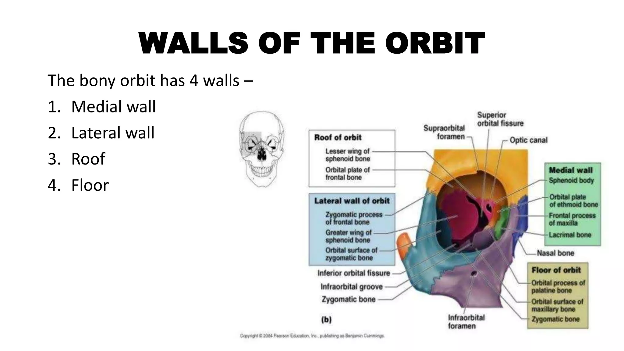
The orbit is a pyramid-shaped cavity in the skull that houses the eyeball and surrounding structures. It is formed by 7 bones and has 4 walls - medial, lateral, roof and floor. The orbit contains the eyeball, extraocular muscles, nerves, vessels, fat and fascia. At the back of the orbit are the optic canal and superior orbital fissure, which transmit nerves and vessels to and from the orbit. The base of the orbit has openings that allow communication between the orbital and eyelid spaces.
Direct Download Link ❤❤https://healthkura.com/eye-ppt/28/❤❤
Dear viewers Check Out my other piece of works at ❤❤❤ https://healthkura.com/eye-ppt/❤❤❤
anatomy of optic nerve and its blood supply and clinical corelation
Presentation Layout: optic nerve anatomy
Embryology of optic nerve
Introduction
Parts of optic nerve
Blood supply
Clinical significance
For Further Reading
Wolff’s Anatomy of the eye and orbit by Bron, Tripathi and Tripathi
Anatomy and Physiology of eye by A.K. Khurana 2nd edition
Comprehensive Ophthalmology by A.K. Khurana 5th edition
AAO- Fundamentals & Principles of Ophthalmology : sec 2
Walsh and Hoyt’s Clinical Ophthalmology
Internet
Glaucoma is not a single disease process but a group of disorders characterized by a progressive optic neuropathy resulting in a irreversible visual field defects that are associated frequently raised intraocular pressure (IOP).
IOP is the most common risk factor but not the only risk factor for development of glaucoma.
Uveal tissue is the middle vascular coat of the eyeball.
From anterior to posterior, it can be divided into 3 parts –
IRIS, CILIARY BODY CHOROID.
Iris is the anterior most part of the uveal tract.
The iris consists of four layers,
Anterior limiting layer
Iris stroma
Anterior epithelial layer
Posterior epithelial layer
The colour of iris depends on Anterior limiting layer.
The eyelids are mobile tissue curtains placed in front of the eyeballs. These act as shutters protecting the eyes from injuries and excessive light. These also perform an important function of spreading the tear film over the cornea and conjunctiva and also help in drainage of tears by lacrimal pump system.
This pdf describes the details of some pathological conditions with their treatment.
some conditions
Albinism,
Aniridia
Coloboma
Corneal dystrophies
Cataract
Dislocated lens
Diabetic retinopathy
Keratoconus
Macular hole
Glaucoma
Myopic degeneration
Nystagmus
Optic trophy
Retnial detachment
Retinopathy of prematurity
Retinitis pigmentosa
Stargardt's disease
About disease of Conjunctiva
1. inflammatory conditions of conjunctiva
2.Symptomatic conditions of conjunctiva
3. degenerative conditions of conjunctiva
4. tumors of conjunctiva
5. cyst of conjunctiva
Amsler grid, to check central visual field about with in 20 degree.
It is diagnostic tool, to use detection of macular diseases, optic nerve and visual pathway.
Management of visual problems with agingMeghna Verma
Aging bring a continuous changes in visual system.
The visual system is also affected by age related ocular pathological conditions.
In it, routine ocular examination is compulsory.
Telescope have a small device but an important place in low vision.
Telescope eyelenses magnify the apparent size of distant objects.
Binoculars placed into eyeglass frame is called telescopic spectacle or spectacle mounted telescope.
TYPES
1. handheld monocular telescope
2. clip on spectacle mounted telescope
3. bioptic telescope
Telescope are of two types of designs-
1. galilean telescope
2. keplerian telescope
These are various structures in an eye , which are changing with age.
# ocular adnexa/ eyelids
# eyelashes / eyelid margin
# tear film
# cornea
# conjunctiva
# anterior chamber
# ciliary body
# pupil /iris
# crystalline lens
# vitreous
# choroid
# retina
Low vision patient have serious visual problems that have caused serious visual loss.
1. Contrast sensitivity testing and visual field testing
2. subjective testing of patients with media loss
# potential acuity meter
# interferometry
# photostress recovery test
# glare test
# color vision test
# dark adaptometry
3. objective testing of retinal loss
# USG
ERG/EOG
Non - surgical treatment of squint i.e. all types of squint have some modalities of treatment [ optical treatment, orthoptic treatment, Prismo-therapy, and pharmacological treatment] except surgical treatment.
1. OPTICAL TREATMENT -
in optical treatment, it should be include correction of refractive error and prismotherapy.
SPECTACLES should be prescribed in every cases.
It may correct to squint partially or completely.
IN PRISMOTHERAPY, for correction of squint, This is light weight, and easy to apply on the back surface of glass.
It is useful in heterophoria, nystagmus, convergence insufficiency, managing diplopia and maintain binocular single vision.
IN PHARMACOLOGICAL TREATMENT, miotics, atropine and botulinum toxin are prescribed in some types of cases of strabismus.
IN ORTHOPTIC TREATMENT, means straight eyes.
It is used as a diagnostic purpose and therapeutic purposes.
- to increase fusion amplitude.
- anti suppression exercises.
- treatment of amblyopia.
- treatment of abnormal retinal correspondance.
- to control deviations.
ORDER OF ORHOPTIC TREATMENT -
. amblyopia is treated firstly.
. anti- suppression therapy.
- diplopia training.
- amplitude improvement.
It is not a refractive error but a physiological insufficiency of accommodation.
It leads to progressive fall in near vision.
Decrease in the accommodative power of crystalline lens due to increasing age related conditions , leads to presbyopia.
1. decrease in the elasticity and plasticity of crystalline lens.
2. decease in the power of ciliary muscles.
Some conditions to generate premature presbyopia -
1 uncorrected hypermetropia
2. premature sclerosis of crystalline lens
3. weakness of ciliary muscles.
4. chronic simple glaucoma
SOME SYMPTOMS -
fever, blurring, asthenopic symptoms, fatigue, illness, diplopia
TREATMENT -
bifocal lens, trifocal lens and progressive addition lens
convex lens should be prescribed to correct presbyopia.
Magnifiers is a type of magnifying glass or lens.
It consists of high convex lens.
It is mounted on a frame with handle or without handle.
Magnifiers mostly used in low vision patients.
Magnifiers are of 5 types available;
1. Hand magnifiers
2. Spectacle magnifier
3. Stand magnifier
4. Telescopic magnifier
5. CCTV
These types of magnifiers details in this pdf.
THANK YOU..
SOFT CONTACT LENS FITTING
1. Alternative names of soft contact lens.
2. Need to know fitting requirement and performance requirements.
3. Centration and decentration of soft contact lens. -- There are cartesian system and binasal system.
4. what governs fitting of lens.
5. There are need to know about physical properties of soft contact lens.
6. Now, what is sag and sagital depth.
7. Finally, SAME SAG AND SAME DIAMETER but DIFFERENT DESIGN AND DIFFERENT BEHAVIOUR.
8. Parameters of soft contact lens -
total diameter
back optic zone radius
centre thickness
front optic zone radius
water content
9. There are two types of prescribing methods -
empirical prescribing
trial fit prescribing
10. Effect of a blink with soft contact lens - too flat and too steep.
11. Requirements of lens movement.
12. Lens lag position - primary gaze, up gaze and lateral gaze position.
13. Compulsory of lower lid push up test.
14. Ranges of fitting of soft contact lens - either too fit or too loose or ideal fitting.
15. All step of soft contact lens fitting is done.
Astigmatism
Definition - It is a type of refractive error where in the refraction varies in the different meridian.
The rays of light entering the eye can not converge to a point focus but form a focal lne.
Types of astigmatism -
1. Regular astigmatism 2. Irregular astigmatism
Etiology of regular astigmatism -
1. corneal astigmatism 2. lenticular astigmatism 3. retinal astigmatism
Types of regular astigmatism -
1. depending upon axis and angle b/w two principal meridian-
-with the rule astigmatism
- against the rule astigmatism
- oblique astigmatism
- bi oblique astigmatism
2. depending upon their position of two focal lines-
- simple astigmatism
- compound astigmatism
- mixed astigmatism
Optics of regular astigmatism
Treatment of regular astigmatism
Irregular astigmatism
Etiological types
Symptoms of astigmatism
Treatment
263778731218 Abortion Clinic /Pills In Harare ,sisternakatoto
263778731218 Abortion Clinic /Pills In Harare ,ABORTION WOMEN’S CLINIC +27730423979 IN women clinic we believe that every woman should be able to make choices in her pregnancy. Our job is to provide compassionate care, safety,affordable and confidential services. That’s why we have won the trust from all generations of women all over the world. we use non surgical method(Abortion pills) to terminate…Dr.LISA +27730423979women Clinic is committed to providing the highest quality of obstetrical and gynecological care to women of all ages. Our dedicated staff aim to treat each patient and her health concerns with compassion and respect.Our dedicated group ABORTION WOMEN’S CLINIC +27730423979 IN women clinic we believe that every woman should be able to make choices in her pregnancy. Our job is to provide compassionate care, safety,affordable and confidential services. That’s why we have won the trust from all generations of women all over the world. we use non surgical method(Abortion pills) to terminate…Dr.LISA +27730423979women Clinic is committed to providing the highest quality of obstetrical and gynecological care to women of all ages. Our dedicated staff aim to treat each patient and her health concerns with compassion and respect.Our dedicated group of receptionists, nurses, and physicians have worked together as a teamof receptionists, nurses, and physicians have worked together as a team wwww.lisywomensclinic.co.za/
NVBDCP.pptx Nation vector borne disease control programSapna Thakur
NVBDCP was launched in 2003-2004 . Vector-Borne Disease: Disease that results from an infection transmitted to humans and other animals by blood-feeding arthropods, such as mosquitoes, ticks, and fleas. Examples of vector-borne diseases include Dengue fever, West Nile Virus, Lyme disease, and malaria.
Flu Vaccine Alert in Bangalore Karnatakaaddon Scans
As flu season approaches, health officials in Bangalore, Karnataka, are urging residents to get their flu vaccinations. The seasonal flu, while common, can lead to severe health complications, particularly for vulnerable populations such as young children, the elderly, and those with underlying health conditions.
Dr. Vidisha Kumari, a leading epidemiologist in Bangalore, emphasizes the importance of getting vaccinated. "The flu vaccine is our best defense against the influenza virus. It not only protects individuals but also helps prevent the spread of the virus in our communities," he says.
This year, the flu season is expected to coincide with a potential increase in other respiratory illnesses. The Karnataka Health Department has launched an awareness campaign highlighting the significance of flu vaccinations. They have set up multiple vaccination centers across Bangalore, making it convenient for residents to receive their shots.
To encourage widespread vaccination, the government is also collaborating with local schools, workplaces, and community centers to facilitate vaccination drives. Special attention is being given to ensuring that the vaccine is accessible to all, including marginalized communities who may have limited access to healthcare.
Residents are reminded that the flu vaccine is safe and effective. Common side effects are mild and may include soreness at the injection site, mild fever, or muscle aches. These side effects are generally short-lived and far less severe than the flu itself.
Healthcare providers are also stressing the importance of continuing COVID-19 precautions. Wearing masks, practicing good hand hygiene, and maintaining social distancing are still crucial, especially in crowded places.
Protect yourself and your loved ones by getting vaccinated. Together, we can help keep Bangalore healthy and safe this flu season. For more information on vaccination centers and schedules, residents can visit the Karnataka Health Department’s official website or follow their social media pages.
Stay informed, stay safe, and get your flu shot today!
Adv. biopharm. APPLICATION OF PHARMACOKINETICS : TARGETED DRUG DELIVERY SYSTEMSAkankshaAshtankar
MIP 201T & MPH 202T
ADVANCED BIOPHARMACEUTICS & PHARMACOKINETICS : UNIT 5
APPLICATION OF PHARMACOKINETICS : TARGETED DRUG DELIVERY SYSTEMS By - AKANKSHA ASHTANKAR
New Drug Discovery and Development .....NEHA GUPTA
The "New Drug Discovery and Development" process involves the identification, design, testing, and manufacturing of novel pharmaceutical compounds with the aim of introducing new and improved treatments for various medical conditions. This comprehensive endeavor encompasses various stages, including target identification, preclinical studies, clinical trials, regulatory approval, and post-market surveillance. It involves multidisciplinary collaboration among scientists, researchers, clinicians, regulatory experts, and pharmaceutical companies to bring innovative therapies to market and address unmet medical needs.
Ozempic: Preoperative Management of Patients on GLP-1 Receptor Agonists Saeid Safari
Preoperative Management of Patients on GLP-1 Receptor Agonists like Ozempic and Semiglutide
ASA GUIDELINE
NYSORA Guideline
2 Case Reports of Gastric Ultrasound
Basavarajeeyam is a Sreshta Sangraha grantha (Compiled book ), written by Neelkanta kotturu Basavaraja Virachita. It contains 25 Prakaranas, First 24 Chapters related to Rogas& 25th to Rasadravyas.
Basavarajeeyam is an important text for ayurvedic physician belonging to andhra pradehs. It is a popular compendium in various parts of our country as well as in andhra pradesh. The content of the text was presented in sanskrit and telugu language (Bilingual). One of the most famous book in ayurvedic pharmaceutics and therapeutics. This book contains 25 chapters called as prakaranas. Many rasaoushadis were explained, pioneer of dhatu druti, nadi pareeksha, mutra pareeksha etc. Belongs to the period of 15-16 century. New diseases like upadamsha, phiranga rogas are explained.
How STIs Influence the Development of Pelvic Inflammatory Disease.pptx
THE ORBIT.pptx
1. THE ORBIT
BY - MS. MEGHNA VERMA
ASSISTANT PROFESSOR
DEPARTMENT OF OPTOMETRY
RAMA UNIVERSITY
2. CONTENTS
• Introduction [size, shape, relations]
• Walls of orbit
• Base or orbit
• Apex of orbit
• Periorbita
• Orbital fascia
• Fascia bulbi
• Orbital fat and reticular tissue
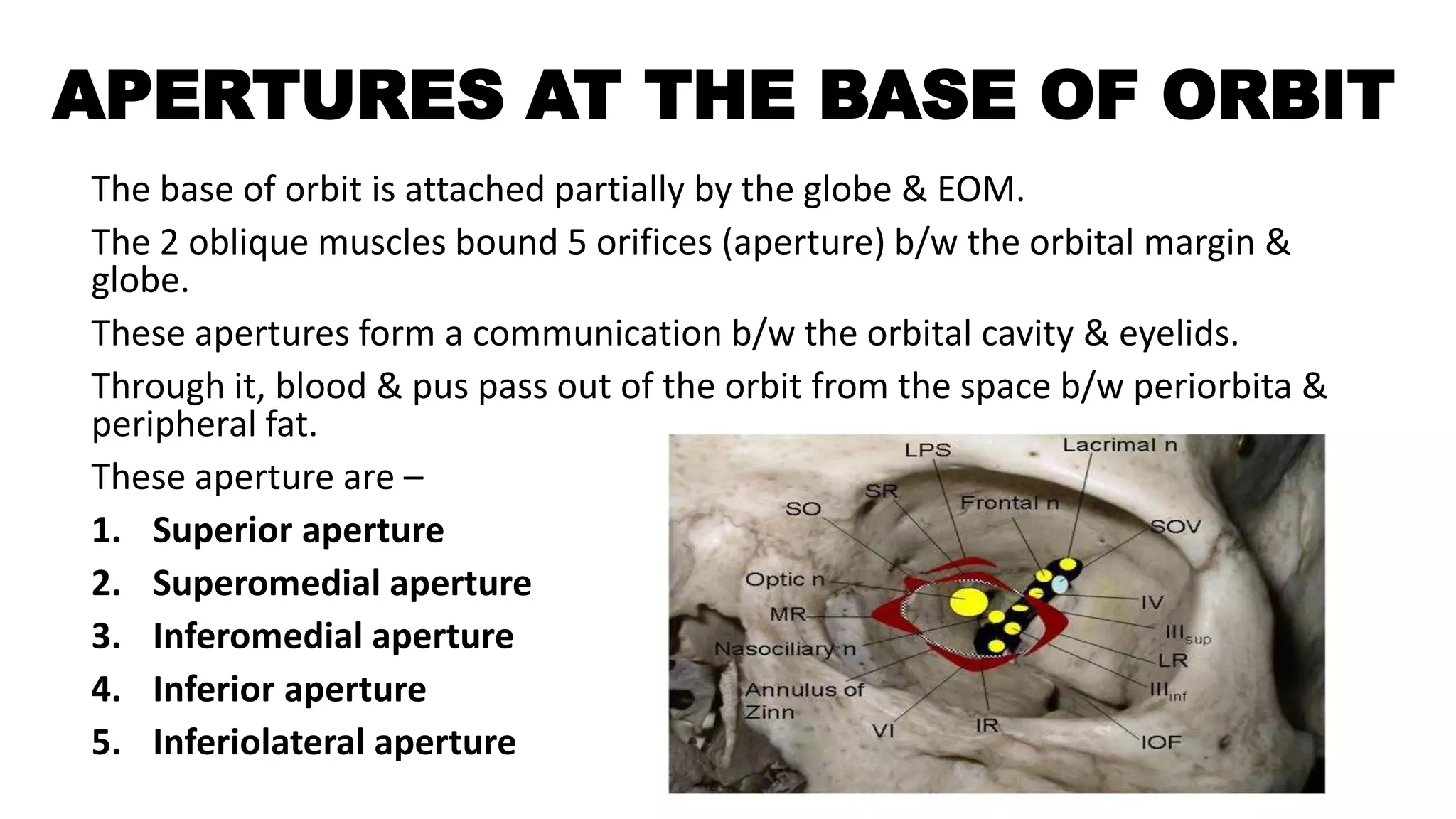
• Aperture at the base of the orbit
• Surgical space in the orbit
• Contents of the orbit
3. INTRODUCTION
The orbits are conical or four-sided pyramidal cavities, which open into
the midline of the face and point back into the head. Each consists of a
base, an apex and four walls.[4]
Each orbit is formed by seven bones –
1) Frontal bone
2) Ethmoidal bone
3) Lacrimal bone
4) Palatine bone
5) Maxilla bone
6) Zygomatic bone
7) Sphenoid bone
4.
5. • The medial walls of two orbit are parallel to each other.
• They are in contact with the sphenoid bone & ethmoid bone.
• The depth of the orbits is 42mm along to medial wall & 50mm
along the lateral wall.
• Base of orbit is 40 mm in width & 35 mm in height.
• The intra orbital width [b/w the medial margin of two orbit] is
25mm.
• The extra orbital width [b/w the lateral margin of two orbit] is
100mm.
6. WALLS OF THE ORBIT
The bony orbit has 4 walls –
1. Medial wall
2. Lateral wall
3. Roof
4. Floor
7. MEDIAL WALL
The medial wall is formed primarily by the orbital plate of ethmoid, as well as
contributions from the frontal process of maxilla, the lacrimal bone, and a small part of the
body of the sphenoid. It is the thinnest wall of the orbit, evidenced by pneumatized
ethmoidal cells.[7]
The anterior part of medial wall – lacrimal sac fossa
Lacrimal fossa attached, inferiorly - naso-lacrimal canal.
anteriorly - anterior lacrimal crest
posteriorly – posterior lacrimal crest.
Behind the posterior lacrimal crest – fibres of orbicularis muscle, orbital septum
and medial rectus muscle ligaments.
Medial part of medial wall – anterior ethmoidal sinus, middle ethmoidal sinus and
posterior ethmoidal sinus and sphenoid sinus.
In it, SO & MR muscle, b/w two muscles situated anterior and posterior ethmoidal
nerve, ophthalmic artery and infratrochlear nerve.
8. LATERAL WALL
It is triangular in shape.
It is bounded anteriorly by zygomatic bone and posteriorly by sphenoid bone.
Anterior part of lateral wall – zygomatic groove.
Posterior part of lateral wall - lateral rectus muscle.
Lateral part of lateral wall - lateral rectus muscle lacrimal nerve and vessels and
zygomatic nerves.
9. SUPERIOR WALL [ROOF]
It is triangular in shape.
The roof (superior wall) is formed primarily by the orbital plate frontal
bone, and also the lesser wing of sphenoid near the apex of the orbit.
The orbital surface presents medially by trochlear fovea and laterally by
lacrimal fossa.[7]
Above the roof – cerebrum and meninges.
Below the roof – periorbita, frontal nerves, LPS muscle, superior rectus,
lacrimal gland, trochlear nerve.
Antero-lateral part of the roof has a fossa, called lacrimal gland fossa.
b/w medial wall and roof has a superior oblique fossa, ethmoidal sinus.
b/w lateral wall and roof has a superior orbital fissure [space].
10. INFERIOR WALL [FLOOR]
It is triangular in shape.
It is formed by 3 bones – medially maxilla bone, laterally zygomatic bone
and posteriorly palatine bone.
Above the floor – inferior rectus muscle, inferior oblique muscle.
Below the floor – maxillary sinus and palatine sinus.
Posterior part – infra orbital foramen [hole] – transmits, infra orbital
artery, infra orbital vein [connects to facial vein].
11. BASE OF THE ORBIT
•Orbital base is the anterior end of the orbit.
•It is bounded by orbital margins.
•The margins are formed by a ring of compact bone.
•It gives attachment to the septum orbit.
It is of four parts –
1. Superior orbital margin
2. Inferior orbital margin
3. Medial orbital margin
4. Lateral orbital margin
12. SUPERIOR ORBITAL MARGIN-
•It is formed by the frontal bone.
•Its lateral 2/3rd part is very sharp & medial 1/3rd is rounded.
•Above the frontal bone - supra orbital notch, bears supra orbital nerve &
artery.
•About 10mm medial to this notch - supra trochlear groove, transmits supra
trochlear nerve & artery.
INFERIOR ORBITAL MARGIN –
•It is formed by zygomatic bone laterally & maxilla medially.
•Medially, it becomes continuous with the anterior lacrimal crest.
•The infra orbital foramen transmitting their nerves & vessels, situated 4-5
mm below the orbital margin.
13. LATERAL ORBITAL MARGIN –
It is the more strong & formed by frontal bone & zygomatic bone.
The anterior half of the globe is not protected by these bone.
MEDIAL ORBITAL MARGIN –
It is formed by the anterior lacrimal crest of the maxilla & above by the
frontal bone.
Its upper part becomes continuous with the posterior lacrimal crest.
14. APEX OF THE ORBIT
Orbital apex is the posterior end of the orbit.
The apex has two orifices (opening)– optical canal & superior orbital
fissure [in sphenoid bone].
OPTICAL CANAL –
• It connects the orbit to the middle cranial fossa.
• It transmits the optic nerve & ophthalmic artery.
• Its normal dimensions, attained by the age of 4-5 years.
• Its average length is 6-11 mm.
• Tumours may lead to unilateral enlargement of optic canal, detected by X
ray.
15.
16. SUPERIOR ORBITAL FISSURE –
• It is a comma shaped aperture in the orbital cavity, and attached by
sphenoid bone.
• It is situated lateral to the optic foramina at the orbital apex.
• The fissure(opening) is divided into upper, middle & lower parts by
common tendinous ring.
• The structures passing through the upper & lateral parts are the
lacrimal & frontal nerves, trochlear nerve, superior ophthalmic vein &
artery.
• The lower & medial parts of fissure - the inferior ophthalmic veins.
• The middle part of fissure transmits the oculomotor nerve, trigeminal
nerve & abducent nerve.
17. PERIORBITA
• The periosteum lining the surface of the orbital bones is called the
periorbita.
• It is loosely adhere with bone.
• It is firmly attached at the orbital margin, superior & inferior orbital
fissures, optic canal, lacrimal fossa & sutures.
• In optic canal, the optic nerve is closely attached to periorbita.
• At the orbital margin, periorbita is thicken & the orbital septum is attached.
• At the posterior lacrimal crest, the periorbita divides into two layers which
reunite at the anterior lacrimal crest.
• These two layers enclose the lacrimal sac.
• At the apex of orbit, the periorbita is thicken to form the common tendinous
ring.
18.
19. ORBITAL FASCIA
•This part has head of fascia bulbi, muscular sheaths, inter-muscular
septum and extra ocular muscles ligaments.
• It is a thin connective tissue membrane lining the various intra orbital
structure.
•In the orbit, the surrounding fascia allows for smooth rotation and
protects the orbital contents. If excessive tissue accumulates behind
the ocular globe, the eye can protrude, or become exophthalmic.[4]
•Graves disease may also cause axial protrusion of the eye, known
as Graves' ophthalmopathy, due to build up of extracellular
matrix proteins and fibrosis in the rectus muscles. Development of
Graves' ophthalmopathy may be independent of thyroid function.[10]
20.
21. FASCIA BULBI/TENON’S CAPSULE
•It covers the globe from the limbus to the optic disc.
•It is situated in close contact with sclera.
•The outer surface – posteriorly - orbital fat, anteriorly – subconjunctival
tissue (limbus).
•Fascia bulbi/ tenon’s capsule is pierced – posteriorly – optic nerve, ciliary
nerves and vessels and anteriorly by the - six extra ocular muscles.
22. SURGICAL SPACE IN THE ORBIT
These are most important as much orbital tumors tend to remain within the
space in which they are performed surgery by surgeon.
It has 4 spaces in the orbit –
1. Subperiosteal space
2. Peripheral orbital space (ant. space)
3. Central space
4. Sub-tenon’s space
23. ORBITAL FAT & RETICULAR TISSUE
•The orbital cavity is occupied by orbital fat, which extends from the optic
nerve to the orbital wall.
•It forms the apex of the orbit to the orbital septum.
•The fat lobules is situated in the interstices of a web of reticular tissue
called orbital reticulum.
•This tissue is the supporting framework of the orbital fat.
•Orbital fat is divided into central & peripheral parts by the intermuscular
septa.
•Posteriorly, where there is no intermuscular septum, the peripheral &
central fat parts are continuous with each other.
•The peripheral orbital fat consists of 4 lobules – superomedial,
superolateral, inferomedial & inferolateral.
24. APERTURES AT THE BASE OF ORBIT
The base of orbit is attached partially by the globe & EOM.
The 2 oblique muscles bound 5 orifices (aperture) b/w the orbital margin &
globe.
These apertures form a communication b/w the orbital cavity & eyelids.
Through it, blood & pus pass out of the orbit from the space b/w periorbita &
peripheral fat.
These aperture are –
1. Superior aperture
2. Superomedial aperture
3. Inferomedial aperture
4. Inferior aperture
5. Inferiolateral aperture
25. SUPERIOR APERTURE –
•This is a comma shaped orifice & situated b/w roof of the orbit &
upper surface of the LPS.
•The head of the comma is situated near the trochlea & the tail reaches
the lacrimal gland.
SUPEROMEDIAL APERTURE –
•It is vertically oval aperture is situated b/w the superior oblique
muscle & the medial ligament.
•The infra trochlear nerve, dorsal nasal artery & angular vein pass
through this aperture.
26. INFERO MEDIAL APERTURE –
•This is vertically oval in shape & is situated b/w the medial check
ligament, origin of inferior oblique & lacrimal sac.
INFERIOR APERTURE –
•This is triangular in shape & is bounded by the inferior oblique muscle,
arcuate expansion of inferior oblique & floor of the orbit.
INFERO LATERAL APERTURE –
•This is small oval aperture situated b/w the arcuate expansion of the
inferior oblique muscle and lateral check ligament.
27. CONTENTS OF THE ORBIT
• Eyeball occupies about 1/5th of the total orbital volume.
• Muscles includes - SR, IR, MR, LR, SO, IO, LPS & muller’s muscles.
• Nerves includes - optic nerve, oculomotor nerve, trochlear nerve,
abducent nerve, branches of ophthalmic nerve, branches of maxillary
division.
• Vessels includes - ophthalmic artery & its branches, infra orbital
vessels, meningeal artery, superior & inferior ophthalmic veins.
• Orbital fat, reticular tissue & orbital fascia.
• Lacrimal gland & lacrimal sac.
28.
29. REFRENCES
• eye, human."Encyclopædia Britannica from Encyclopædia Britannica
2006 Ultimate Reference Suite DVD 2009.
• Moore, Keith L. (2010). Clinically Oriented Anatomy (6th ed.).
Lippincott Williams & Wilkins. ISBN 978-07817-7525-0.
• Hatton, M. P.; Rubin, P. A. (2002). "The pathophysiology of thyroid-
associated ophthalmopathy". Ophthalmol Clin North Am. 15 (1): 113–
119. doi:10.1016/S0896-1549(01)00004-9. PMID 12064074