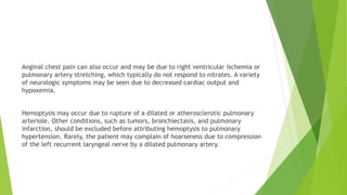
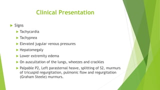
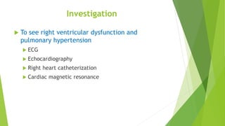
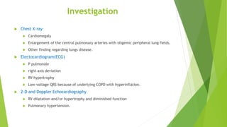
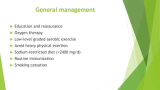
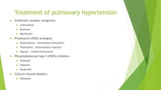
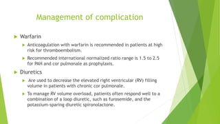
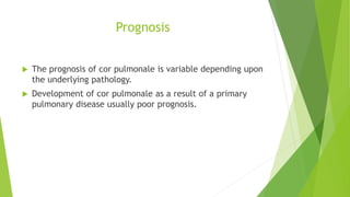
Cor Pulmonale
Definition
 Cor pulmonale is defined as an alteration in the
structure and function of the right ventricle (RV)
of the heart caused by a primary disorder of the
respiratory system.
 Right-sided ventricular disease caused by a
primary abnormality of the left side of the heart
or congenital heart disease is not considered cor
pulmonale.
Epidemiology
 Cor pulmonale is estimated to account for 6-7% of all
types of adult heart disease in the United States.
 chronic obstructive pulmonary disease (COPD) is the
causative factor in more than 50% of cases.
 50,000 deaths in the United States are estimated to occur
per year from pulmonary emboli and about half occur
within the first hour due to acute right heart failure.
 Globally, the incidence of cor pulmonale varies widely
among countries, depending on the prevalence of
cigarette smoking, air pollution, and other risk factors for
various lung diseases.
Classification
1. Acute Cor Pulmonale
2. Chronic Cor pulmonale
Pathophysiology
 Common pathophysiologic mechanism is pulmonary
hypertension that is sufficient to alter RV structure (i.e.,
dilation with or without hypertrophy) and function.
 parenchymal lung diseases, primary pulmonary vascular
disorders, or chronic (alveolar) hypoxia, the circulatory
bed undergoes varying degrees of vascular remodeling,
vasoconstriction, and destruction. As a result, pulmonary
artery pressures and RV afterload increase.
Pathophysiology
 The response of the RV to pulmonary hypertension
depends on the acuteness and severity of the pressure
overload.
 Acute cor pulmonale occurs after a sudden and severe
stimulus (e.g.massive pulmonary embolus), with RV
dilatation and failure but no RV hypertrophy.
 Chronic cor pulmonale, however, is associated with a
more slowly evolving and progressive pulmonary
hypertension that leads to initial modest RV hypertrophy
and subsequent RV dilation.
Pathophysiology
 systemic consequences of cor pulmonale relate to
alterations in cardiac output as well as salt and water
homeostasis.
 Triggers include worsening hypoxia from any cause (e.g.,
pneumonia), acidemia (e.g., exacerbation of COPD),
acute pulmonary embolus, atrial tachyarrhythmia,
hypervolemia and mechanical ventilation that leads to
compressive forces on alveolar blood vessels.
 The World Health Organization (WHO) has five classifications for pulmonary
hypertension, and all except one of these groups can result in cor pulmonale
(WHO Classification group 2 is pulmonary artery hypertension due to left
ventricular [LV] dysfunction).
 Group 1: Pulmonary artery hypertension, including heritable causes;
connective-tissue disorders, including scleroderma; and other idiopathic
causes.
 Group 3: Pulmonary hypertension due to lung disease and/or hypoxia; these
disorders include chronic obstructive pulmonary disease (COPD), which is the
most common cause of for pulmonale. There have been studies correlating
the degree of hypoxia with the severity of cor pulmonale. Other disorders
that can result in cor pulmonale in this group include interstitial lung disease
(ILD) and obstructive sleep apnea (OSA)
 Group 4: Chronic thromboembolic pulmonary hypertension; blood clots that
form in the lungs can lead to increased resistance, pulmonary hypertension
and, subsequently, cor pulmonale

 Group 5: Pulmonary hypertension caused by other diseases or conditions,
including sarcoidosis, polycythemia vera (which can lead to increased blood
viscosity and, subsequently, pulmonary hypertension), vasculitis, and other
disorders.
 The end result of the above mechanisms is increased pulmonary arterial
pressure and resistance.
 RV and LV output.
 The RV is a thin-walled chamber that is a better volume pump than a pressure
pump. It is better suited to adapt to changing preload than afterload. With an
increase in afterload, the RV systolic pressure is increased to maintain the
circulatory gradient. At a critical point, a further increase in pulmonary
arterial pressure and resistance produces significant RV dilatation, an
increase in RV end-diastolic pressure, and RV circulatory failure.
 A decrease in RV output leads to a decrease in LV filling, which results in
decreased cardiac output. Because the right coronary artery originates from
the aorta, decreased LV output causes decreased right coronary blood flow
and ischemia to the RV wall. What ensues is a vicious cycle between
decreases in LV and RV output.
 RV overload
 RV pressure and volume overload is associated with septal displacement
toward the left ventricle. Septal displacement, which can be visualized on
echocardiography, is an additional factor that decreases LV filling and output
in the setting of cor pulmonale and RV enlargement.
Etiology of Acute Cor Pulmonale
 massive pulmonary embolism
 acute respiratory distress syndrome (ARDS)
Etiology of Chronic Cor Pulmonale
 Diseases of the Lung Parenchyma
 Chronic obstructive pulmonary disease
 Emphysema
 Chronic bronchitis
 Cystic fibrosis
 Idiopathic interstitial pneumonitis
 Idiopathic pulmonary fibrosis
 Nonspecific interstitial pneumonitis
 Sarcoidosis
 Bronchiectasis
 Pulmonary Langerhans cell histiocytosis
 Lymphangioleiomyomatosis
Etiology of Chronic Cor Pulmonale
 Diseases of the Pulmonary Vasculature
 Pulmonary arterial hypertension (PAH)
Idiopathic PAH
Heritable PAH
Associated PAH
 Venoocclusive disease
 Chronic thromboembolic pulmonary hypertension
 Pulmonary tumor thrombotic microangiopathy
Etiology of Chronic Cor Pulmonale
 Disorders of Chronic (Alveolar) Hypoxia
 Alveolar hypoventilation syndromes
 Obesity hypoventilation syndrome
 Central hypoventilation syndrome
 Neuromuscular respiratory failure
 Chest wall disorders
Kyphoscoliosis
 Living at high altitude
Clinical Presentation
 Symptoms
 Dyspnea
 Cough
 Lower-extremity edema
 Abdominal pain and ascites
 Rarely Orthopnea and PND
 nonspecific symptoms
 Fatigue,
 palpitations,
 atypical chest pain,
 dizziness,
 syncope
Anginal chest pain can also occur and may be due to right ventricular ischemia or
pulmonary artery stretching, which typically do not respond to nitrates. A variety
of neurologic symptoms may be seen due to decreased cardiac output and
hypoxemia.
Hemoptysis may occur due to rupture of a dilated or atherosclerotic pulmonary
arteriole. Other conditions, such as tumors, bronchiectasis, and pulmonary
infarction, should be excluded before attributing hemoptysis to pulmonary
hypertension. Rarely, the patient may complain of hoarseness due to compression
of the left recurrent laryngeal nerve by a dilated pulmonary artery.
Clinical Presentation
 Signs
 Tachycardia
 Tachypnea
 Elevated jugular venous pressures
 Hepatomegaly
 Lower extremity edema
 On auscultation of the lungs, wheezes and crackles
 Palpable P2, Left parasternal heave, splitting of S2, murmurs
of tricuspid regurgitation, pulmonic flow and regurgitation
(Graham Steele) murmurs.
Investigation
 To see right ventricular dysfunction and
pulmonary hypertension
 ECG
 Echocardiography
 Right heart catheterization
 Cardiac magnetic resonance
Investigation
 To identify the primary cause
 Complete blood count – Polycythemia, increase
haematocrit.
 Chest X-ray
 Pulmonary function test
 Alpha 1- antitrypsin
 ANA
 anticentromere antibodies
 Protein S and C, antithrombin III,
 Arterial blood gas analysis
Investigation
 Chest X-ray
 Cardiomegaly
 Enlargement of the central pulmonary arteries with oligemic peripheral lung fields.
 Other finding regarding lungs disease.
 Electocardiogram(ECG)
 P pulmonale
 right axis deviation
 RV hypertrophy
 Low-voltage QRS because of underlying COPD with hyperinflation.
 2-D and Doppler Echocardiography
 RV dilatation and/or hypertrophy and diminished function
 Pulmonary hypertension.
Management
 General management
 Specific management
 Management of complication
 Management of acute exacerbation
General management
 Education and reassurance
 Oxygen therapy
 Low-level graded aerobic exercise
 Avoid heavy physical exertion
 Sodium-restricted diet (<2400 mg/d)
 Routine immunization
 Smoking cessation
Specific management
 Treatment of underlying cause.
 Treatment of pulmonary hypertension.
Treatment of pulmonary hypertension
 Endothelin receptor antagonists
 Ambrisentan
 Bosentan
 Macitentan
 Prostacyclin (PGI2) analogues
 Epoprostenol - intravenous formulation
 Treprostinil - subcutaneous injection
 Iloprost – inhaled formulation
 Phosphodiesterase type 5 (PDE5) inhibitors
 Sildenafil
 Tadalafil
 Vardenafil
 Calcium channel blockers
 Diltiazem
Management of complication
 Warfarin
 Anticoagulation with warfarin is recommended in patients at high
risk for thromboembolism.
 Recommended international normalized ratio range is 1.5 to 2.5
for PAH and cor pulmonale as prophylaxis.
 Diuretics
 Are used to decrease the elevated right ventricular (RV) filling
volume in patients with chronic cor pulmonale.
 To manage RV volume overload, patients often respond well to a
combination of a loop diuretic, such as furosemide, and the
potassium-sparing diuretic spironolactone.
Surgical management
 Phlebotomy is indicated in patients with chronic cor pulmonale and chronic
hypoxia causing severe polycythemia
 Single-lung, double-lung, and heart-lung transplantation
Management of acute exacerbation
 Hospitalization
 Oxygen therapy
 Control of heart failure
 Control of cardiac arrhythmia
 Control of infection
 Treatment of lung disease accordingly
Prognosis
 The prognosis of cor pulmonale is variable depending upon
the underlying pathology.
 Development of cor pulmonale as a result of a primary
pulmonary disease usually poor prognosis.
 1. **What is cor pulmonale?**
 - a) A heart condition caused by systemic hypertension
 - b) Right ventricular enlargement due to lung pathology
 - c) Left ventricular failure
 - d) Coronary artery disease
 Which of the following is the most common cause of cor pulmonale?**
 - a) Pulmonary embolism
 - b) Chronic obstructive pulmonary disease (COPD)
 - c) Congenital heart defects
 - d) Systemic hypertension
 3. **What is the primary physiological mechanism behind cor pulmonale?**
 - a) Increased left atrial pressure
 - b) Increased right ventricular afterload
 - c) Decreased systemic vascular resistance
 - d) Decreased pulmonary artery pressure

 4. **Which symptom is commonly associated with cor pulmonale?**
 - a) Cyanosis
 - b) Bradycardia
 - c) Hyperlipidemia
 - d) Polyuria
 5. **Which diagnostic tool is most useful in confirming the presence of cor pulmonale?**
 - a) Electrocardiogram (ECG)
 - b) Chest X-ray
 - c) Echocardiogram
 - d) Blood pressure measurement

 6. **In cor pulmonale, what change is typically seen on an ECG?**
 - a) Left axis deviation
 - b) T-wave inversion in the lateral leads
 - c) Right axis deviation
 - d) ST-segment elevation
 7. **What physical sign might be evident on examination of a patient with cor pulmonale?**
 - a) Jugular venous distension
 - b) Elevated blood pressure
 - c) Bradycardia
 - d) Exophthalmos

 8. **Which treatment is most appropriate for managing cor pulmonale secondary to COPD?**
 - a) Beta-blockers
 - b) Supplemental oxygen
 - c) ACE inhibitors
 - d) Statins
 9. **What is a common complication of untreated cor pulmonale?**
 - a) Left ventricular hypertrophy
 - b) Right heart failure
 - c) Atrial fibrillation
 - d) Pulmonary embolism

 10. **Which of the following is a potential long-term outcome for patients with cor
pulmonale?**
 - a) Complete recovery with no residual effects
 - b) Progressive heart failure
 - c) Myocardial infarction
 - d) Systemic hypertension
 Answers

 1. b) Right ventricular enlargement due to lung pathology
 2. b) Chronic obstructive pulmonary disease (COPD)
 3. b) Increased right ventricular afterload
 4. a) Cyanosis
 5. c) Echocardiogram
 6. c) Right axis deviation
 7. a) Jugular venous distension
 8. b) Supplemental oxygen
 9. b) Right heart failure
 10. b) Progressive heart failure
Seminar presentation on cor pulmonale ..pptx

Seminar presentation on cor pulmonale ..pptx

  • 1.
  • 2.
    Definition  Cor pulmonaleis defined as an alteration in the structure and function of the right ventricle (RV) of the heart caused by a primary disorder of the respiratory system.  Right-sided ventricular disease caused by a primary abnormality of the left side of the heart or congenital heart disease is not considered cor pulmonale.
  • 3.
    Epidemiology  Cor pulmonaleis estimated to account for 6-7% of all types of adult heart disease in the United States.  chronic obstructive pulmonary disease (COPD) is the causative factor in more than 50% of cases.  50,000 deaths in the United States are estimated to occur per year from pulmonary emboli and about half occur within the first hour due to acute right heart failure.  Globally, the incidence of cor pulmonale varies widely among countries, depending on the prevalence of cigarette smoking, air pollution, and other risk factors for various lung diseases.
  • 4.
    Classification 1. Acute CorPulmonale 2. Chronic Cor pulmonale
  • 5.
    Pathophysiology  Common pathophysiologicmechanism is pulmonary hypertension that is sufficient to alter RV structure (i.e., dilation with or without hypertrophy) and function.  parenchymal lung diseases, primary pulmonary vascular disorders, or chronic (alveolar) hypoxia, the circulatory bed undergoes varying degrees of vascular remodeling, vasoconstriction, and destruction. As a result, pulmonary artery pressures and RV afterload increase.
  • 6.
    Pathophysiology  The responseof the RV to pulmonary hypertension depends on the acuteness and severity of the pressure overload.  Acute cor pulmonale occurs after a sudden and severe stimulus (e.g.massive pulmonary embolus), with RV dilatation and failure but no RV hypertrophy.  Chronic cor pulmonale, however, is associated with a more slowly evolving and progressive pulmonary hypertension that leads to initial modest RV hypertrophy and subsequent RV dilation.
  • 7.
    Pathophysiology  systemic consequencesof cor pulmonale relate to alterations in cardiac output as well as salt and water homeostasis.  Triggers include worsening hypoxia from any cause (e.g., pneumonia), acidemia (e.g., exacerbation of COPD), acute pulmonary embolus, atrial tachyarrhythmia, hypervolemia and mechanical ventilation that leads to compressive forces on alveolar blood vessels.
  • 8.
     The WorldHealth Organization (WHO) has five classifications for pulmonary hypertension, and all except one of these groups can result in cor pulmonale (WHO Classification group 2 is pulmonary artery hypertension due to left ventricular [LV] dysfunction).  Group 1: Pulmonary artery hypertension, including heritable causes; connective-tissue disorders, including scleroderma; and other idiopathic causes.  Group 3: Pulmonary hypertension due to lung disease and/or hypoxia; these disorders include chronic obstructive pulmonary disease (COPD), which is the most common cause of for pulmonale. There have been studies correlating the degree of hypoxia with the severity of cor pulmonale. Other disorders that can result in cor pulmonale in this group include interstitial lung disease (ILD) and obstructive sleep apnea (OSA)
  • 9.
     Group 4:Chronic thromboembolic pulmonary hypertension; blood clots that form in the lungs can lead to increased resistance, pulmonary hypertension and, subsequently, cor pulmonale   Group 5: Pulmonary hypertension caused by other diseases or conditions, including sarcoidosis, polycythemia vera (which can lead to increased blood viscosity and, subsequently, pulmonary hypertension), vasculitis, and other disorders.
  • 10.
     The endresult of the above mechanisms is increased pulmonary arterial pressure and resistance.  RV and LV output.  The RV is a thin-walled chamber that is a better volume pump than a pressure pump. It is better suited to adapt to changing preload than afterload. With an increase in afterload, the RV systolic pressure is increased to maintain the circulatory gradient. At a critical point, a further increase in pulmonary arterial pressure and resistance produces significant RV dilatation, an increase in RV end-diastolic pressure, and RV circulatory failure.
  • 11.
     A decreasein RV output leads to a decrease in LV filling, which results in decreased cardiac output. Because the right coronary artery originates from the aorta, decreased LV output causes decreased right coronary blood flow and ischemia to the RV wall. What ensues is a vicious cycle between decreases in LV and RV output.  RV overload  RV pressure and volume overload is associated with septal displacement toward the left ventricle. Septal displacement, which can be visualized on echocardiography, is an additional factor that decreases LV filling and output in the setting of cor pulmonale and RV enlargement.
  • 12.
    Etiology of AcuteCor Pulmonale  massive pulmonary embolism  acute respiratory distress syndrome (ARDS)
  • 13.
    Etiology of ChronicCor Pulmonale  Diseases of the Lung Parenchyma  Chronic obstructive pulmonary disease  Emphysema  Chronic bronchitis  Cystic fibrosis  Idiopathic interstitial pneumonitis  Idiopathic pulmonary fibrosis  Nonspecific interstitial pneumonitis  Sarcoidosis  Bronchiectasis  Pulmonary Langerhans cell histiocytosis  Lymphangioleiomyomatosis
  • 14.
    Etiology of ChronicCor Pulmonale  Diseases of the Pulmonary Vasculature  Pulmonary arterial hypertension (PAH) Idiopathic PAH Heritable PAH Associated PAH  Venoocclusive disease  Chronic thromboembolic pulmonary hypertension  Pulmonary tumor thrombotic microangiopathy
  • 15.
    Etiology of ChronicCor Pulmonale  Disorders of Chronic (Alveolar) Hypoxia  Alveolar hypoventilation syndromes  Obesity hypoventilation syndrome  Central hypoventilation syndrome  Neuromuscular respiratory failure  Chest wall disorders Kyphoscoliosis  Living at high altitude
  • 16.
    Clinical Presentation  Symptoms Dyspnea  Cough  Lower-extremity edema  Abdominal pain and ascites  Rarely Orthopnea and PND  nonspecific symptoms  Fatigue,  palpitations,  atypical chest pain,  dizziness,  syncope
  • 17.
    Anginal chest paincan also occur and may be due to right ventricular ischemia or pulmonary artery stretching, which typically do not respond to nitrates. A variety of neurologic symptoms may be seen due to decreased cardiac output and hypoxemia. Hemoptysis may occur due to rupture of a dilated or atherosclerotic pulmonary arteriole. Other conditions, such as tumors, bronchiectasis, and pulmonary infarction, should be excluded before attributing hemoptysis to pulmonary hypertension. Rarely, the patient may complain of hoarseness due to compression of the left recurrent laryngeal nerve by a dilated pulmonary artery.
  • 18.
    Clinical Presentation  Signs Tachycardia  Tachypnea  Elevated jugular venous pressures  Hepatomegaly  Lower extremity edema  On auscultation of the lungs, wheezes and crackles  Palpable P2, Left parasternal heave, splitting of S2, murmurs of tricuspid regurgitation, pulmonic flow and regurgitation (Graham Steele) murmurs.
  • 19.
    Investigation  To seeright ventricular dysfunction and pulmonary hypertension  ECG  Echocardiography  Right heart catheterization  Cardiac magnetic resonance
  • 20.
    Investigation  To identifythe primary cause  Complete blood count – Polycythemia, increase haematocrit.  Chest X-ray  Pulmonary function test  Alpha 1- antitrypsin  ANA  anticentromere antibodies  Protein S and C, antithrombin III,  Arterial blood gas analysis
  • 21.
    Investigation  Chest X-ray Cardiomegaly  Enlargement of the central pulmonary arteries with oligemic peripheral lung fields.  Other finding regarding lungs disease.  Electocardiogram(ECG)  P pulmonale  right axis deviation  RV hypertrophy  Low-voltage QRS because of underlying COPD with hyperinflation.  2-D and Doppler Echocardiography  RV dilatation and/or hypertrophy and diminished function  Pulmonary hypertension.
  • 22.
    Management  General management Specific management  Management of complication  Management of acute exacerbation
  • 23.
    General management  Educationand reassurance  Oxygen therapy  Low-level graded aerobic exercise  Avoid heavy physical exertion  Sodium-restricted diet (<2400 mg/d)  Routine immunization  Smoking cessation
  • 24.
    Specific management  Treatmentof underlying cause.  Treatment of pulmonary hypertension.
  • 25.
    Treatment of pulmonaryhypertension  Endothelin receptor antagonists  Ambrisentan  Bosentan  Macitentan  Prostacyclin (PGI2) analogues  Epoprostenol - intravenous formulation  Treprostinil - subcutaneous injection  Iloprost – inhaled formulation  Phosphodiesterase type 5 (PDE5) inhibitors  Sildenafil  Tadalafil  Vardenafil  Calcium channel blockers  Diltiazem
  • 26.
    Management of complication Warfarin  Anticoagulation with warfarin is recommended in patients at high risk for thromboembolism.  Recommended international normalized ratio range is 1.5 to 2.5 for PAH and cor pulmonale as prophylaxis.  Diuretics  Are used to decrease the elevated right ventricular (RV) filling volume in patients with chronic cor pulmonale.  To manage RV volume overload, patients often respond well to a combination of a loop diuretic, such as furosemide, and the potassium-sparing diuretic spironolactone.
  • 27.
    Surgical management  Phlebotomyis indicated in patients with chronic cor pulmonale and chronic hypoxia causing severe polycythemia  Single-lung, double-lung, and heart-lung transplantation
  • 28.
    Management of acuteexacerbation  Hospitalization  Oxygen therapy  Control of heart failure  Control of cardiac arrhythmia  Control of infection  Treatment of lung disease accordingly
  • 29.
    Prognosis  The prognosisof cor pulmonale is variable depending upon the underlying pathology.  Development of cor pulmonale as a result of a primary pulmonary disease usually poor prognosis.
  • 30.
     1. **Whatis cor pulmonale?**  - a) A heart condition caused by systemic hypertension  - b) Right ventricular enlargement due to lung pathology  - c) Left ventricular failure  - d) Coronary artery disease  Which of the following is the most common cause of cor pulmonale?**  - a) Pulmonary embolism  - b) Chronic obstructive pulmonary disease (COPD)  - c) Congenital heart defects  - d) Systemic hypertension
  • 31.
     3. **Whatis the primary physiological mechanism behind cor pulmonale?**  - a) Increased left atrial pressure  - b) Increased right ventricular afterload  - c) Decreased systemic vascular resistance  - d) Decreased pulmonary artery pressure   4. **Which symptom is commonly associated with cor pulmonale?**  - a) Cyanosis  - b) Bradycardia  - c) Hyperlipidemia  - d) Polyuria
  • 32.
     5. **Whichdiagnostic tool is most useful in confirming the presence of cor pulmonale?**  - a) Electrocardiogram (ECG)  - b) Chest X-ray  - c) Echocardiogram  - d) Blood pressure measurement   6. **In cor pulmonale, what change is typically seen on an ECG?**  - a) Left axis deviation  - b) T-wave inversion in the lateral leads  - c) Right axis deviation  - d) ST-segment elevation
  • 33.
     7. **Whatphysical sign might be evident on examination of a patient with cor pulmonale?**  - a) Jugular venous distension  - b) Elevated blood pressure  - c) Bradycardia  - d) Exophthalmos   8. **Which treatment is most appropriate for managing cor pulmonale secondary to COPD?**  - a) Beta-blockers  - b) Supplemental oxygen  - c) ACE inhibitors  - d) Statins
  • 34.
     9. **Whatis a common complication of untreated cor pulmonale?**  - a) Left ventricular hypertrophy  - b) Right heart failure  - c) Atrial fibrillation  - d) Pulmonary embolism   10. **Which of the following is a potential long-term outcome for patients with cor pulmonale?**  - a) Complete recovery with no residual effects  - b) Progressive heart failure  - c) Myocardial infarction  - d) Systemic hypertension
  • 35.
     Answers   1.b) Right ventricular enlargement due to lung pathology  2. b) Chronic obstructive pulmonary disease (COPD)  3. b) Increased right ventricular afterload  4. a) Cyanosis  5. c) Echocardiogram  6. c) Right axis deviation  7. a) Jugular venous distension  8. b) Supplemental oxygen  9. b) Right heart failure  10. b) Progressive heart failure