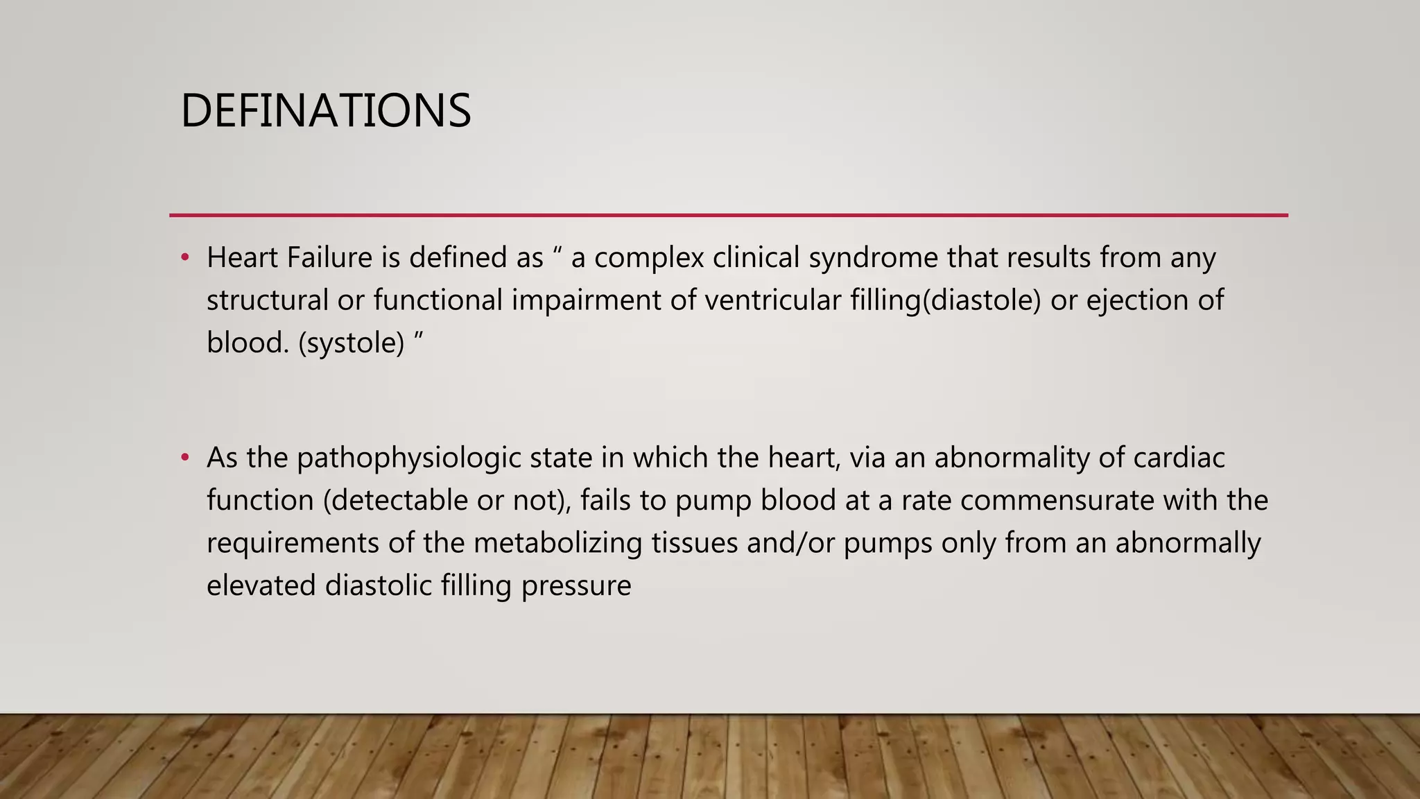
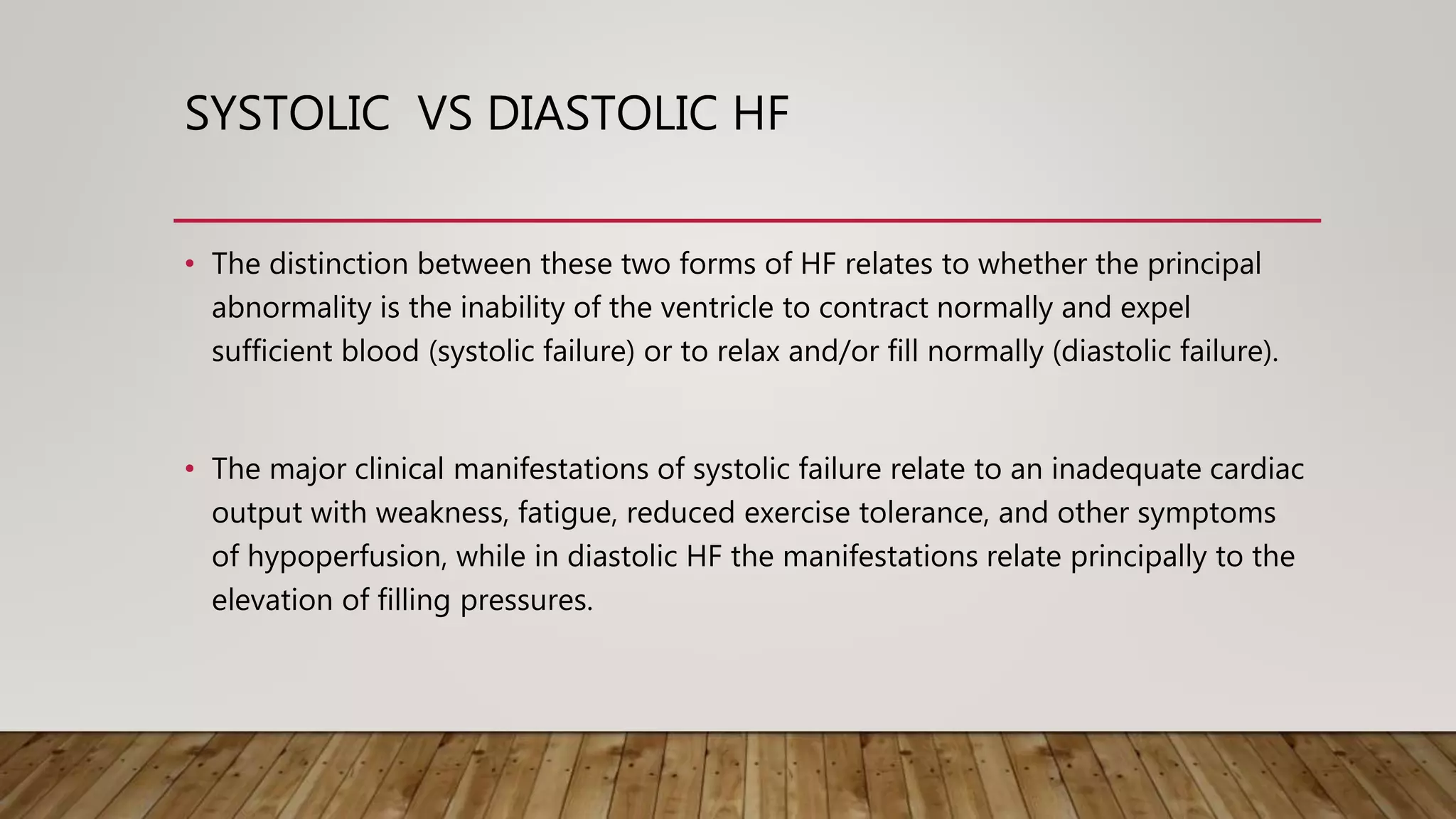
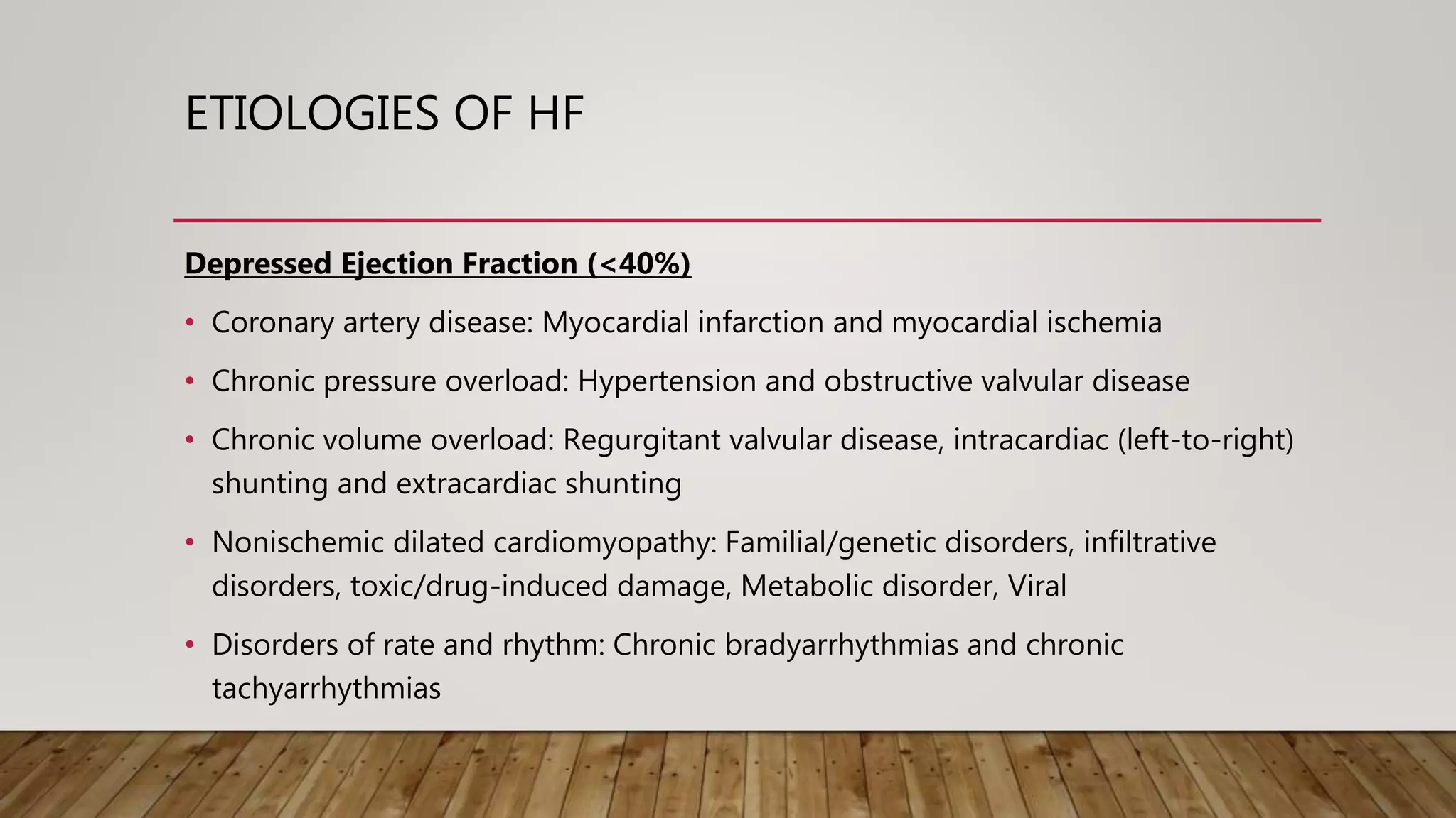
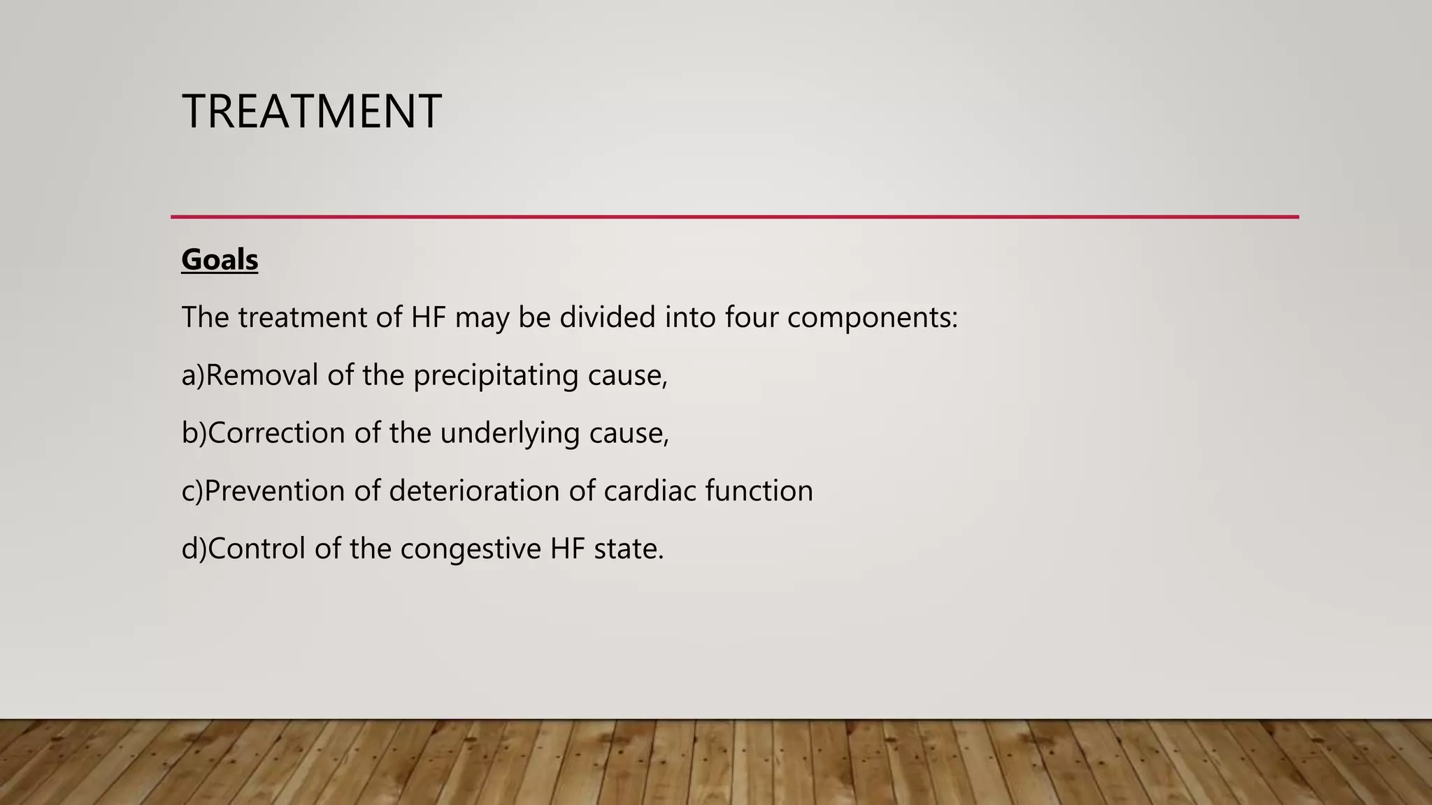
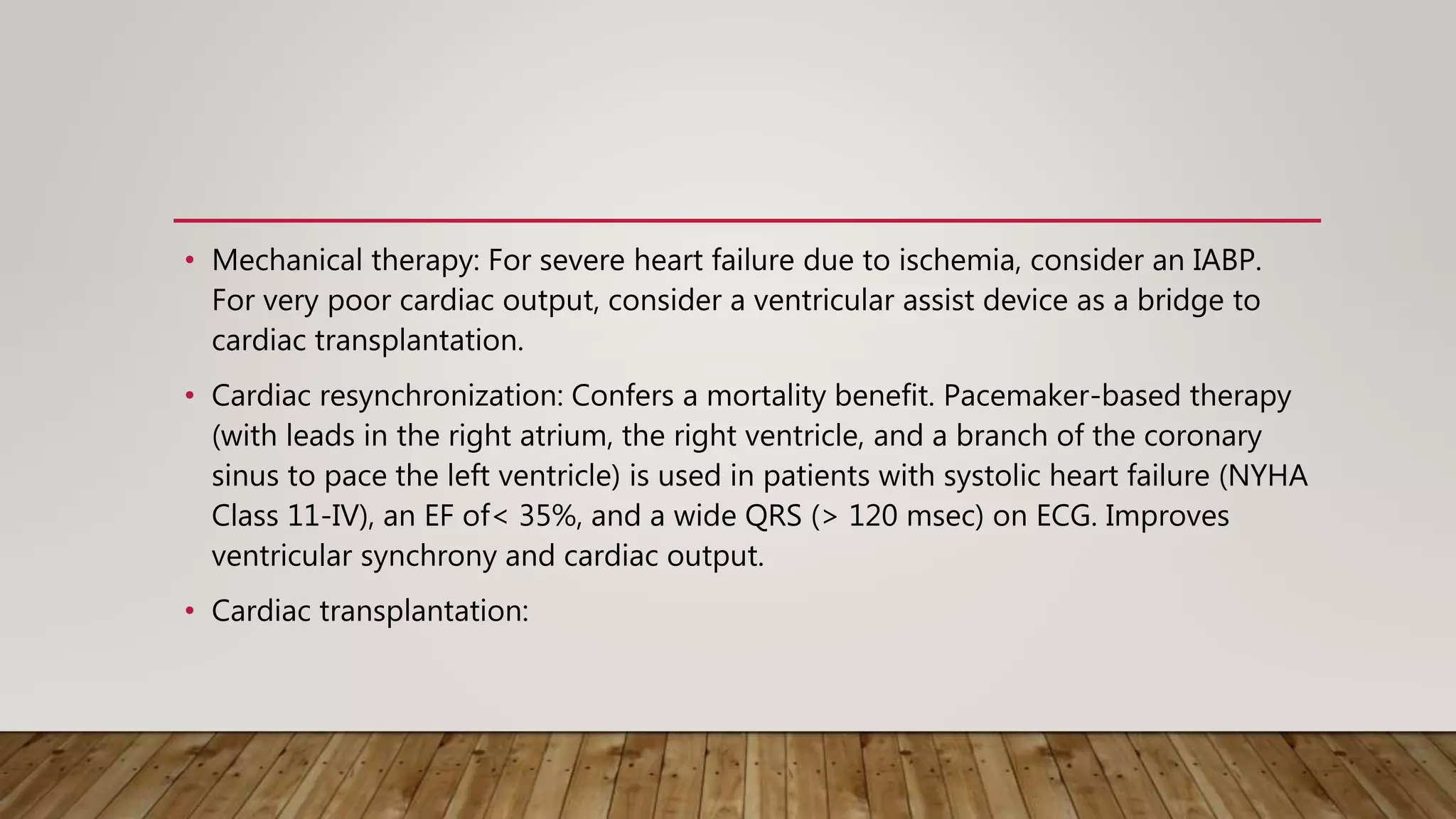
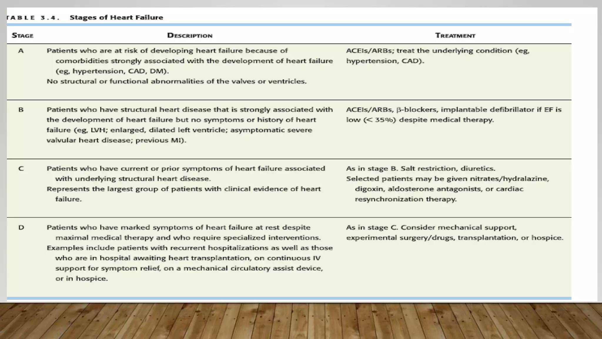
HEART FAILURE
DR. WILSON MTWANA
DEFINATIONS
• Heart Failure is defined as “ a complex clinical syndrome that results from any
structural or functional impairment of ventricular filling(diastole) or ejection of
blood. (systole) ”
• As the pathophysiologic state in which the heart, via an abnormality of cardiac
function (detectable or not), fails to pump blood at a rate commensurate with the
requirements of the metabolizing tissues and/or pumps only from an abnormally
elevated diastolic filling pressure
EPIDEMIOLOGY
• Approximate 2 % prevalence in developed countries. Women have better survival
than men.
• Coronary artery disease is the major cause for heart failure. (60 – 75%)
• HF prevalence follows an exponential pattern, rising with age, and affects 6–10%
of people over the age of 65
• The overall prevalence of HF is thought to be increasing, in part because current
therapies of cardiac disorders, such as myocardial infarction (MI), valvular heart
disease, and arrhythmias, are allowing patients to survive longer.
EPIDEMIOLOGY
• Rheumatic heart disease remains a major cause of HF in Africa and Asia, especially in
the young.
• Hypertension is an important cause of HF in the African and African-American
populations.
• Chagas' disease is still a major cause of HF in South America.
• Not surprisingly, anemia is a frequent concomitant factor in HF in many developing
nations.
• As developing nations undergo socioeconomic development, the epidemiology of HF
is becoming similar to that of Western Europe and North America, with CAD emerging
as the single most common cause of HF.
EPIDEMIOLOGY
• Although the contribution of diabetes mellitus to HF is not well understood,
diabetes accelerates atherosclerosis and is often associated with hypertension
• In industrialized countries, coronary artery disease (CAD) has become the
predominant cause in men and women and is responsible for 60–75% of cases of
HF.
• Hypertension contributes to the development of HF in 75% of patients, including
most patients with CAD.
• Both CAD and hypertension interact to augment the risk of HF, as does diabetes
mellitus
CLASSIFICATION
HF may be described as
• Systolic or diastolic,
• High-output or low-output,
• Acute or chronic,
• Right-sided or left-sided and
• Forward or backward.
SYSTOLIC VS DIASTOLIC HF
• The distinction between these two forms of HF relates to whether the principal
abnormality is the inability of the ventricle to contract normally and expel
sufficient blood (systolic failure) or to relax and/or fill normally (diastolic failure).
• The major clinical manifestations of systolic failure relate to an inadequate cardiac
output with weakness, fatigue, reduced exercise tolerance, and other symptoms
of hypoperfusion, while in diastolic HF the manifestations relate principally to the
elevation of filling pressures.
HIGH OUTPUT VS LOW OUTPUT
• It is useful to classify patients with HF into those with a low cardiac output, i.e.,
low-output HF, and those with an elevated cardiac output, i.e., high-output HF.
• The former occurs secondary to ischemic heart disease, hypertension, dilated
cardiomyopathy, and valvular and pericardial disease, while the latter is seen in
patients with HF and hyperthyroidism, anemia, pregnancy, arteriovenous fistulas,
beriberi, and Paget's disease.
ACUTE VS CHRONIC HF
• The prototype of acute HF is the sudden development of a large myocardial
infarction or rupture of a cardiac valve in a patient who previously was entirely
well.
• Chronic HF is typically observed in patients with dilated cardiomyopathy or
multivalvular heart disease that develops or progresses slowly.
RIGHT VS LEFT SIDED HF
• Many of the clinical manifestations of HF result from the accumulation of excess
fluid behind either one or both ventricles
• This fluid usually localizes upstream to (behind) the ventricle that is initially
affected.
• For example, patients in whom the left ventricle is hemodynamically overloaded
(e.g., aortic stenosis) or weakened (e.g., post myocardial infarction) develop
dyspnea and orthopnea as a result of pulmonary congestion, a condition referred
to as left-sided HF.
ETIOLOGIES OF HF
Depressed Ejection Fraction (<40%)
• Coronary artery disease: Myocardial infarction and myocardial ischemia
• Chronic pressure overload: Hypertension and obstructive valvular disease
• Chronic volume overload: Regurgitant valvular disease, intracardiac (left-to-right)
shunting and extracardiac shunting
• Nonischemic dilated cardiomyopathy: Familial/genetic disorders, infiltrative
disorders, toxic/drug-induced damage, Metabolic disorder, Viral
• Disorders of rate and rhythm: Chronic bradyarrhythmias and chronic
tachyarrhythmias
ETIOLOGY OF HF
Preserved Ejection Fraction (>40–50%)
• Pathological hypertrophy: Primary (hypertrophic cardiomyopathies) and Secondary
(hypertension)
• Restrictive cardiomyopathy: Infiltrative disorders (amyloidosis, sarcoidosis), Storage
diseases (hemochromatosis)
• Aging
• Fibrosis
• Endomyocardial disorders
WHAT CAUSES DECOMPENSATION ?
• Infection.
• Anemia.
• Thyrotoxicosis and pregnancy.
• Arrhythmias.
• Systemic hypertension.
• Infective endocarditis.
• Physical, dietary, fluid, environmental, and emotional excesses.
• The inappropriate discontinuation of pharmaceuticals to treat HF, blood
transfusions,
• Myocardial infarction.
• Pulmonary embolism.
• Rheumatic, viral, and other forms of myocarditis.
PATHOPHYSIOLOGY
Inadequate adaptation of the cardiac myocytes to increased wall stress in order to
maintain adequate cardiac output following myocardial injury is the inciting event
in CHF.
Most important among these adaptations are the
• Frank-Starling mechanism, in which an increased preload helps to sustain cardiac
performance
• Myocardial hypertrophy with or without cardiac chamber dilatation, in which the
mass of contractile tissue is augmented
• Activation of neurohumoral systems, especially the release of norepinephrine (NE)
by adrenergic cardiac nerves, which augments myocardial contractility and the
activation of the renin-angiotensin-aldosterone system (RAAS) and other
neurohumoral adjustments that act to maintain arterial pressure and perfusion of
vital organs.
CLINICAL MANIFESTATIONS
History
Breathlessness, a cardinal sign of LV failure, may manifest with progressively
increasing severity as
• exertional dyspnea,
• orthopnea,
• paroxysmal nocturnal dyspnea,
• dyspnea at rest.
• Fatigue and weakness:
• Nocturia
• Oliguria
• Cerebral symptoms: Confusion, memory impairment, anxiety, headaches,
insomnia, bad dreams or nightmares, and rarely, psychosis with disorientation,
delirium, or hallucinations may occur in elderly patients with advanced heart
failure, particularly in those with cerebrovascular atherosclerosis.
• Predominant right-sided heart failure: Ascites, congestive hepatomegaly, and
anasarca due to elevated right-sided heart pressures transmitted backward into
the portal vein circulation may result in increased abdominal girth and epigastric
and right upper quadrant (RUQ) abdominal pain.
• Other gastrointestinal symptoms, owing to congestion of the hepatic and
gastrointestinal venous circulation, include anorexia, bloating, nausea, and
constipation.
• In preterminal heart failure, inadequate bowel perfusion can cause abdominal
pain, distention, and bloody stools.
PHYSICAL FEARTURES
• General appearance: Patients with mild heart failure appear to be in no distress after a
few minutes of rest, but they may be obviously dyspneic during and immediately after
moderate activity.
• Patients with LV failure may be dyspneic when lying flat without elevation of the head
for more than a few minutes.
• Those with severe heart failure appear anxious and may exhibit signs of air hunger in
this position.
• Patients with recent onset of heart failure are generally well nourished, but those with
chronic severe heart failure are often malnourished and sometimes even cachectic
• Chronic marked elevation of systemic venous pressure may produce
exophthalmos and severe tricuspid regurgitation and may lead to visible
pulsation of the eyes and of the neck veins.
• Central cyanosis, icterus, and malar flush may be evident in patients with severe
heart failure.
• In mild or moderate heart failure, stroke volume is normal at rest; in severe heart
failure, it is reduced, as reflected by a diminished pulse pressure and a dusky
discoloration of the skin.
• The pulse may be weak, rapid, and thready; the proportional pulse pressure (pulse
pressure/systolic pressure) may be markedly reduced.
• The proportional pulse pressure correlates reasonably well with cardiac output.
• Evidence of increased adrenergic activity: Increased adrenergic activity is
manifested by tachycardia, diaphoresis, pallor, peripheral cyanosis with pallor and
coldness of the extremities, and obvious distention of the peripheral veins
secondary to venoconstriction.
• Systemic venous hypertension: This is manifested by jugular venous distention
(normal ≤ 8 cm).
• Pulmonary rales: Rales heard over the lung bases are characteristic of CHF of at least
moderate severity
• Hepatojugular reflux: This is found in patients with right-sided heart failure and is
helpful in differentiating hepatic enlargement due to heart failure from that caused by
other conditions.
• Edema: Although a cardinal manifestation of CHF, edema does not correlate well with
the level of systemic venous pressure.
• Hepatomegaly: Hepatomegaly is prominent in patients with chronic right-sided heart
failure, but it may occur rapidly in acute heart failure.
• Ascites: This finding occurs in patients with increased pressure in the hepatic veins and
in the veins draining into the peritoneum.
• Protodiastolic (S3) gallop: This is the earliest cardiac physical finding in
decompensated heart failure in the absence of severe mitral or tricuspid regurgitation
or left-to-right shunts.
• Cardiomegaly: A nonspecific finding, cardiomegaly nonetheless occurs in most
patients with chronic heart failure.
DIAGNOSIS
Modified Framingham Criteria for Diagnosis of Congestive Heart Failure
Major criteria ;-
1. Paroxysmal nocturnal dyspnea
2. Orthopnea
3. Elevated jugular venous pressure
4. Pulmonary rales
5. Third heart sound
6. Cardiomegaly on chest x-ray
7. Pulmonary edema on chest x-ray
8. Weight loss ≥ 4.5 kg in five days in response to treatment of presumed heart failure Rales
Minor criteria
1. Bilateral leg edema
2. Nocturnal cough
3. Dyspnea on ordinary exertion
4. Hepatomegaly
5. Pleural effusion
6. Tachycardia (heart rate 120 beats/min)
7. Weight loss ≥ 4.5 kg in five days
• To establish a clinical diagnosis of congestive heart failure by these criteria, at
least one major and two minor criteria are required.
• FHG,LFT,EUCs
• Echocardiography
• Ecg
• Chest xray
DDX
• HF is sometimes difficult to distinguish from pulmonary disease
• Pulmonary embolism also presents many of the manifestations of HF, but hemoptysis, pleuritic
chest pain, a right ventricular lift, and the characteristic mismatch between ventilation and
perfusion on lung scan should point to this diagnosis
• Ankle edema may be due to varicose veins, cyclic edema, or gravitational effects, but in these
patients there is no jugular venous hypertension at rest or with pressure over the abdomen.
• Edema secondary to renal disease can usually be recognized by appropriate renal function tests
and urinalysis and is rarely associated with elevation of venous pressure.
• Enlargement of the liver and ascites occur in patients with hepatic cirrhosis and also may be
distinguished from HF by normal jugular venous pressure and absence of a positive
abdominojugular reflux.
TREATMENT
Goals
The treatment of HF may be divided into four components:
a)Removal of the precipitating cause,
b)Correction of the underlying cause,
c)Prevention of deterioration of cardiac function
d)Control of the congestive HF state.
NON PHARMACOLOGICAL
• Activity
Although heavy physical labor is not recommended in HF, routine modest exercise
has been shown to be beneficial in patients with NYHA class I–III HF.
• Diet
Dietary restriction of sodium (2–3 g daily) is recommended in all patients with HF
and preserved or depressed EF.
Further restriction (<2 g daily) may be considered in moderate to severe HF.
• Alcohol and Cigarrete cessation.
• Lifestyle modification.
PHARMACOLOGICAL
Systolic Heart Failure
• Diuretics: Acutely used to decrease symptoms of pulmonary edema; confer no mortality benefit.
• ACEis: Have a proven mortality benefit. If ACEis are not tolerated because of cough, switch to an
ARB.
• Hydralazine with nitrates: Useful in an acute setting to decrease Pulmonary edema by decreasing
preload and afterload. Associated with a mortality benefit when used, but confers less benefit
than ACEis. In African American patients, a greater mortality benefit is gained than that provided
by ACEI/ARB therapy.
• Spironolactone: Yields a mortality benefit in class III-IV heart failure.
• B-blockers: Confer a mortality benefit in all classes of heart failure. However, they
should not be initiated in the setting of acutely decompensated heart failure.
Proven benefit is limited to carvedilol, bisoprolol, and long acting metoprolol.
• B-type natriuretic peptide (nesiritide): Acts primarily as a vasodilator. Can be
considered in severe heart failure requiring vasodilator therapy when
nitroprusside and nitroglycerin are contraindicated. May provide additional
improvement in symptoms, but may be associated with increase renal dysfunction
and possibly increase mortality.
• Mechanical therapy: For severe heart failure due to ischemia, consider an IABP.
For very poor cardiac output, consider a ventricular assist device as a bridge to
cardiac transplantation.
• Cardiac resynchronization: Confers a mortality benefit. Pacemaker-based therapy
(with leads in the right atrium, the right ventricle, and a branch of the coronary
sinus to pace the left ventricle) is used in patients with systolic heart failure (NYHA
Class 11-IV), an EF of< 35%, and a wide QRS (> 120 msec) on ECG. Improves
ventricular synchrony and cardiac output.
• Cardiac transplantation:
Diastolic Heart Failure
• No treatment has been convincingly shown to decrease mortality in patients
with diastolic heart failure.
• Avoid exacerbating factors such as AF, tachycardia, ischemia, hypertension,
fluid overload, and anemia. Aggressively control hypertension.
• ACEis and ARBs may aid in cardiac remodeling.
• Diuresis and slowing the heart rate: B-blockers and nondihydropyridine CCBs (eg,
diltiazem, verapamil) may help symptoms but have not been proven to decrease
mortality. Use caution in patients with advanced diastolic heart failure, who may
need preload and/or chronotropy (relatively high heart rates) to maintain cardiac
output.
THANK YOU 

Heart failure 2019

  • 1.
  • 2.
    DEFINATIONS • Heart Failureis defined as “ a complex clinical syndrome that results from any structural or functional impairment of ventricular filling(diastole) or ejection of blood. (systole) ” • As the pathophysiologic state in which the heart, via an abnormality of cardiac function (detectable or not), fails to pump blood at a rate commensurate with the requirements of the metabolizing tissues and/or pumps only from an abnormally elevated diastolic filling pressure
  • 3.
    EPIDEMIOLOGY • Approximate 2% prevalence in developed countries. Women have better survival than men. • Coronary artery disease is the major cause for heart failure. (60 – 75%) • HF prevalence follows an exponential pattern, rising with age, and affects 6–10% of people over the age of 65 • The overall prevalence of HF is thought to be increasing, in part because current therapies of cardiac disorders, such as myocardial infarction (MI), valvular heart disease, and arrhythmias, are allowing patients to survive longer.
  • 4.
    EPIDEMIOLOGY • Rheumatic heartdisease remains a major cause of HF in Africa and Asia, especially in the young. • Hypertension is an important cause of HF in the African and African-American populations. • Chagas' disease is still a major cause of HF in South America. • Not surprisingly, anemia is a frequent concomitant factor in HF in many developing nations. • As developing nations undergo socioeconomic development, the epidemiology of HF is becoming similar to that of Western Europe and North America, with CAD emerging as the single most common cause of HF.
  • 5.
    EPIDEMIOLOGY • Although thecontribution of diabetes mellitus to HF is not well understood, diabetes accelerates atherosclerosis and is often associated with hypertension • In industrialized countries, coronary artery disease (CAD) has become the predominant cause in men and women and is responsible for 60–75% of cases of HF. • Hypertension contributes to the development of HF in 75% of patients, including most patients with CAD. • Both CAD and hypertension interact to augment the risk of HF, as does diabetes mellitus
  • 6.
    CLASSIFICATION HF may bedescribed as • Systolic or diastolic, • High-output or low-output, • Acute or chronic, • Right-sided or left-sided and • Forward or backward.
  • 7.
    SYSTOLIC VS DIASTOLICHF • The distinction between these two forms of HF relates to whether the principal abnormality is the inability of the ventricle to contract normally and expel sufficient blood (systolic failure) or to relax and/or fill normally (diastolic failure). • The major clinical manifestations of systolic failure relate to an inadequate cardiac output with weakness, fatigue, reduced exercise tolerance, and other symptoms of hypoperfusion, while in diastolic HF the manifestations relate principally to the elevation of filling pressures.
  • 8.
    HIGH OUTPUT VSLOW OUTPUT • It is useful to classify patients with HF into those with a low cardiac output, i.e., low-output HF, and those with an elevated cardiac output, i.e., high-output HF. • The former occurs secondary to ischemic heart disease, hypertension, dilated cardiomyopathy, and valvular and pericardial disease, while the latter is seen in patients with HF and hyperthyroidism, anemia, pregnancy, arteriovenous fistulas, beriberi, and Paget's disease.
  • 9.
    ACUTE VS CHRONICHF • The prototype of acute HF is the sudden development of a large myocardial infarction or rupture of a cardiac valve in a patient who previously was entirely well. • Chronic HF is typically observed in patients with dilated cardiomyopathy or multivalvular heart disease that develops or progresses slowly.
  • 10.
    RIGHT VS LEFTSIDED HF • Many of the clinical manifestations of HF result from the accumulation of excess fluid behind either one or both ventricles • This fluid usually localizes upstream to (behind) the ventricle that is initially affected. • For example, patients in whom the left ventricle is hemodynamically overloaded (e.g., aortic stenosis) or weakened (e.g., post myocardial infarction) develop dyspnea and orthopnea as a result of pulmonary congestion, a condition referred to as left-sided HF.
  • 11.
    ETIOLOGIES OF HF DepressedEjection Fraction (<40%) • Coronary artery disease: Myocardial infarction and myocardial ischemia • Chronic pressure overload: Hypertension and obstructive valvular disease • Chronic volume overload: Regurgitant valvular disease, intracardiac (left-to-right) shunting and extracardiac shunting • Nonischemic dilated cardiomyopathy: Familial/genetic disorders, infiltrative disorders, toxic/drug-induced damage, Metabolic disorder, Viral • Disorders of rate and rhythm: Chronic bradyarrhythmias and chronic tachyarrhythmias
  • 12.
    ETIOLOGY OF HF PreservedEjection Fraction (>40–50%) • Pathological hypertrophy: Primary (hypertrophic cardiomyopathies) and Secondary (hypertension) • Restrictive cardiomyopathy: Infiltrative disorders (amyloidosis, sarcoidosis), Storage diseases (hemochromatosis) • Aging • Fibrosis • Endomyocardial disorders
  • 13.
    WHAT CAUSES DECOMPENSATION? • Infection. • Anemia. • Thyrotoxicosis and pregnancy. • Arrhythmias. • Systemic hypertension.
  • 14.
    • Infective endocarditis. •Physical, dietary, fluid, environmental, and emotional excesses. • The inappropriate discontinuation of pharmaceuticals to treat HF, blood transfusions, • Myocardial infarction. • Pulmonary embolism. • Rheumatic, viral, and other forms of myocarditis.
  • 15.
    PATHOPHYSIOLOGY Inadequate adaptation ofthe cardiac myocytes to increased wall stress in order to maintain adequate cardiac output following myocardial injury is the inciting event in CHF. Most important among these adaptations are the • Frank-Starling mechanism, in which an increased preload helps to sustain cardiac performance • Myocardial hypertrophy with or without cardiac chamber dilatation, in which the mass of contractile tissue is augmented
  • 16.
    • Activation ofneurohumoral systems, especially the release of norepinephrine (NE) by adrenergic cardiac nerves, which augments myocardial contractility and the activation of the renin-angiotensin-aldosterone system (RAAS) and other neurohumoral adjustments that act to maintain arterial pressure and perfusion of vital organs.
  • 17.
    CLINICAL MANIFESTATIONS History Breathlessness, acardinal sign of LV failure, may manifest with progressively increasing severity as • exertional dyspnea, • orthopnea, • paroxysmal nocturnal dyspnea, • dyspnea at rest.
  • 18.
    • Fatigue andweakness: • Nocturia • Oliguria • Cerebral symptoms: Confusion, memory impairment, anxiety, headaches, insomnia, bad dreams or nightmares, and rarely, psychosis with disorientation, delirium, or hallucinations may occur in elderly patients with advanced heart failure, particularly in those with cerebrovascular atherosclerosis.
  • 19.
    • Predominant right-sidedheart failure: Ascites, congestive hepatomegaly, and anasarca due to elevated right-sided heart pressures transmitted backward into the portal vein circulation may result in increased abdominal girth and epigastric and right upper quadrant (RUQ) abdominal pain. • Other gastrointestinal symptoms, owing to congestion of the hepatic and gastrointestinal venous circulation, include anorexia, bloating, nausea, and constipation. • In preterminal heart failure, inadequate bowel perfusion can cause abdominal pain, distention, and bloody stools.
  • 20.
    PHYSICAL FEARTURES • Generalappearance: Patients with mild heart failure appear to be in no distress after a few minutes of rest, but they may be obviously dyspneic during and immediately after moderate activity. • Patients with LV failure may be dyspneic when lying flat without elevation of the head for more than a few minutes. • Those with severe heart failure appear anxious and may exhibit signs of air hunger in this position. • Patients with recent onset of heart failure are generally well nourished, but those with chronic severe heart failure are often malnourished and sometimes even cachectic
  • 21.
    • Chronic markedelevation of systemic venous pressure may produce exophthalmos and severe tricuspid regurgitation and may lead to visible pulsation of the eyes and of the neck veins. • Central cyanosis, icterus, and malar flush may be evident in patients with severe heart failure. • In mild or moderate heart failure, stroke volume is normal at rest; in severe heart failure, it is reduced, as reflected by a diminished pulse pressure and a dusky discoloration of the skin.
  • 22.
    • The pulsemay be weak, rapid, and thready; the proportional pulse pressure (pulse pressure/systolic pressure) may be markedly reduced. • The proportional pulse pressure correlates reasonably well with cardiac output. • Evidence of increased adrenergic activity: Increased adrenergic activity is manifested by tachycardia, diaphoresis, pallor, peripheral cyanosis with pallor and coldness of the extremities, and obvious distention of the peripheral veins secondary to venoconstriction.
  • 23.
    • Systemic venoushypertension: This is manifested by jugular venous distention (normal ≤ 8 cm). • Pulmonary rales: Rales heard over the lung bases are characteristic of CHF of at least moderate severity • Hepatojugular reflux: This is found in patients with right-sided heart failure and is helpful in differentiating hepatic enlargement due to heart failure from that caused by other conditions. • Edema: Although a cardinal manifestation of CHF, edema does not correlate well with the level of systemic venous pressure.
  • 24.
    • Hepatomegaly: Hepatomegalyis prominent in patients with chronic right-sided heart failure, but it may occur rapidly in acute heart failure. • Ascites: This finding occurs in patients with increased pressure in the hepatic veins and in the veins draining into the peritoneum. • Protodiastolic (S3) gallop: This is the earliest cardiac physical finding in decompensated heart failure in the absence of severe mitral or tricuspid regurgitation or left-to-right shunts. • Cardiomegaly: A nonspecific finding, cardiomegaly nonetheless occurs in most patients with chronic heart failure.
  • 26.
    DIAGNOSIS Modified Framingham Criteriafor Diagnosis of Congestive Heart Failure Major criteria ;- 1. Paroxysmal nocturnal dyspnea 2. Orthopnea 3. Elevated jugular venous pressure 4. Pulmonary rales 5. Third heart sound 6. Cardiomegaly on chest x-ray 7. Pulmonary edema on chest x-ray 8. Weight loss ≥ 4.5 kg in five days in response to treatment of presumed heart failure Rales
  • 27.
    Minor criteria 1. Bilateralleg edema 2. Nocturnal cough 3. Dyspnea on ordinary exertion 4. Hepatomegaly 5. Pleural effusion 6. Tachycardia (heart rate 120 beats/min) 7. Weight loss ≥ 4.5 kg in five days
  • 28.
    • To establisha clinical diagnosis of congestive heart failure by these criteria, at least one major and two minor criteria are required.
  • 29.
  • 30.
    DDX • HF issometimes difficult to distinguish from pulmonary disease • Pulmonary embolism also presents many of the manifestations of HF, but hemoptysis, pleuritic chest pain, a right ventricular lift, and the characteristic mismatch between ventilation and perfusion on lung scan should point to this diagnosis • Ankle edema may be due to varicose veins, cyclic edema, or gravitational effects, but in these patients there is no jugular venous hypertension at rest or with pressure over the abdomen. • Edema secondary to renal disease can usually be recognized by appropriate renal function tests and urinalysis and is rarely associated with elevation of venous pressure. • Enlargement of the liver and ascites occur in patients with hepatic cirrhosis and also may be distinguished from HF by normal jugular venous pressure and absence of a positive abdominojugular reflux.
  • 31.
    TREATMENT Goals The treatment ofHF may be divided into four components: a)Removal of the precipitating cause, b)Correction of the underlying cause, c)Prevention of deterioration of cardiac function d)Control of the congestive HF state.
  • 32.
    NON PHARMACOLOGICAL • Activity Althoughheavy physical labor is not recommended in HF, routine modest exercise has been shown to be beneficial in patients with NYHA class I–III HF. • Diet Dietary restriction of sodium (2–3 g daily) is recommended in all patients with HF and preserved or depressed EF. Further restriction (<2 g daily) may be considered in moderate to severe HF.
  • 33.
    • Alcohol andCigarrete cessation. • Lifestyle modification.
  • 35.
    PHARMACOLOGICAL Systolic Heart Failure •Diuretics: Acutely used to decrease symptoms of pulmonary edema; confer no mortality benefit. • ACEis: Have a proven mortality benefit. If ACEis are not tolerated because of cough, switch to an ARB. • Hydralazine with nitrates: Useful in an acute setting to decrease Pulmonary edema by decreasing preload and afterload. Associated with a mortality benefit when used, but confers less benefit than ACEis. In African American patients, a greater mortality benefit is gained than that provided by ACEI/ARB therapy. • Spironolactone: Yields a mortality benefit in class III-IV heart failure.
  • 36.
    • B-blockers: Confera mortality benefit in all classes of heart failure. However, they should not be initiated in the setting of acutely decompensated heart failure. Proven benefit is limited to carvedilol, bisoprolol, and long acting metoprolol. • B-type natriuretic peptide (nesiritide): Acts primarily as a vasodilator. Can be considered in severe heart failure requiring vasodilator therapy when nitroprusside and nitroglycerin are contraindicated. May provide additional improvement in symptoms, but may be associated with increase renal dysfunction and possibly increase mortality.
  • 37.
    • Mechanical therapy:For severe heart failure due to ischemia, consider an IABP. For very poor cardiac output, consider a ventricular assist device as a bridge to cardiac transplantation. • Cardiac resynchronization: Confers a mortality benefit. Pacemaker-based therapy (with leads in the right atrium, the right ventricle, and a branch of the coronary sinus to pace the left ventricle) is used in patients with systolic heart failure (NYHA Class 11-IV), an EF of< 35%, and a wide QRS (> 120 msec) on ECG. Improves ventricular synchrony and cardiac output. • Cardiac transplantation:
  • 38.
    Diastolic Heart Failure •No treatment has been convincingly shown to decrease mortality in patients with diastolic heart failure. • Avoid exacerbating factors such as AF, tachycardia, ischemia, hypertension, fluid overload, and anemia. Aggressively control hypertension. • ACEis and ARBs may aid in cardiac remodeling.
  • 39.
    • Diuresis andslowing the heart rate: B-blockers and nondihydropyridine CCBs (eg, diltiazem, verapamil) may help symptoms but have not been proven to decrease mortality. Use caution in patients with advanced diastolic heart failure, who may need preload and/or chronotropy (relatively high heart rates) to maintain cardiac output.
  • 43.