Downloaded 243 times


This document provides an overview of congestive heart failure (CHF), including its definition, causes, pathophysiology, clinical manifestations, diagnostic evaluation, and management. CHF is defined as a clinical syndrome where the heart cannot pump enough blood to meet the body's needs. It is most commonly caused by conditions that overload or damage the heart such as hypertension, heart attacks, and cardiomyopathy. Clinically, it presents with symptoms of fluid backup like dyspnea, edema, and fatigue. Diagnostic tests include chest x-rays, EKGs, blood tests like BNP, and echocardiography. Treatment focuses on managing symptoms, addressing the underlying cause, and preventing complications through medications, lifestyle changes, and potentially devices


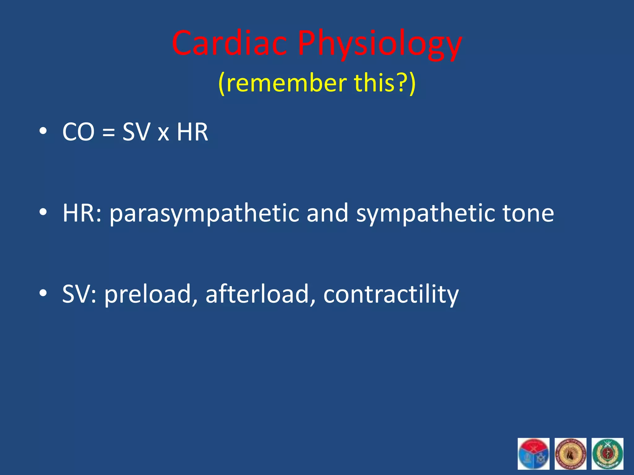
![Acute heart failure [MBBS]](https://cdn.slidesharecdn.com/ss_thumbnails/acuteheartfailure-170323061012-thumbnail.jpg?width=640&height=640&fit=bounds)


