Download as PDF, PPTX
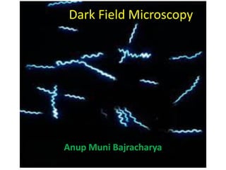
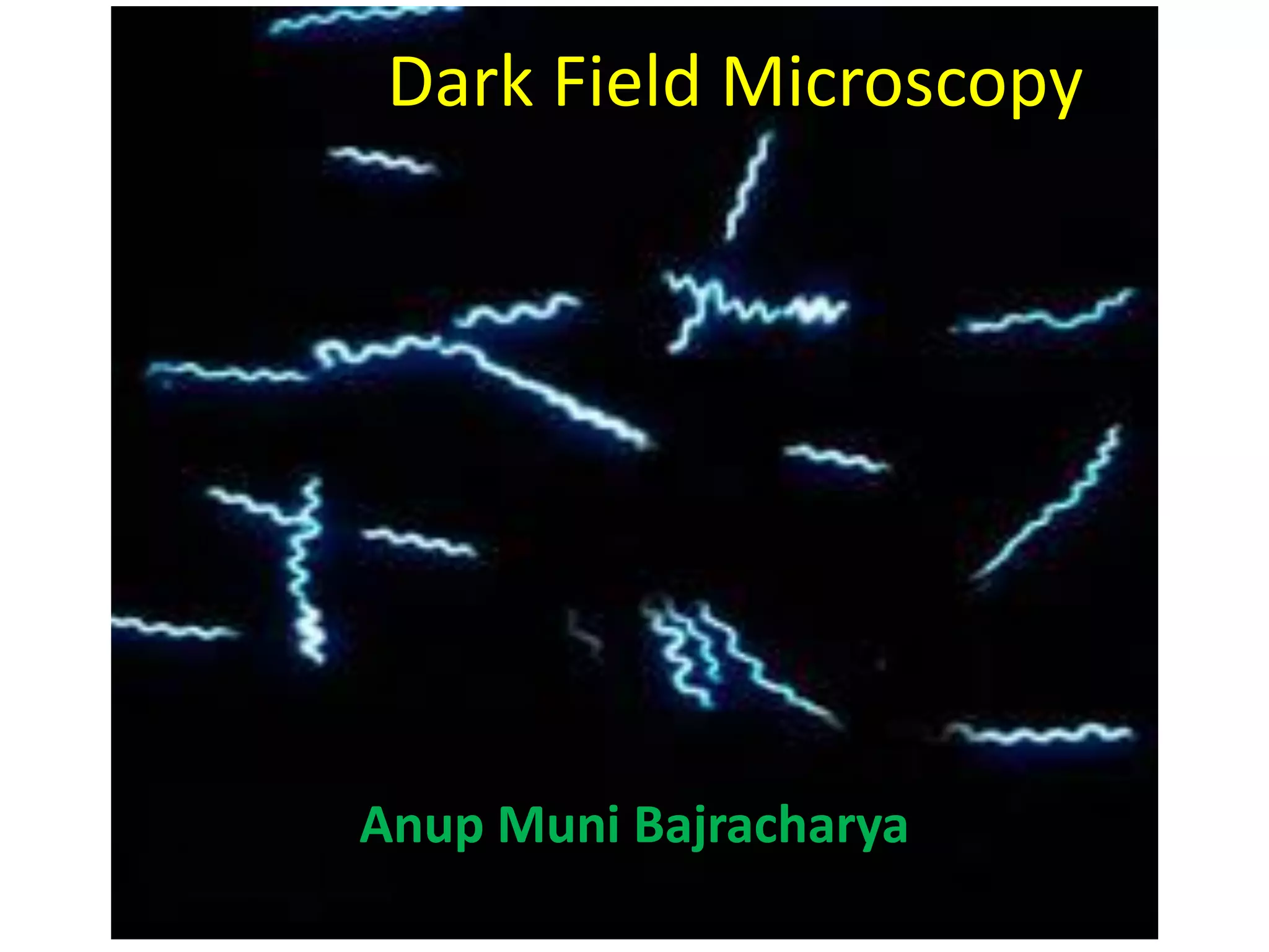
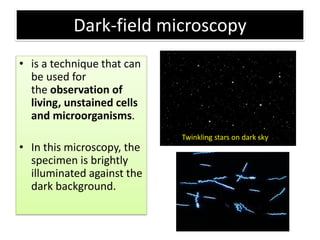
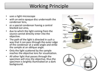
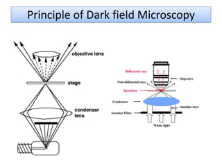
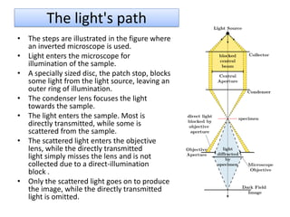
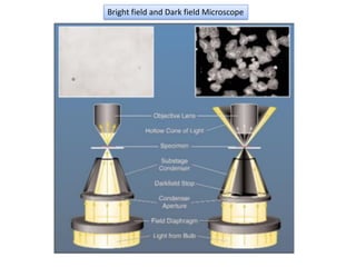
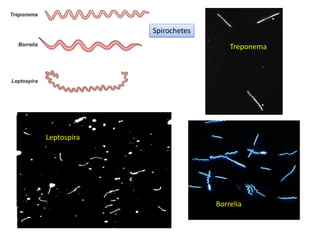
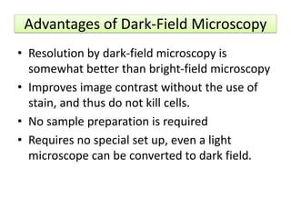
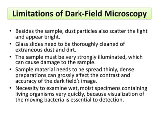


Dark-field microscopy is a technique for observing living, unstained cells against a dark background, allowing for enhanced visualization of microorganisms like spirochetes. It differs from bright-field microscopy by using a special condenser to illuminate samples at oblique angles, improving image contrast without the need for staining. While it offers better resolution and requires minimal sample preparation, it also has limitations such as potential interference from dust and the necessity for strong illumination.