Download as PDF, PPTX


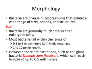


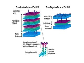


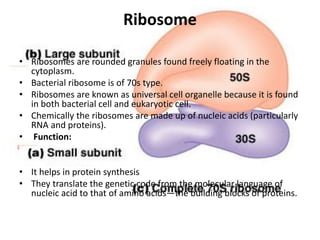


Bacteria are unicellular prokaryotic microorganisms with a simple cellular structure lacking a membrane-bound nucleus. They vary in size, shape, and morphology, primarily classified into cocci, bacilli, and spirilla, and reproduce asexually through binary fission. Their cell structure includes a cell wall made of peptidoglycan, a plasma membrane, and ribosomes, while features like flagella and pili assist in motility and adhesion.