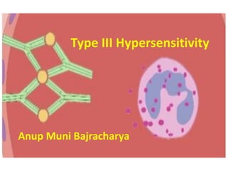
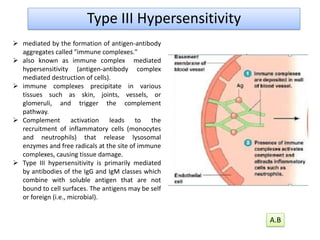
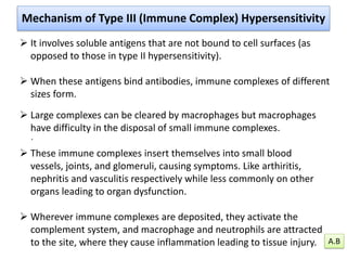
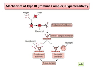
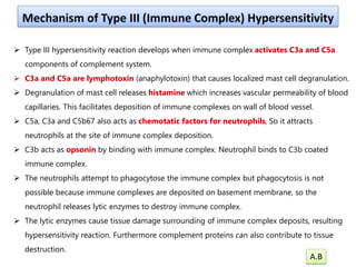
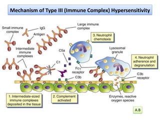
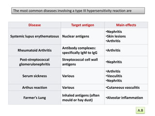
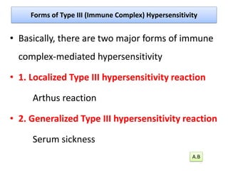
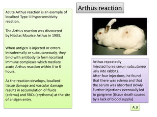
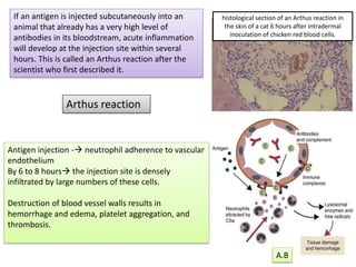
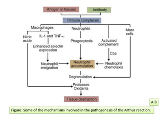
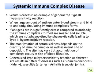
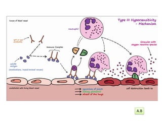
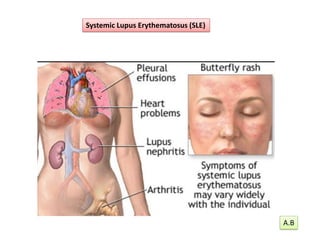
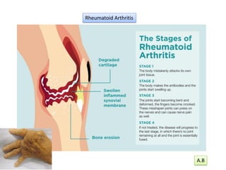
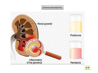
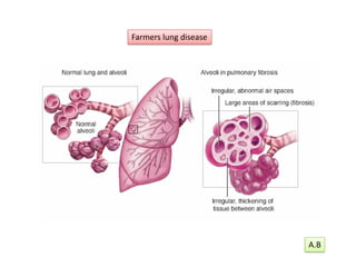
Type III Hypersensitivity
Anup Muni Bajracharya
➢ mediated by the formation of antigen-antibody
aggregates called "immune complexes."
➢ also known as immune complex mediated
hypersensitivity (antigen-antibody complex
mediated destruction of cells).
➢ immune complexes precipitate in various
tissues such as skin, joints, vessels, or
glomeruli, and trigger the complement
pathway.
➢ Complement activation leads to the
recruitment of inflammatory cells (monocytes
and neutrophils) that release lysosomal
enzymes and free radicals at the site of immune
complexes, causing tissue damage.
➢ Type III hypersensitivity is primarily mediated
by antibodies of the IgG and IgM classes which
combine with soluble antigen that are not
bound to cell surfaces. The antigens may be self
or foreign (i.e., microbial).
Type III Hypersensitivity
A.B
.
➢ It involves soluble antigens that are not bound to cell surfaces (as
opposed to those in type II hypersensitivity).
➢ When these antigens bind antibodies, immune complexes of different
sizes form.
➢ Large complexes can be cleared by macrophages but macrophages
have difficulty in the disposal of small immune complexes.
➢ These immune complexes insert themselves into small blood
vessels, joints, and glomeruli, causing symptoms. Like arthiritis,
nephritis and vasculitis respectively while less commonly on other
organs leading to organ dysfunction.
➢ Wherever immune complexes are deposited, they activate the
complement system, and macrophage and neutrophils are attracted
to the site, where they cause inflammation leading to tissue injury.
Mechanism of Type III (Immune Complex) Hypersensitivity
A.B
Mechanism of Type III (Immune Complex) Hypersensitivity
A.B
➢ Type III hypersensitivity reaction develops when immune complex activates C3a and C5a
components of complement system.
➢ C3a and C5a are lymphotoxin (anaphylotoxin) that causes localized mast cell degranulation.
➢ Degranulation of mast cell releases histamine which increases vascular permeability of blood
capillaries. This facilitates deposition of immune complexes on wall of blood vessel.
➢ C5a, C3a and C5b67 also acts as chemotatic factors for neutrophils, So it attracts
neutrophils at the site of immune complex deposition.
➢ C3b acts as opsonin by binding with immune complex. Neutrophil binds to C3b coated
immune complex.
➢ The neutrophils attempt to phagocytose the immune complex but phagocytosis is not
possible because immune complexes are deposited on basement membrane, so the
neutrophil releases lytic enzymes to destroy immune complex.
➢ The lytic enzymes cause tissue damage surrounding of immune complex deposits, resulting
hypersensitivity reaction. Furthermore complement proteins can also contribute to tissue
destruction.
Mechanism of Type III (Immune Complex) Hypersensitivity
A.B
Mechanism of Type III (Immune Complex) Hypersensitivity
A.B
Disease Target antigen Main effects
Systemic lupus erythematosus Nuclear antigens
•Nephritis
•Skin lesions
•Arthritis
Rheumatoid Arthritis
Antibody complexes:
specifically IgM to IgG
•Arthritis
Post-streptococcal
glomerulonephritis
Streptococcal cell wall
antigens
•Nephritis
Serum sickness Various
•Arthritis
•Vasculitis
•Nephritis
Arthus reaction Various •Cutaneous vasculitis
Farmer's Lung
Inhaled antigens (often
mould or hay dust)
•Alveolar inflammation
The most common diseases involving a type III hypersensitivity reaction are
A.B
Forms of Type III (Immune Complex) Hypersensitivity
• Basically, there are two major forms of immune
complex-mediated hypersensitivity
• 1. Localized Type III hypersensitivity reaction
Arthus reaction
• 2. Generalized Type III hypersensitivity reaction
Serum sickness
A.B
Arthus reaction
Acute Arthus reaction is an example of
localized Type III hypersensitivity
reaction.
The Arthus reaction was discovered
by Nicolas Maurice Arthus in 1903.
When antigen is injected or enters
intradermally or subcutaneously, they
bind with antibody to form localized
immune complexes which mediate
acute Arthus reaction within 4 to 8
hours.
As the reaction develops, localized
tissue damage and vascular damage
results in accumulation of fluids
(edema) and RBCs (erythema) at the site
of antigen entry.
Arthus repeatedly
Injected horse serum subcutaneo
usly into rabbits.
After four injections, he found
that there was edema and that
the serum was absorbed slowly.
Further injections eventually led
to gangrene (tissue death caused
by a lack of blood supply)
A.B
If an antigen is injected subcutaneously into an
animal that already has a very high level of
antibodies in its bloodstream, acute inflammation
will develop at the injection site within several
hours. This is called an Arthus reaction after the
scientist who first described it.
Antigen injection -→ neutrophil adherence to vascular
endothelium
By 6 to 8 hours→ the injection site is densely
infiltrated by large numbers of these cells.
Destruction of blood vessel walls results in
hemorrhage and edema, platelet aggregation, and
thrombosis.
Arthus reaction
histological section of an Arthus reaction in
the skin of a cat 6 hours after intradermal
inoculation of chicken red blood cells.
A.B
Figure: Some of the mechanisms involved in the pathogenesis of the Arthus reaction.
A.B
Hypersensitivity Pneumonitis
If cattle are fed moldy hay for long periods, constant inhalation of S.
rectivirgula spores will result in sensitization and in the development of high-
titered antibodies to S. rectivirgula antigens in serum.
Eventually inhaled spore antigens will encounter antibodies within the alveolar
walls, and the resulting immune complexes and complement activation will
cause a pneumonia (or pneumonitis), the basis of which is a type III
hypersensitivity reaction. A.B
Type Specific antigen Exposure
Bird fancier's lung
Also called bird breeder's lung,
pigeon breeder's lung, and poultry
worker's lung
Avian proteins
Feathers and bird
droppings
Bagassosis
Exposure to moldy molasses
Thermophilic actinomycetes
Moldy bagasse
(pressed
sugarcane)
Farmer's lung
•The molds Aspergillus species
The bacteria
•Thermophilic actinomycetes
•Thermoactinomyces vulgaris
•Saccharopolyspora rectivirgula
•Absidia corymbifera
•Eurotium amstelodami
Moldy hay
Cheese-washer's lung Penicillum casei or P. roqueforti Cheese casings
Coffee worker's lung Coffee bean protein Coffee bean dust
Compost lung Aspergillus Compost
Detergent worker's disease Bacillus subtilis enzymes Detergent
Hypersensitivity pneumonitis may also be called many different names, based on
the provoking antigen These include:
A.B
Systemic Immune Complex Disease
• Serum sickness is an example of generalized Type III
hypersensitivity reaction.
• When large amount of antigen enter blood stream and bind
to antibody, circulating immune complexes forms.
• If antigens are in significantly excess compared to antibody,
the immune complexes formed are smaller and soluble
which are not phagocytized by phagocytic cells leading to
Type III hypersensitivity reaction.
• The manifestation of serum sickness depends on the
quantity of immune complex as well as overall site of
deposition. The site may vary but accumulation of
complexes occurs at site of blood filtration.
• Generalized Type III hypersensitivity reaction at different
site results in different diseases such as Glomerulonephritis
(Kideny), vasculitis (arteries), Arthritis (synovial joints).
A.B
A.B
Systemic Lupus Erythematosus (SLE)
A.B
Rheumatoid Arthritis
A.B
Glomerulonephritis
A.B
Farmers lung disease
A.B
Type III Hypersensitivity

Type III Hypersensitivity

  • 1.
  • 2.
    ➢ mediated bythe formation of antigen-antibody aggregates called "immune complexes." ➢ also known as immune complex mediated hypersensitivity (antigen-antibody complex mediated destruction of cells). ➢ immune complexes precipitate in various tissues such as skin, joints, vessels, or glomeruli, and trigger the complement pathway. ➢ Complement activation leads to the recruitment of inflammatory cells (monocytes and neutrophils) that release lysosomal enzymes and free radicals at the site of immune complexes, causing tissue damage. ➢ Type III hypersensitivity is primarily mediated by antibodies of the IgG and IgM classes which combine with soluble antigen that are not bound to cell surfaces. The antigens may be self or foreign (i.e., microbial). Type III Hypersensitivity A.B
  • 3.
    . ➢ It involvessoluble antigens that are not bound to cell surfaces (as opposed to those in type II hypersensitivity). ➢ When these antigens bind antibodies, immune complexes of different sizes form. ➢ Large complexes can be cleared by macrophages but macrophages have difficulty in the disposal of small immune complexes. ➢ These immune complexes insert themselves into small blood vessels, joints, and glomeruli, causing symptoms. Like arthiritis, nephritis and vasculitis respectively while less commonly on other organs leading to organ dysfunction. ➢ Wherever immune complexes are deposited, they activate the complement system, and macrophage and neutrophils are attracted to the site, where they cause inflammation leading to tissue injury. Mechanism of Type III (Immune Complex) Hypersensitivity A.B
  • 4.
    Mechanism of TypeIII (Immune Complex) Hypersensitivity A.B
  • 5.
    ➢ Type IIIhypersensitivity reaction develops when immune complex activates C3a and C5a components of complement system. ➢ C3a and C5a are lymphotoxin (anaphylotoxin) that causes localized mast cell degranulation. ➢ Degranulation of mast cell releases histamine which increases vascular permeability of blood capillaries. This facilitates deposition of immune complexes on wall of blood vessel. ➢ C5a, C3a and C5b67 also acts as chemotatic factors for neutrophils, So it attracts neutrophils at the site of immune complex deposition. ➢ C3b acts as opsonin by binding with immune complex. Neutrophil binds to C3b coated immune complex. ➢ The neutrophils attempt to phagocytose the immune complex but phagocytosis is not possible because immune complexes are deposited on basement membrane, so the neutrophil releases lytic enzymes to destroy immune complex. ➢ The lytic enzymes cause tissue damage surrounding of immune complex deposits, resulting hypersensitivity reaction. Furthermore complement proteins can also contribute to tissue destruction. Mechanism of Type III (Immune Complex) Hypersensitivity A.B
  • 6.
    Mechanism of TypeIII (Immune Complex) Hypersensitivity A.B
  • 7.
    Disease Target antigenMain effects Systemic lupus erythematosus Nuclear antigens •Nephritis •Skin lesions •Arthritis Rheumatoid Arthritis Antibody complexes: specifically IgM to IgG •Arthritis Post-streptococcal glomerulonephritis Streptococcal cell wall antigens •Nephritis Serum sickness Various •Arthritis •Vasculitis •Nephritis Arthus reaction Various •Cutaneous vasculitis Farmer's Lung Inhaled antigens (often mould or hay dust) •Alveolar inflammation The most common diseases involving a type III hypersensitivity reaction are A.B
  • 8.
    Forms of TypeIII (Immune Complex) Hypersensitivity • Basically, there are two major forms of immune complex-mediated hypersensitivity • 1. Localized Type III hypersensitivity reaction Arthus reaction • 2. Generalized Type III hypersensitivity reaction Serum sickness A.B
  • 9.
    Arthus reaction Acute Arthusreaction is an example of localized Type III hypersensitivity reaction. The Arthus reaction was discovered by Nicolas Maurice Arthus in 1903. When antigen is injected or enters intradermally or subcutaneously, they bind with antibody to form localized immune complexes which mediate acute Arthus reaction within 4 to 8 hours. As the reaction develops, localized tissue damage and vascular damage results in accumulation of fluids (edema) and RBCs (erythema) at the site of antigen entry. Arthus repeatedly Injected horse serum subcutaneo usly into rabbits. After four injections, he found that there was edema and that the serum was absorbed slowly. Further injections eventually led to gangrene (tissue death caused by a lack of blood supply) A.B
  • 10.
    If an antigenis injected subcutaneously into an animal that already has a very high level of antibodies in its bloodstream, acute inflammation will develop at the injection site within several hours. This is called an Arthus reaction after the scientist who first described it. Antigen injection -→ neutrophil adherence to vascular endothelium By 6 to 8 hours→ the injection site is densely infiltrated by large numbers of these cells. Destruction of blood vessel walls results in hemorrhage and edema, platelet aggregation, and thrombosis. Arthus reaction histological section of an Arthus reaction in the skin of a cat 6 hours after intradermal inoculation of chicken red blood cells. A.B
  • 11.
    Figure: Some ofthe mechanisms involved in the pathogenesis of the Arthus reaction. A.B
  • 12.
    Hypersensitivity Pneumonitis If cattleare fed moldy hay for long periods, constant inhalation of S. rectivirgula spores will result in sensitization and in the development of high- titered antibodies to S. rectivirgula antigens in serum. Eventually inhaled spore antigens will encounter antibodies within the alveolar walls, and the resulting immune complexes and complement activation will cause a pneumonia (or pneumonitis), the basis of which is a type III hypersensitivity reaction. A.B
  • 13.
    Type Specific antigenExposure Bird fancier's lung Also called bird breeder's lung, pigeon breeder's lung, and poultry worker's lung Avian proteins Feathers and bird droppings Bagassosis Exposure to moldy molasses Thermophilic actinomycetes Moldy bagasse (pressed sugarcane) Farmer's lung •The molds Aspergillus species The bacteria •Thermophilic actinomycetes •Thermoactinomyces vulgaris •Saccharopolyspora rectivirgula •Absidia corymbifera •Eurotium amstelodami Moldy hay Cheese-washer's lung Penicillum casei or P. roqueforti Cheese casings Coffee worker's lung Coffee bean protein Coffee bean dust Compost lung Aspergillus Compost Detergent worker's disease Bacillus subtilis enzymes Detergent Hypersensitivity pneumonitis may also be called many different names, based on the provoking antigen These include: A.B
  • 14.
    Systemic Immune ComplexDisease • Serum sickness is an example of generalized Type III hypersensitivity reaction. • When large amount of antigen enter blood stream and bind to antibody, circulating immune complexes forms. • If antigens are in significantly excess compared to antibody, the immune complexes formed are smaller and soluble which are not phagocytized by phagocytic cells leading to Type III hypersensitivity reaction. • The manifestation of serum sickness depends on the quantity of immune complex as well as overall site of deposition. The site may vary but accumulation of complexes occurs at site of blood filtration. • Generalized Type III hypersensitivity reaction at different site results in different diseases such as Glomerulonephritis (Kideny), vasculitis (arteries), Arthritis (synovial joints). A.B
  • 15.
  • 16.
  • 17.
  • 18.
  • 19.