Download to read offline
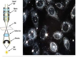
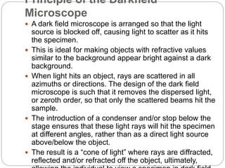
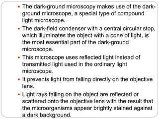
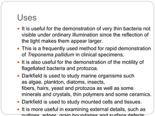


Darkfield microscopy uses oblique illumination to make specimens appear bright against a dark background. Light is directed around the specimen so that it is scattered and refracted off of it. This allows thin structures like bacteria to be seen more easily compared to brightfield microscopy. Some applications of darkfield microscopy include viewing unstained live samples, motile organisms, fibers, and external surface details of cells. It has advantages like simplicity, quality images, and lack of artifacts, though light levels are lower.


![谷歌留痕技术 [ 𝙩𝙤𝙥 𝟮𝟯𝟯. 𝙘 𝙤𝙢 ]](https://cdn.slidesharecdn.com/ss_thumbnails/top233-260130174328-3833018c-thumbnail.jpg?width=640&height=640&fit=bounds)
