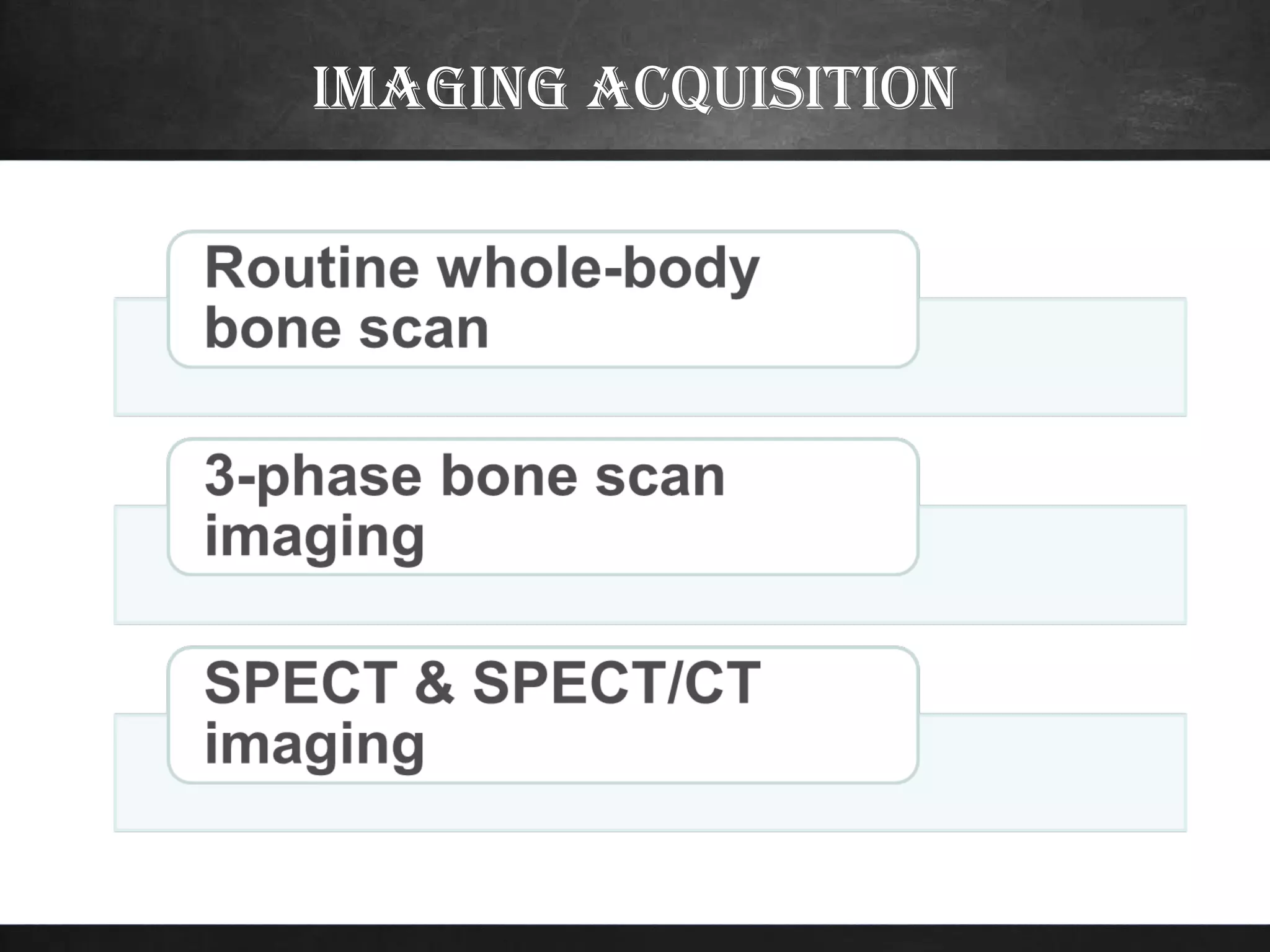
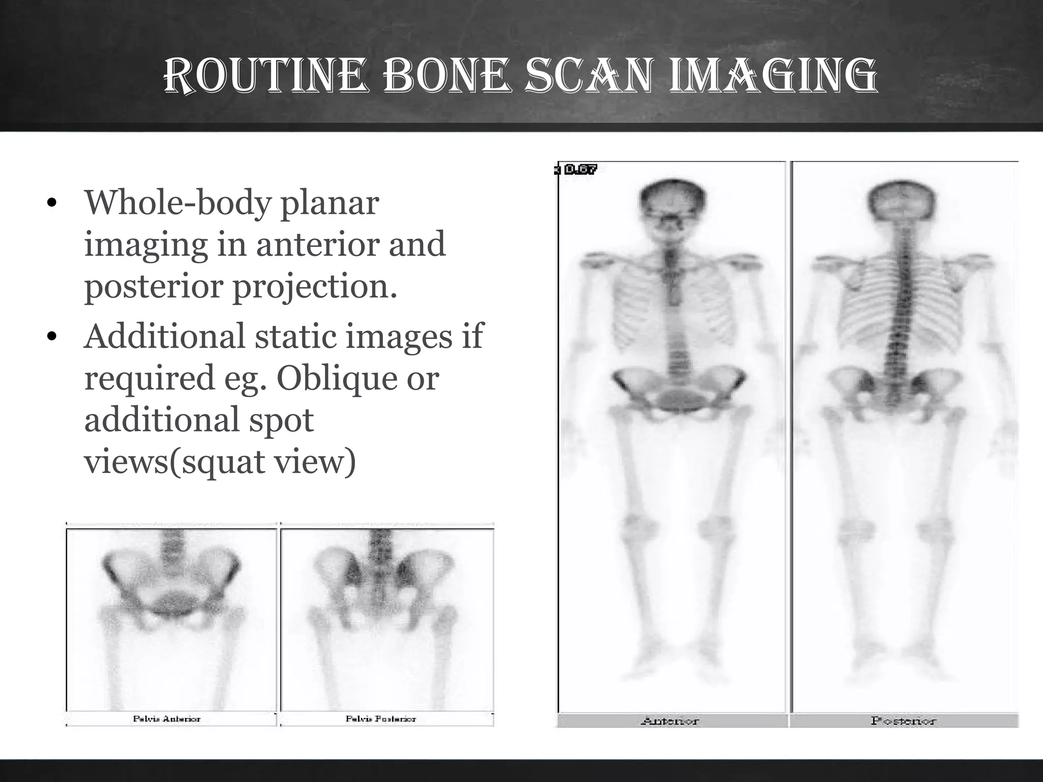
Definition
• A bone scan is a test that detects areas of
increased or decreased bone activity by
injecting a certain radiopharmaceutical i.e.
Tc-99m MDP. This may indicate bone
injury or disease.
SenSitivity anD
Specificity
• Bone scan is very sensitive study but it is not
specific.
• Although findings on bone scan are non-specific,
its monostotic or polyostotic status and
anatomical distribution can provide important
clues to the differential diagnosis.
aDvantageS vS DiSaDvantageS
Advantages Disadvantages
 Needs radiopharmaceuticals
& gamma camera not widely
available.
 Low specificity.
 Cost is high.
 Whole-body evaluation in one
test/ same rad exposure.
 Low radiation exposure.
 Sensitive evaluation
anatomy
RadiophaRmaceuticals
 Bone-seeking agents are analogs of calcium,
phosphates.
Tc-99m Methylene diphosphonate
(Tc-99m MDP)
Tc-99m MDP
The most widely used
Radiopharmaceuticals is
Tc-99m labeled
diphosphonates
tc-99m mdp
• Used within 4 hrs after preparation.
• High target/bcg ratio within 2-3 hrs. with
• Rapid renal clearance
• Renal Impairment: increased soft tissue
activity, poor image quality, delayed excretion,
higher radiation exposure due to retained high
activity
technique
• Preparation: None
• Injection of Tc-99m 20-25
mCi IV, (250 uCi/Kg) for
children, good hydration
afterwards & frequent
voiding
• Wait for about 2-3 hrs to
start imaging, avoid
contamination
• Empty bladder prior to
scanning
• Change the cloth and remove
things likely cause artifact.
pooR image quality
• Renal impairment
• Decreased cardiac output
• Dehydration
• Extravasation of the radiopharamceutical
• Aging.
• Underdosage of the radiopharmacetical.
• Obesity.
imaging acquisition
Routine Bone scan imaging
• Whole-body planar
imaging in anterior and
posterior projection.
• Additional static images if
required eg. Oblique or
additional spot
views(squat view)
• This is the bony phase of bone scan.
• Inject radiopharmaceutical and image in 2-
4 hours.
• When we say bone scan, we usually mean
whole body bone scan.
3-phase bone scan imaging
• Phase 1; Vascular phase: 60 s
dynamic immediately post
injection
• Phase 2; Soft-tissue (blood-
pool) phase: 5 min post
injection.
• Phase 3; Delayed (bone)
phase: 2-3 hr post injection.
Soft-tissue delayed 2-3hr
Vascular phase
First phase
• 30-60 dynamic images are usually obtained
over 1 minute immediately after injection.
• This is radionuclide angiography and gives an
idea about the local vasculature. During the
first minute after injection, injected dose is
still intravascular.
second phase
• Static image is obtained in 5 minutes after
dose injection.
• Within 5 minutes post injection,
radiopharmaceutical moves from
intravascular space to extravascular space
(soft tissue). It gives idea about soft tissue
edema.
blood pool
Hyperemia
• If there is focal increased activity in
the first and second phases,
hyperemia or acute inflammatory
process is present.
THird pHase
• It is the bony phase image obtained in
2-4 hours post injection.
• It is the same as whole body bone
scan.
indicaTions
 Neoplastic disease: Primary bone tumors & Bone metastasis
– Staging for malignancies that have high incidence of bone
metastasis eg. cancers of prostate, breast, lung,
neuroblastoma.
– Unexplained bone pain in a patient with known malignancy (neg X-ray)
– Unexplained bone pain in a patient with no history of
malignancy.
 Trauma: Fracture/ Stress injuries (eg, stress fracture, shin splints).
 Infection:(osteomyelitis)
 Avascular necrosis
 Arthritis:Degenerative changes
 Metabolic & Endocrine disorders: (eg, Paget’s Disease,
Hyperparathyroidism)
 Bone marrow diseases: Sickle cell disease
clinical applicaTions
• Malignancy
1- Primary bone cancer
2- Secondary metastatic bone disease
• Osteomyelitis
• Stress fractures
THANK YOU

Bone scan

  • 3.
    Definition • A bonescan is a test that detects areas of increased or decreased bone activity by injecting a certain radiopharmaceutical i.e. Tc-99m MDP. This may indicate bone injury or disease.
  • 4.
    SenSitivity anD Specificity • Bonescan is very sensitive study but it is not specific. • Although findings on bone scan are non-specific, its monostotic or polyostotic status and anatomical distribution can provide important clues to the differential diagnosis.
  • 5.
    aDvantageS vS DiSaDvantageS AdvantagesDisadvantages  Needs radiopharmaceuticals & gamma camera not widely available.  Low specificity.  Cost is high.  Whole-body evaluation in one test/ same rad exposure.  Low radiation exposure.  Sensitive evaluation
  • 6.
  • 7.
    RadiophaRmaceuticals  Bone-seeking agentsare analogs of calcium, phosphates. Tc-99m Methylene diphosphonate (Tc-99m MDP) Tc-99m MDP The most widely used Radiopharmaceuticals is Tc-99m labeled diphosphonates
  • 8.
    tc-99m mdp • Usedwithin 4 hrs after preparation. • High target/bcg ratio within 2-3 hrs. with • Rapid renal clearance • Renal Impairment: increased soft tissue activity, poor image quality, delayed excretion, higher radiation exposure due to retained high activity
  • 9.
    technique • Preparation: None •Injection of Tc-99m 20-25 mCi IV, (250 uCi/Kg) for children, good hydration afterwards & frequent voiding • Wait for about 2-3 hrs to start imaging, avoid contamination • Empty bladder prior to scanning • Change the cloth and remove things likely cause artifact.
  • 10.
    pooR image quality •Renal impairment • Decreased cardiac output • Dehydration • Extravasation of the radiopharamceutical • Aging. • Underdosage of the radiopharmacetical. • Obesity.
  • 11.
  • 12.
    Routine Bone scanimaging • Whole-body planar imaging in anterior and posterior projection. • Additional static images if required eg. Oblique or additional spot views(squat view)
  • 13.
    • This isthe bony phase of bone scan. • Inject radiopharmaceutical and image in 2- 4 hours. • When we say bone scan, we usually mean whole body bone scan.
  • 15.
    3-phase bone scanimaging • Phase 1; Vascular phase: 60 s dynamic immediately post injection • Phase 2; Soft-tissue (blood- pool) phase: 5 min post injection. • Phase 3; Delayed (bone) phase: 2-3 hr post injection. Soft-tissue delayed 2-3hr Vascular phase
  • 16.
    First phase • 30-60dynamic images are usually obtained over 1 minute immediately after injection. • This is radionuclide angiography and gives an idea about the local vasculature. During the first minute after injection, injected dose is still intravascular.
  • 17.
    second phase • Staticimage is obtained in 5 minutes after dose injection. • Within 5 minutes post injection, radiopharmaceutical moves from intravascular space to extravascular space (soft tissue). It gives idea about soft tissue edema.
  • 18.
  • 19.
    Hyperemia • If thereis focal increased activity in the first and second phases, hyperemia or acute inflammatory process is present.
  • 20.
    THird pHase • Itis the bony phase image obtained in 2-4 hours post injection. • It is the same as whole body bone scan.
  • 21.
    indicaTions  Neoplastic disease:Primary bone tumors & Bone metastasis – Staging for malignancies that have high incidence of bone metastasis eg. cancers of prostate, breast, lung, neuroblastoma. – Unexplained bone pain in a patient with known malignancy (neg X-ray) – Unexplained bone pain in a patient with no history of malignancy.  Trauma: Fracture/ Stress injuries (eg, stress fracture, shin splints).  Infection:(osteomyelitis)  Avascular necrosis  Arthritis:Degenerative changes  Metabolic & Endocrine disorders: (eg, Paget’s Disease, Hyperparathyroidism)  Bone marrow diseases: Sickle cell disease
  • 22.
    clinical applicaTions • Malignancy 1-Primary bone cancer 2- Secondary metastatic bone disease • Osteomyelitis • Stress fractures
  • 23.