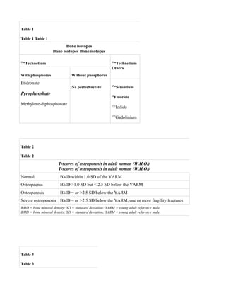
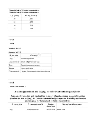
This document discusses various medical applications of radioisotopes and isotope scans, including salivary gland scanning, Meckel's diverticulum scans, carotid body tumor scans, bone scans, osteoporosis scans using gadolinium or x-rays, venous thrombosis scans using fibrinogen or albumin isotopes, and mediastinal scans. It provides technical details on the tracers and principles used in each type of scan.
An overview of Renography - the medical imaging of kidneys using Nuclear Medicine - including its advantages and disadvantages over other Radiographic imaging modalities.
The subtotal laparoscopic pancreatic resection can safely be performed. The da Vinci robotic system allowed for technical refinements of laparoscopic pancreatic resection. Robotic assistance improved the dissection and control of major blood vessels due to three-dimensional visualization of the operative field and instruments with wrist-type end-effectors.
Progress in liver surgery has enabled hepatectomy with concomitant venous resection for liver malignancies involving the inferior vena cava (IVC). The authors describe an alternative technique for IVC reconstruction without the need of graft.
http://www.drmarcel.com.br
Laparoscopic intrahepatic Glissonian technique for liver surgery. Hepatectomi...Marcel Autran Machado
The main advantage of the intrahepatic Glissonian procedure over other techniques is the possibility of gaining a rapid and precise access to the left Glissonian sheaths facilitating left hemihepatectomy, bisegmentectomy 2-3, and individual resections of segments 2, 3, and 4. The authors believe that the intrahepatic Glissonian technique facilitates laparoscopic liver resection and may increase the development of segment-based laparoscopic liver resection.
Laparoscopic adrenalectomy in patients with subclinical cushing syndrome | γι...Γιώργος Ζωγράφος
Abstract:
Background Subclinical Cushing syndrome in patients with adrenal incidentalomas has been associated with an increased prevalence of the metabolic syndrome and car- diovascular risk. The management of these patients, be it conservative or surgical, is still debated, but there is accumulating evidence that surgery is best and that lapa- roscopic adrenalectomy, when possible, is the most pre- ferred procedure. Here we present the short- and long-term results of laparoscopic adrenalectomy for subclinical Cushing syndrome and determine the effect of this proce- dure on components of the metabolic syndrome.
Methods Twenty-nine patients, 8 men and 21 women with adrenal incidentalomas and subclinical Cushing syn- drome who underwent laparoscopic adrenalectomy, were studied retrospectively. They had undergone postoperative follow-up for improvement or worsening of their arterial blood pressure, body weight, and fasting glucose level for a mean period of 77 months.
Results:
Preoperatively, 17 patients (58.6 %) had arterial hypertension, 14 (48.3%) had a body mass index exceeding 27 kg/m2, and 12 (41.4 %) had diabetes melli- tus. Postoperatively, a decrease in mean arterial pressure was found in 12 patients (70.6 %), a decrease in body mass index in 6 patients (42.9 %), and an improvement in gly- cemic control in 5 patients (41.7 %).
Conclusions Laparoscopic adrenalectomy is beneficial in many patients with subclinical Cushing syndrome because it reduces arterial blood pressure, body weight, and fasting glucose levels. Prospective randomized studies are needed to compare laparoscopic adrenalectomy with a conserva- tive approach and to confirm these results.
The IOSR Journal of Pharmacy (IOSRPHR) is an open access online & offline peer reviewed international journal, which publishes innovative research papers, reviews, mini-reviews, short communications and notes dealing with Pharmaceutical Sciences( Pharmaceutical Technology, Pharmaceutics, Biopharmaceutics, Pharmacokinetics, Pharmaceutical/Medicinal Chemistry, Computational Chemistry and Molecular Drug Design, Pharmacognosy & Phytochemistry, Pharmacology, Pharmaceutical Analysis, Pharmacy Practice, Clinical and Hospital Pharmacy, Cell Biology, Genomics and Proteomics, Pharmacogenomics, Bioinformatics and Biotechnology of Pharmaceutical Interest........more details on Aim & Scope).
All manuscripts are subject to rapid peer review. Those of high quality (not previously published and not under consideration for publication in another journal) will be published without delay
Undoubtedly, the use of radiographic imaging has entirely revolutionized diagnosis and treatment planning in medical sciences. The role of imaging in oral malignancies can be broadly grouped into those used to evaluate primary disease and those to evaluate metastatic disease.
It is a useful tool for staging and management planning in oral cancers. Awareness of the presence of cervical node metastasis is important in treatment planning and in prognostic prediction for patients with head and neck cancer (HNC).
. Panoramic radiography (also called pan tomography or rotational radiography) is a radiographic technique for producing a single image of the facial structures that include both maxillary and mandibular arches and their supporting structures.
Undoubtedly, the use of radiographic imaging has entirely revolutionized the diagnosis and treatment planning in medical sciences. The role of imaging in oral malignancies can be broadly grouped in those used to evaluate primary disease and those to evaluate metastatic disease.
Magnetic Resonance Imaging is a technique that uses the magnetic field and radio waves to create detail images of the organs and tissues of the human body
Skull Metastasis From Papillary Thyroid Carcinoma : Case Report and Literatur...komalicarol
Although papillary thyroid carcinoma is a relatively common form of malignancy, metastatic spread to the skull
is exceptional. Here, we report a case of papillary thyroid carcinoma revealed by frontal skull metastasis.
Skull Metastasis from Papillary Thyroid Carcinoma: Case Report and Literature...semualkaira
Although papillary thyroid carcinoma is a relatively common form of malignancy, metastatic spread to the skull
is exceptional. Here, we report a case of papillary thyroid carcinoma revealed by frontal skull metastasis.
Application Brief: Tumor Microenvironment Imaging with Photoacoustic TechnologyFUJIFILM VisualSonics Inc.
Photoacoustics (PA) combines optical contrast with the high spatial resolution and deep tissue penetration offered by ultrasound. Such applications are especially beneficial for monitoring tumor development, measuring blood concentration changes within it, and quantifying networks of vasculature formation and carcinoma growth over time.
Description (with pictures) of successful (and failed) lunar landings - supplemented with numerous pictures of Lunar Surfaces, with Craters and all.
Disclaimer and Credits
All material (Text, Images, Graphics etc.) in these slides have been procured from publicly available sources of ISRO and other agencies, which includes ISRO partners. There are selected images from NASA and Wikipedia also, where relevant, also procured from freely available public resources, with attribution. Some images and text have been individually acknowledged. Others have been collectively credited through this Disclaimer page. The author makes no copyright claims on any material.
They have been sorted, edited as relevant, collated, compiled and inserted to align them with the sequence of the slides, as deemed fit by the author. They have been posted with academic altruism in mind, for those interested in Astrophysics and Astronomy and related technology, like the author. There is no commercial or promotional motivation involved anywhere.
The author is not an Astrophysicist or an Astronomer. The author does not work for ISRO, NASA or any tech company. The author is a nerd who loves technology, Astrophysics and Astronomy and who dabbles in related developments of ISRO, NASA etc. during his spare time, as an intellectual hobby. Thus, he satiates his academic appetite, learns in the process and wishes to share them with like-minded people.
At the time of publication, all material has been updated and is deemed to be accurate. If any errors are detected by the reader(s) the author will be happy to be corrected. The responsibility for any errors are solely the author’s and not that of the parent organization(s).
Here’s is wishing everyone a happy armchair space exploration on this occasion of New Year 2024!
Updated as on 31 January 2024
JAXA being an ethical Space Agency disclosed the real reason why SLIM Lander could not communicate for 1st week after soft-landing on Moon.
Picture released by JAXA
A. SLIM lost one of its Engines during descent
B. Because of the resultant asymmetric Thrust, SLIM landed on Lunar Surface upside down
C. Resulting in its Solar Panels facing away from Sun (See the direction of shadows to determine relative position of Sun and Solar Panels)
MARS Images ISRO-NASA-Compiled by Sanjoy SanyalSanjoy Sanyal
MCC Imaging Timeline
28 September 2014 – MOM (Mars Orbiter Mission) controllers published the spacecraft's first global view of Mars. The image was captured by the Mars Color Camera (MCC)
4 March 2015 – MCC was returning new images of Martian surface
24 September 2015 – ISRO released ‘Mars Atlas’, a 120-page scientific atlas containing images and data from MOM’s 1st year in orbit
19 May 2017 – MOM reached 1,000 days (973 sols (Martian Days)) in orbit around Mars. In that time, the spacecraft completed 388 orbits of the planet and relayed > 715 images back to Earth
24 September 2018 – MOM completed 4 years in its orbit around Mars, although the designed mission life was only 6 months. Over these years, MOM’s MCC captured > 980 images that were released to the public
24 September 2019 – MOM completed 5 years in orbit around Mars, sending 2 TB of imaging data
1 July 2020 – MOM captured a photo of Mars satellite Phobos from 4,200 km away
18 July 2021 – MCC captured full disc image of Mars from an altitude of 75,000 km with spatial resolution about 3.7 km
October 2022 – MCC produced 1,100+ images before retirement
DISCLAIMER
All material (Text, Images, Graphics etc.) in these slides have been procured from publicly available sources of ISRO and other agencies, which includes ISRO partners. There are selected images from NASA also, where relevant, also procured from freely available public resources. Instead of individually acknowledging each image they have been collectively credited through this Disclaimer page. The author makes no copyright claims on any material.
They have been sorted, edited as relevant, collated and inserted to align them with the sequence of the slides, as deemed fit by the author. They have been posted with academic altruism in mind, for those interested in Astrophysics and Astronomy and related technology, like the author. There is no commercial or promotional motivation involved anywhere.
The author is not an Astrophysicist or an Astronomer. The author does not work for ISRO, NASA or any tech company. The author is a nerd who loves technology, Astrophysics and Astronomy and who dabbles in related developments of ISRO, NASA etc. during his spare time as an intellectual hobby. Thus, he satiates his academic appetite, learns in the process and wishes to share them with like-minded people.
At the time of publication, all material has been updated and is deemed to be accurate. If any errors are detected by the reader(s), I shall be happy to be corrected. The responsibility for any errors are solely mine and not that of the parent organizations.
Here’s is wishing everyone a happy armchair space exploration on this occasion of New Year 2024!
Aditya-L1 Suit Images ISRO - Compiled by Sanjoy Sanyal.pptxSanjoy Sanyal
Solar Ultraviolet Imaging Telescope (SUIT):
Instrument on board Aditya-L1 Spacecraft has successfully captured the first full-disk images of the Sun in 200-400 nm Wavelength range
SUIT captures images of the Sun's Photosphere and Chromosphere in this wavelength range using various Filters
20 November 2023: SUIT payload was powered ON
Successful pre-commissioning phase
6 December 2023: SUIT captured its first light science images
Images: Taken using 11 different Filters (Slide 4), include the first-ever full-disk representations of the Sun in Wavelengths ranging from 200 to 400 nm, excluding Ca II h
Notable Features: Sunspots, Plage (Chromosphere variant of Faculae), Limb Darkening, and Quiet Regions of Sun, as marked in Mg II h image (Slide 7), provide scientists with insights into the intricate details of Sun's Photosphere and Chromosphere
SUIT observations will help scientists study the dynamic coupling of magnetized solar atmosphere and assist them in determining the effects of solar radiation on Earth's climate
DISCLAIMER
All material (Text, Images, Graphics etc.) in these slides have been procured from publicly available sources of ISRO and other agencies, which includes ISRO partners. There are selected images from NASA also, where relevant, also procured from freely available public resources. Instead of individually acknowledging each image they have been collectively credited through this Disclaimer page. The author makes no copyright claims on any material.
They have been sorted, edited as relevant, collated and inserted to align them with the sequence of the slides, as deemed fit by the author. They have been posted with academic altruism in mind, for those interested in Astrophysics and Astronomy and related technology, like the author. There is no commercial or promotional motivation involved anywhere.
The author is not an Astrophysicist or an Astronomer. The author does not work for ISRO, NASA or any tech company. The author is a nerd who loves technology, Astrophysics and Astronomy and who dabbles in related developments of ISRO, NASA etc. during his spare time as an intellectual hobby. Thus, he satiates his academic appetite, learns in the process and wishes to share them with like-minded people.
At the time of publication, all material has been updated and is deemed to be accurate. If any errors are detected by the reader(s), I shall be happy to be corrected. The responsibility for any errors are solely mine and not that of the parent organizations.
Here’s is wishing everyone a happy armchair space exploration on this occasion of New Year 2024!
Charting Neural Pathways in Schizophrenia and BPD-Chicago Conference 2016 - S...Sanjoy Sanyal
This was presented by Dr. Sanjoy Sanyal, Professor, Surgeon, Neuroscientist, Informatician, at 2nd International Conference on Brain Disorders and Therapeutics, Chicago, USA, October 26-28, 2016
Types of Schizophrenia
Types of Bipolar Disorder (BPD)
DTI Findings in Schizophrenia / BPD
Videos of White Matter Affected in Psychosis
Brain Network Concepts
Basal Forebrain Components and VTA
Videos of Meso-limbic / Meso-cortical Tracts
Receptors in Psychotic Disorders
Videos of Pathophysiology in Schizophrenia 1 and 2 – Rx Principles
Future Research Possibilities
Summary and Conclusion
Thank you for watching.
Aorta–IVC–Kidney Dissection and Surgical Correlations - Dr Sanjoy SanyalSanjoy Sanyal
Educational PPTX created by Dr. Sanjoy Sanyal; Professor, Department Chair, Surgeon, Neuroscientist and Medical Informatician
It shows the surgical anatomy of the posterior abdominal contents, with special emphasis on the aorta, IVC and Kidney-Ureters. The specimen was harvested from a cadaver.
With real-time narration and relevant captions, it enhances the learning experience by means of a trimodal learning style approach - Visual, Auditory, Textual.
Thank you for watching. If you have any questions or comments, please put them in the comments section below. Have a nice day!
Educational Video created by Dr Sanjoy Sanyal; Professor, Surgeon, Medical Informatician and Department Chair in the Western Hemisphere.
A section of the anterior chest wall from a cadaver is described - the Bones, Muscles, Vessels, and some Clinical Correlations
Camera person is Ms. Selvie Krishna, an enthusiastic student with her Blackberry.
Errata Corrigendum: There is an inadvertent error in my narration, where I mentioned Sternum instead of Vertebral Column. The same was corrected immediately thereafter in my narration, and the error and correction have also been captioned in the body of the video.
Thank you for watching. If there are questions or comments, put them in the comments section below.
Dissections of the calf and its functional and surgical aspects have been discussed in real time by Dr. Sanjoy Sanyal, Professor, Surgeon, Neuroscientist and Medical Informatician.
Important points discussed are: Triceps surae, Gastrocnemius, Soleus, Plantaris, Tendo calcaneus, Love and Whelan classification, Plantar reflex, calcaneal bursitis, calcaneal tendinitis, calcaneal tendon rupture, Gastrocnemius strain, tennis leg, PAES, accessory soleus
Educational importance lies in the following aspects: Combination of audio, video, graphics and textual description in real time
Surgical Aspects of Popliteal Fossa - Dr. Sanjoy SanyalSanjoy Sanyal
Dissection of the popliteal fossa and its surgical aspects has been discussed in real time by Dr. Sanjoy Sanyal, Professor, Surgeon and Neuroscientist.
Important points discussed are: Popliteal fossa, Palpation, Popliteal artery entrapment, PAES, Popliteal aneurysm, Popliteal AV fistula, Popliteal hemorrhage, Genicular anastomosis, Popliteal cyst, Baker cyst, Morrant baker cyst, Heidelberg classification, Love and Whelan classification
Educational importance lies in the following aspects: Combination of audio, video, graphics and textual description in real time
Surgical Anatomy of Cadaveric Abdominal Viscera - Dr Sanjoy SanyalSanjoy Sanyal
Educational Video created by Dr Sanjoy Sanyal; Professor, Surgeon, Neuroscientist and Medical Informatician
A section of abdominal viscera from a cadaver has been described - Stomach, Spleen, Colon, Greater Omenutm, with some clinical and surgical correlations
Corrigendum: Please disregard the inadvertent error when the Gastrosplenic ligament is being described - GS ligament contains Gastro-epiploic vessels; Spleno-renal ligament contains the Splenic vessels
This was presented by Dr Sanjoy Sanyal at the 2016 International Education Conference in Orlando, FL on 4 January 2016 in Disney's Boardwalk Inn.
It was voted the best paper presentation of the session by the attendees.
Educational Video created by Dr Sanjoy Sanyal; Professor, Surgeon and Medical Informatician
Deals with Blended / Hybrid Learning, Rotation Model, Flipped Classroom, Student responses, Audience Response System Clicker,
Abnormal Right Vertebral Artery MRA Sequence - Sanjoy SanyalSanjoy Sanyal
This is an MR Angiography sequence of a 46-year old male patient who was being investigated for TIA. The image sequence shows 3-D Time of Flight (TOF) Spoiled Gradient Recall (SPGR) Echo Acquisition images. It shows the Vertebrobasilar and Carotid systems of Cerebral circulation. An incidental finding was abnormal Right Vertebral artery - Narrow, Double, Accessory, Communication with Right Internal Carotid. The best way to visualize the image is by slideshow - imagine the head is rotating clockwise. There are plenty of labels in the images to guide the viewer.
Ionizing Radiation in Surgery - Sanjoy SanyalSanjoy Sanyal
Ionizing radiations exert their biological effects by excitation and ionization of molecules within cells. In terms of energy deposited within cells, ionizing radiations are the most potent of all physical and chemical agents
Lasers in Surgery Systemic Applications Part-III - Sanjoy SanyalSanjoy Sanyal
Applications of lasers in organ-systems of the body are at the cross-roads today, with limitless horizon ahead of it. The authors dwell upon the applications in important oragns of the human body.
Illustrated Surgical GI Endoscopy - Sanjoy SanyalSanjoy Sanyal
Therapeutic endoscopy has made considerable inroads in the treatment of surgical disorders of the GI tract. This has been brought about by technological innovations in the hardware and the ingenuity of the clinician in accessing the lesion.
Lasers in Surgery Specific Applications Part-II - Sanjoy SanyalSanjoy Sanyal
Experiments with lasers are going on a hectic pace in order to improve upon the existing applications of lasers in surgery. However it behoves the surgeon to be cognizant of its potential hazards and to take appropriate precautions.
Automatic Physiological Assessment in Surgery Computer Program - Sanjoy SanyalSanjoy Sanyal
Computer programs for automatic interpretation of physiological variables in critically ill surgical patients are quick and efficient decision-making aids to the clinician.
Surgical Aspects of Colorectal Endoscopy Part-IV - Sanjoy SanyalSanjoy Sanyal
Colorectal endoscopy differs significantly from UGI endoscopy. The authors describe some differential aspects, some common and some exotic coloscopic findings.
Local Advanced Lung Cancer: Artificial Intelligence, Synergetics, Complex Sys...Oleg Kshivets
Overall life span (LS) was 1671.7±1721.6 days and cumulative 5YS reached 62.4%, 10 years – 50.4%, 20 years – 44.6%. 94 LCP lived more than 5 years without cancer (LS=2958.6±1723.6 days), 22 – more than 10 years (LS=5571±1841.8 days). 67 LCP died because of LC (LS=471.9±344 days). AT significantly improved 5YS (68% vs. 53.7%) (P=0.028 by log-rank test). Cox modeling displayed that 5YS of LCP significantly depended on: N0-N12, T3-4, blood cell circuit, cell ratio factors (ratio between cancer cells-CC and blood cells subpopulations), LC cell dynamics, recalcification time, heparin tolerance, prothrombin index, protein, AT, procedure type (P=0.000-0.031). Neural networks, genetic algorithm selection and bootstrap simulation revealed relationships between 5YS and N0-12 (rank=1), thrombocytes/CC (rank=2), segmented neutrophils/CC (3), eosinophils/CC (4), erythrocytes/CC (5), healthy cells/CC (6), lymphocytes/CC (7), stick neutrophils/CC (8), leucocytes/CC (9), monocytes/CC (10). Correct prediction of 5YS was 100% by neural networks computing (error=0.000; area under ROC curve=1.0).
NVBDCP.pptx Nation vector borne disease control programSapna Thakur
NVBDCP was launched in 2003-2004 . Vector-Borne Disease: Disease that results from an infection transmitted to humans and other animals by blood-feeding arthropods, such as mosquitoes, ticks, and fleas. Examples of vector-borne diseases include Dengue fever, West Nile Virus, Lyme disease, and malaria.
263778731218 Abortion Clinic /Pills In Harare ,sisternakatoto
263778731218 Abortion Clinic /Pills In Harare ,ABORTION WOMEN’S CLINIC +27730423979 IN women clinic we believe that every woman should be able to make choices in her pregnancy. Our job is to provide compassionate care, safety,affordable and confidential services. That’s why we have won the trust from all generations of women all over the world. we use non surgical method(Abortion pills) to terminate…Dr.LISA +27730423979women Clinic is committed to providing the highest quality of obstetrical and gynecological care to women of all ages. Our dedicated staff aim to treat each patient and her health concerns with compassion and respect.Our dedicated group ABORTION WOMEN’S CLINIC +27730423979 IN women clinic we believe that every woman should be able to make choices in her pregnancy. Our job is to provide compassionate care, safety,affordable and confidential services. That’s why we have won the trust from all generations of women all over the world. we use non surgical method(Abortion pills) to terminate…Dr.LISA +27730423979women Clinic is committed to providing the highest quality of obstetrical and gynecological care to women of all ages. Our dedicated staff aim to treat each patient and her health concerns with compassion and respect.Our dedicated group of receptionists, nurses, and physicians have worked together as a teamof receptionists, nurses, and physicians have worked together as a team wwww.lisywomensclinic.co.za/
These simplified slides by Dr. Sidra Arshad present an overview of the non-respiratory functions of the respiratory tract.
Learning objectives:
1. Enlist the non-respiratory functions of the respiratory tract
2. Briefly explain how these functions are carried out
3. Discuss the significance of dead space
4. Differentiate between minute ventilation and alveolar ventilation
5. Describe the cough and sneeze reflexes
Study Resources:
1. Chapter 39, Guyton and Hall Textbook of Medical Physiology, 14th edition
2. Chapter 34, Ganong’s Review of Medical Physiology, 26th edition
3. Chapter 17, Human Physiology by Lauralee Sherwood, 9th edition
4. Non-respiratory functions of the lungs https://academic.oup.com/bjaed/article/13/3/98/278874
ABDOMINAL TRAUMA in pediatrics part one.drhasanrajab
Abdominal trauma in pediatrics refers to injuries or damage to the abdominal organs in children. It can occur due to various causes such as falls, motor vehicle accidents, sports-related injuries, and physical abuse. Children are more vulnerable to abdominal trauma due to their unique anatomical and physiological characteristics. Signs and symptoms include abdominal pain, tenderness, distension, vomiting, and signs of shock. Diagnosis involves physical examination, imaging studies, and laboratory tests. Management depends on the severity and may involve conservative treatment or surgical intervention. Prevention is crucial in reducing the incidence of abdominal trauma in children.
Tom Selleck Health: A Comprehensive Look at the Iconic Actor’s Wellness Journeygreendigital
Tom Selleck, an enduring figure in Hollywood. has captivated audiences for decades with his rugged charm, iconic moustache. and memorable roles in television and film. From his breakout role as Thomas Magnum in Magnum P.I. to his current portrayal of Frank Reagan in Blue Bloods. Selleck's career has spanned over 50 years. But beyond his professional achievements. fans have often been curious about Tom Selleck Health. especially as he has aged in the public eye.
Follow us on: Pinterest
Introduction
Many have been interested in Tom Selleck health. not only because of his enduring presence on screen but also because of the challenges. and lifestyle choices he has faced and made over the years. This article delves into the various aspects of Tom Selleck health. exploring his fitness regimen, diet, mental health. and the challenges he has encountered as he ages. We'll look at how he maintains his well-being. the health issues he has faced, and his approach to ageing .
Early Life and Career
Childhood and Athletic Beginnings
Tom Selleck was born on January 29, 1945, in Detroit, Michigan, and grew up in Sherman Oaks, California. From an early age, he was involved in sports, particularly basketball. which played a significant role in his physical development. His athletic pursuits continued into college. where he attended the University of Southern California (USC) on a basketball scholarship. This early involvement in sports laid a strong foundation for his physical health and disciplined lifestyle.
Transition to Acting
Selleck's transition from an athlete to an actor came with its physical demands. His first significant role in "Magnum P.I." required him to perform various stunts and maintain a fit appearance. This role, which he played from 1980 to 1988. necessitated a rigorous fitness routine to meet the show's demands. setting the stage for his long-term commitment to health and wellness.
Fitness Regimen
Workout Routine
Tom Selleck health and fitness regimen has evolved. adapting to his changing roles and age. During his "Magnum, P.I." days. Selleck's workouts were intense and focused on building and maintaining muscle mass. His routine included weightlifting, cardiovascular exercises. and specific training for the stunts he performed on the show.
Selleck adjusted his fitness routine as he aged to suit his body's needs. Today, his workouts focus on maintaining flexibility, strength, and cardiovascular health. He incorporates low-impact exercises such as swimming, walking, and light weightlifting. This balanced approach helps him stay fit without putting undue strain on his joints and muscles.
Importance of Flexibility and Mobility
In recent years, Selleck has emphasized the importance of flexibility and mobility in his fitness regimen. Understanding the natural decline in muscle mass and joint flexibility with age. he includes stretching and yoga in his routine. These practices help prevent injuries, improve posture, and maintain mobilit
These lecture slides, by Dr Sidra Arshad, offer a quick overview of the physiological basis of a normal electrocardiogram.
Learning objectives:
1. Define an electrocardiogram (ECG) and electrocardiography
2. Describe how dipoles generated by the heart produce the waveforms of the ECG
3. Describe the components of a normal electrocardiogram of a typical bipolar lead (limb II)
4. Differentiate between intervals and segments
5. Enlist some common indications for obtaining an ECG
6. Describe the flow of current around the heart during the cardiac cycle
7. Discuss the placement and polarity of the leads of electrocardiograph
8. Describe the normal electrocardiograms recorded from the limb leads and explain the physiological basis of the different records that are obtained
9. Define mean electrical vector (axis) of the heart and give the normal range
10. Define the mean QRS vector
11. Describe the axes of leads (hexagonal reference system)
12. Comprehend the vectorial analysis of the normal ECG
13. Determine the mean electrical axis of the ventricular QRS and appreciate the mean axis deviation
14. Explain the concepts of current of injury, J point, and their significance
Study Resources:
1. Chapter 11, Guyton and Hall Textbook of Medical Physiology, 14th edition
2. Chapter 9, Human Physiology - From Cells to Systems, Lauralee Sherwood, 9th edition
3. Chapter 29, Ganong’s Review of Medical Physiology, 26th edition
4. Electrocardiogram, StatPearls - https://www.ncbi.nlm.nih.gov/books/NBK549803/
5. ECG in Medical Practice by ABM Abdullah, 4th edition
6. Chapter 3, Cardiology Explained, https://www.ncbi.nlm.nih.gov/books/NBK2214/
7. ECG Basics, http://www.nataliescasebook.com/tag/e-c-g-basics
micro teaching on communication m.sc nursing.pdfAnurag Sharma
Microteaching is a unique model of practice teaching. It is a viable instrument for the. desired change in the teaching behavior or the behavior potential which, in specified types of real. classroom situations, tends to facilitate the achievement of specified types of objectives.
Title: Sense of Taste
Presenter: Dr. Faiza, Assistant Professor of Physiology
Qualifications:
MBBS (Best Graduate, AIMC Lahore)
FCPS Physiology
ICMT, CHPE, DHPE (STMU)
MPH (GC University, Faisalabad)
MBA (Virtual University of Pakistan)
Learning Objectives:
Describe the structure and function of taste buds.
Describe the relationship between the taste threshold and taste index of common substances.
Explain the chemical basis and signal transduction of taste perception for each type of primary taste sensation.
Recognize different abnormalities of taste perception and their causes.
Key Topics:
Significance of Taste Sensation:
Differentiation between pleasant and harmful food
Influence on behavior
Selection of food based on metabolic needs
Receptors of Taste:
Taste buds on the tongue
Influence of sense of smell, texture of food, and pain stimulation (e.g., by pepper)
Primary and Secondary Taste Sensations:
Primary taste sensations: Sweet, Sour, Salty, Bitter, Umami
Chemical basis and signal transduction mechanisms for each taste
Taste Threshold and Index:
Taste threshold values for Sweet (sucrose), Salty (NaCl), Sour (HCl), and Bitter (Quinine)
Taste index relationship: Inversely proportional to taste threshold
Taste Blindness:
Inability to taste certain substances, particularly thiourea compounds
Example: Phenylthiocarbamide
Structure and Function of Taste Buds:
Composition: Epithelial cells, Sustentacular/Supporting cells, Taste cells, Basal cells
Features: Taste pores, Taste hairs/microvilli, and Taste nerve fibers
Location of Taste Buds:
Found in papillae of the tongue (Fungiform, Circumvallate, Foliate)
Also present on the palate, tonsillar pillars, epiglottis, and proximal esophagus
Mechanism of Taste Stimulation:
Interaction of taste substances with receptors on microvilli
Signal transduction pathways for Umami, Sweet, Bitter, Sour, and Salty tastes
Taste Sensitivity and Adaptation:
Decrease in sensitivity with age
Rapid adaptation of taste sensation
Role of Saliva in Taste:
Dissolution of tastants to reach receptors
Washing away the stimulus
Taste Preferences and Aversions:
Mechanisms behind taste preference and aversion
Influence of receptors and neural pathways
Impact of Sensory Nerve Damage:
Degeneration of taste buds if the sensory nerve fiber is cut
Abnormalities of Taste Detection:
Conditions: Ageusia, Hypogeusia, Dysgeusia (parageusia)
Causes: Nerve damage, neurological disorders, infections, poor oral hygiene, adverse drug effects, deficiencies, aging, tobacco use, altered neurotransmitter levels
Neurotransmitters and Taste Threshold:
Effects of serotonin (5-HT) and norepinephrine (NE) on taste sensitivity
Supertasters:
25% of the population with heightened sensitivity to taste, especially bitterness
Increased number of fungiform papillae
1. Isotope scan in surgical diseases: Part 5
Sanjoy Sanyal MS (sanyal.sanjoy8@gmail.com)
Sr. Lecturer, National University of Rwanda
I. Nyaruhirira
Directeur,
Centre Hospitalier de Kigali, "C.H.K.",
Kigali, Rwanda.
In this concluding issue of the series in SURGERY journal (1999) on the use of radioisotopes in
surgery, the authors present a few miscellaneous applications
of radio-nuclides in bone and other minor organ systems.
Published in 2001 in SURGERY journal (Passi Publications), New Delhi, India
Salivary scintigraphy
As mentioned in the previous issue, 99mTcO4- (pertechnetate ion) is distributed like Cl- and is
concentrated in the salivary glands. Therefore sodium pertechnetate (Na99mTcO4) is used to ; (a) scan
the salivary glands, especially the parotids; and (b) assess salivary gland function.
Salivary tumours: All salivary tumours produce a ‘cold’ spot on 99mTc (technetium) scan except
adenolymphoma (Warthin’s tumour), which is a form of monomorphic adenoma. The latter takes up
more isotope than the rest of the gland to give a ‘hot’ spot on scan, so that a definite pre-operative
diagnosis can be made without biopsy.
Xerostomia: Dry mouth (xerostomia) occurs in; (a) Sjogren’s syndrome (keratoconjunctivitis sicca,
xerostomia and rheumatoid arthritis / SLE / scleroderma); (b) sicca syndrome (first 2 components of
Sjogren’s syndrome); (c) Mikulicz syndrome (leukaemic infiltration) or disease (Sjogren’s variant);
and (d) post-radiation. In these conditions 99mTc is used to quantify salivary gland function. Sequential
salivary scintigraphy with 99mTc pertechnetate confirms lack of salivary secretion and xerostomia.
2. Meckel’s diverticulum scan
Approximately half of the patients with Meckel’s diverticulum (MD) have ectopic gastric mucosa. If
the quantum of gastric mucosa is > 2 cm 3 (as in less than half of symptomatic patients), it can be
demonstrated by 99mTc pertechnetate scan. The ectopic gastric mucosa traps 99mTc just like it does Cl-.
In acute situations (Meckel's perforation, haemorrhage, diverticulitis), 30-100 µ Ci of 99mTc IV may
localise the gastric mucosa in 90% of cases. The uptake can be increased by administering pentagastrin
0.6 µg kg -1 SC. Administration of potassium perchlorate prior to injection of technetium reduces
uptake of the latter by the ectopic mucosa by virtue of competitive inhibition. 99mTc scanning may thus
be a promising diagnostic aid for MD, but instances of false-positive and false-negative results have
been reported.
Carotid body scan
Carotid body tumour (CBT) in the neck is the most common chemodectoma (nonchromaffin
paraganglioma) among those encountered from the base of skull to the thorax. It is situated in the
arterial adventitia (tunica media is hardly ever involved) at the carotid bifurcation (causing their
separation), and is fed by the external carotid artery. The differential diagnosis includes; (a) carotid
aneurysm; (b) branchial cyst; (c) neurofibroma; and (d) metastatic node.
99m
Tc pertechnetate scan: Peters et al described CBT scanning in 1979. It involves perfusion of 99mTc
pertechnetate, accompanied by dynamic angioscanning, that depicts blood flow through the tumour. It
is a useful screening procedure to differentiate it from the other lesions mentioned above, and is usually
performed prior to angiography. The latter and spiral CT are the most important diagnostic modalities.
Bone scan
Radio-nuclide skeletal scan is usually done to; (a) diagnose specific diseases of bone and joints; and (b)
elucidate the skeletal manifestations of disease elsewhere.
Isotopes
Technetium: 99mTc-{etidronate, methylene-diphosphonate(MDP), pyrophosphate(PYP)} are all
stannous (tin) chelates of 99mTc and the corresponding phosphorus-bearing compound (table 1).The first
two are diphosphonates and the last is a linear polyphosphate. The first is not hydrolysed by enzymes,
unlike polyphosphates. All have high affinity for Ca2+ and are highly specific bone-seeking compounds,
selectively localising in bone with kinetics similar to PO43-. 99mTc-pertechnetate ion also localises in
joints.
Miscellaneous: 87mSr(strontium) and 18F(fluorine) have been used for detecting skeletal metastases
from breast cancer and 131I (iodine) for detecting same from thyroid cancer. 87mSr (T1/2=2.8 hours) is
suitable for clinical scanning. The importance of 85, 89, 90Sr (T1/2=65 days, 51 days and 28 years,
respectively) lies in bone accumulation and cumulative toxicity following nuclear fall-out.
Specificity: Any process associated with increased bone metabolism, vascularity, osteogenesis or high
bone turnover, increases tracer accumulation. Therefore, except multiple myeloma, all neoplastic
{benign, malignant (primary or secondary)}, traumatised and inflamed areas (osteomyelitis, arthritis,
3. infarction, Paget's disease) non-specifically take up increased quantities of the tracer and appear as 'hot'
spots. This lack of specificity of bone scan makes it essential to correlate the same with x-ray images.
Sensitivity: Scan is highly sensitive, being able to detect bone changes (except multiple myeloma)
much before they are visible in x-ray. The calcium content of bone must be altered by about 50%
before a lesion is apparent on x-ray. Thus the latter is highly insensitive in detecting early metastases to
the bone.
Skeletal manifestations
Bone metastases: Radio-nuclide scanning is particularly useful in detecting metastases from carcinoma
of prostate, breast, lung, kidney, stomach and thyroid to the bones. However, excretion of isotope in the
urinary bladder should not be confused with a 'hot' spot of a secondary deposit.
Scanning frequently diagnoses skeletal metastases at the time of initial diagnosis of the primary itself.
The extent of metastases documented by scan frequently exceeds the involvement suggested by
symptoms.
Scanning often diagnoses metastases when x-rays are normal or detects more deposits than by x-rays.
These facts enable selection of the appropriate treatment at an early stage and also guide the surgeon to
the optimum site to take a biopsy.
Thyroid cancer: The sites of metastases, total body scan with 131I, skeletal scan with 99mTc-PYP and the
therapeutic aspects have been detailed in the previous issue.
Breast cancer: A lot of isotope studies (87mSr, 18F, 99mTc) of bone metastases from breast cancer have
been conducted and they have been correlated with clinical and x-ray findings.
The risk of occult bone disease has been correlated with clinical bone disease, based on scan findings.
87m
Sr studies have shown that 10% of women with otherwise operable (early) breast cancer have occult
bone disease (i.e. not detected by x-rays and not producing skeletal symptoms) at the time of first
diagnosis.
A significant correlation has been found between positive bone scans and the presence of > 6 involved
axillary nodes.
Superiority of scan over x-ray notwithstanding, the latter should be done when scan is positive, to avoid
erroneous interpretation due to benign disease.
The accuracy of 99mTc-PYP scan is highest (99%) and twice that of 18F scan, and the latter is twice as
accurate as x-ray in detecting bone metastases.
Multiple myeloma: It is the only malignancy in which scan is less sensitive than x-ray. The multiple
lytic myeloma lesions are missed by the scanning isotopes because there are very few reactive cells
surrounding the lesions which can take up the isotope.
Osteomyelitis: This is not apparent in x-ray before 3 weeks after onset. Bone scan is very useful in
detecting early osteomyelitis (OM), several weeks before x-ray.
4. MPUO: The sensitivity of tracer accumulation in areas of increased bone metabolism (Paget's disease,
OM, trauma, arthritis etc) has made bone scanning a useful procedure in cases of musculoskeletal pain
of uncertain origin (MPUO).
Scanning in osteoporosis
Osteoporosis: Whether senile, post-menopausal or steroid-induced, this is characterised by; (a)
generalised reduction in bone mineral density and content (BMD, BMC); (b) increased incidence of
'fragility fractures' (spinal compression, femoral neck, Colles etc); and (c) non-specific features
(reduction in height, skeletal pain ).
Diagnosis: The investigations are; (1) quantitative computed tomography (100-300 m rads of
exposure); (2) bone densitometry. This is the best method for diagnosing osteoporosis, assessing the
risk of osteoporotic fractures and evaluating the response to treatment by the exact gain in BMD.
Densitometry can be performed by; (a) ultrasound (non-ionising, safe, inexpensive but with low
precision and reproducibility); and (b) dual photon absorptiometry (x-ray or 153Gadolinium (Gd) γ
emission).
Gadolinium: Gd is a rare element (AN=64, AW=157.25) whose chelated form is used as a
paramagnetic contrast in MRI scan (see "Brain scan"). 153Gd is an artificial isotope (T1/2 = 241.6 days),
it decays by electron capture and emission of 70, 97 and 103 keV γ rays, and is used in dual photon
absorptiometry (DPA). Absorptiometry is the measurement of the degree to which the radiation is
completely dissipated within a tissue, and DPA is the measurement of the BMC in the axial skeleton
(especially lumbar spine), by comparing the transmission through both soft tissue and bone of 2
separate photoelectric energy peaks emitted by 153Gd.
Principle of DPA: It requires (a) a dual energy-producing photon source (153Gd or dual-energy x-ray
machine); (b) photon detector; and (c) interface with a computer system for imaging the scanned area
and for calculating the BMC and BMD. After photon beams of 2 different energies are passed through
the site their differential absorption by bone and soft tissue is analysed. From the amount of reduction
in the intensity of the dual-energy photon rays, one can calculate the amount of soft tissue, fat and
BMD without the need to have constant body part thickness. This allows determination of whole body
composition and BMC of multiple skeletal sites.
Application: Primary fracture sites may be evaluated by DPA. The BMD is determined a 2-
dimensional (A-P) image of; (a) the lumbar spine, (L1-4 including the discs, most commonly); or (b)
other sites (femoral neck, forearm, whole body and skeletal segments). The time interval of BMD
measurements are variable; (1) post-menopausal osteoporosis: 1-2 year intervals; (2) steroid-induced
osteoporosis: ½-1 year intervals, because of rapidity of bone loss; and (3) senile osteoporosis (>65
years): single measurement of the femoral neck.
Results: The results of DPA are quantitative and are expressed as; (1) T-scores (patient's BMD
compared to the peak bone mass of the normal young adult reference population, using standard
deviation; table 2); and (2) Z-scores (patient's BMD compared to mean BMD of people of same age;
table 3). A Z-score within one standard deviation of normal indicates no bone loss beyond that
anticipated for the patient's chronological age.
5. BMD in IBD: Patients with inflammatory bowel disease (IBD) are at risk of developing metabolic
bone disease. Using DPA it has been found that Z-scores are significantly lower in patients with
Crohn's disease than in those with ulcerative colitis (UC) and healthy subjects. The BMD are similar in
the latter two groups. Use of corticosteroids is the single most important factor responsible for the
reduction in BMD in Crohn's disease. Body mass index and sex are also significant predictor variables
of BMD in both Crohn's disease and UC. Disease localisation and small bowel resection have no
influence on BMD in Crohn's disease.1
153
Gd vs. x-ray: Both are examples of DPA because both use ionising photon radiation (γ or x-rays).
Dual energy x-ray absorptiometry (DEXA) is more expensive than 153Gd DPA. However, DEXA is
considered the gold standard for the diagnosis of osteoporosis. It is more precise (detects minute BMD
changes), versatile and reproducible (making it useful in assessing therapeutic response). It gives; (a)
far better image resolution (due to much better photon flux); (b) faster scan (5 minutes instead of 20 for
lumbar spine; due to high beam intensity); and (c) low radiation (1-3 m rad / sec). These characteristics
make it very safe.
Venous scan
This pertains to deep vein thrombosis (DVT). The investigations include; (a) invasive tests {contrast
phlebography (ascending, exercise, cine and intra-osseous phlebography; ortho-/retrograde
catheterisation and direct injection)}; and (b) non-invasive tests (Doppler sonography, impedance
plethysmography, and 125I-fibrinogen scan). The sensitivity and reliability of the last two combined are
equal to that of contrast phlebography.
99m
Tc-MAA radio-nuclide phlebography
Labelled macro-aggregated albumin (MAA) is injected into a distal lower limb vein as a preliminary
screening examination prior to lung scan for pulmonary embolism (PE). An obstructing thrombus is
indicated in the gamma image of the proximal deep veins. It is not as accurate as contrast
phlebography.
125
I-fibrinogen scan
Principle: Once injected IV, 125I (T1/2=60 days; only γ emission) is progressively incorporated into a
developing thrombus. Daily measurements of γ emissions over selected sites on the lower limb, using a
hand-held γ detector and rate meter, indicate; (a) asymmetry in the γ emissions of the two limbs; or (b)
rising γ emission at one point compared to the praecordial count.
Sensitivity: This test is extremely sensitive in detecting a new lower limb thrombus, reveals many
clinically non-apparent thrombi and shows excellent correlation with contrast phlebography and is also
non-invasive, unlike the latter. However, it is only applicable to thrombi below the mid-thigh level. Its
low accuracy in detecting iliofemoral thrombi is due to background radioactivity in the urinary bladder
and also due to relatively poor penetration of 125I. Thus, in situ iliofemoral thrombus (rather than one
that has propagated from the calf) may remain undetected.
Evolution of DVT: 125I-fibrinogen testing has revealed that 50% of calf DVT actually commence in the
OT, and the remainder develop in the first few post-operative days. Prospective studies with the isotope
have shown that only 20% of calf DVT propagate variably along the axial leg-thigh veins, cause fatal
6. PE or, more commonly, permanent vein damage, and thus require treatment. The rest 80% either lyse
spontaneously or remain localised in the soleal venous sinuses.
Mediastinal scan
Radio-nuclide mediastinal scanning is done for 3 purposes; (a) to evaluate the cause of mediastinal
masses (e.g. intra-thoracic thyroid); (b) to localise and study mediastinal organs (e.g. thymus, intra-
thoracic parathyroid, heterotopic marrow); and (c) for diagnostic work-up of PUO and malignancies
(Hodgkin's disease, malignant lymphoma, lung cancer).
Thyroid: 131I scanning for mediastinal thyroid is important because; (a) it may be the only functioning
thyroid tissue present; (b) its continuity (or otherwise) with the cervical thyroid may be demonstrated;
(c) it may be non-functioning thyroid tissue in a mediastinal mass; and (d) the surgical technique for its
removal may be decided (see "Thyroid scan" in previous issue).
Parathyroid: 75Se-methionine scan has been used for mediastinal parathyroid and other mediastinal
masses (e.g. thymus) and also to localise intra-thymic parathyroid. The role of selective venous
sampling and radioimmunoassay of parathormone have been elaborated under "Parathyroid scan" in a
previous issue.
Heterotopic marrow: Haemolytic anaemia causes compensatory normoblastic marrow hyperplasia
with the extension of red marrow in the mid-shafts of long bones and occasionally with extramedullary
haematopoiesis, at times leading to the formation of radiographically visible paravertebral masses.
Some tumours can also stimulate intra-thoracic extramedullary haematopoiesis. Techniques have been
reported for radioisotope scanning of heterotopic marrow in the mediastinum though they are not in
routine clinical use.
Malignancy: 67Gallium (Ga) citrate is a soft tissue-specific gamma emitting radio-nuclide (see "Liver
scan"; 2nd issue). Following IV injection it demonstrates a special affinity for inflammatory and
neoplastic tissues and is thus invaluable in detecting these pathologies in the lungs and mediastinal
lymph nodes. 67Ga scan is used in; (a) Hodgkin's disease and malignant lymphoma (clinical staging and
follow-up); (b) lung cancer, where it is very sensitive and specific; and (c) 'metastatic work-up' of
malignancies for most sites.
Myasthenia gravis: Acetyl choline (ACh) receptor antibodies are found in the serum of 90% of
patients with myasthenia gravis (MG), with a concomitant reduction in the number of functioning
receptors. The antibodies are measured by radioimmunoassay with 125I-labelled α-bungarotoxin. This
snake venom* binds specifically to the ACh receptor antigen in muscle preparations, usually obtained
from human donors. When serum from the patient is added to the labelled antigen preparation, ACh
receptor antibodies, if present in the serum, bind to the receptor antigens, but at a different locus from
the 125I-labelled α-bungarotoxin. The addition of anti-human globulin antibody results in precipitation
of the 125I-α-bungarotoxin-receptor antigen-receptor antibody complex. The amount of ACh receptor
antibody can then be calculated as a function of the radioactivity in the precipitant. There is a tendency
for the ACh receptor antibodies to decrease following thymectomy.
{*Use of labelled snake venom is not a unique concept. 131I-labelled snake venom has been used by Snyder in experimental
animals to show that uptake of venom is reduced by 2/3 after the application of a tourniquet.}
7. Isotopes in GIT
Gastro-oesophageal reflux: 99mTc dissolved in water is swallowed by the patient. Gamma images are
taken to detect gastro-oesophageal reflux (GER) while abdominal pressure is increased stepwise to 35
mm Hg. It is simple and non-invasive (unlike manometry or pH recording which require endoscopy or
intubation), but requires costly equipment and radioisotope for a relatively straightforward clinical
situation. It is specially suitable for children. One minute of study with 0.74 x 10 7 Bq 99mTc delivers a
radiation dose equal to minute of fluoroscopy.2
H. pylori: This bacteria, known for its association with dyspepsia, gastritis, peptic ulcer, gastric cancer,
MALT (Mucosa-Associated Lymphoid Tissue) lymphoma etc., produces urease in large amounts
which converts urea into carbon dioxide and ammonia. Thus: CO(NH2)2 + H2O = CO2 + 2NH3. The
CO2 is absorbed in the body and excreted via the lungs. This is the basis of urea breath test for H.
pylori in the stomach. A solution of 13 or 14C-urea is given orally. In H. pylori infected patients the
labelled CO2 appears in the breath within 20 minutes and remains for > 100 minutes (normally 14CO2
does not appear in the breath). Studies have shown that this is a powerful predictor of the presence of
underlying ulcer disease in a dyspeptic patient, especially in smokers, and that a negative test is a
powerful predictor of absence of same. It also indicates that a negative a negative upper GI
investigation does not preclude the subsequent presentation with ulcer disease.3
GI bleeding: 99mTc-labelled RBC is used in scintigraphy to detect gastrointestinal (GI) bleeding. In the
technique called sequential subtraction scintigraphy (SSS), following IV administration of 99mTc-RBC,
5-minute interval images are subtracted from each other using a digital camera. Compared to
conventional non-subtraction scintigraphy (CNS) this technique detects bleeding site and calculates
bleeding rate earlier. The minimum detectable rate by SSS is 0.05 ml min.-1 and the minimum
detectable activity volume within 10 minutes is 0.5 ml (vs. 3 ml by CNS).4
Malabsorption: Vitamin B12 deficiency is almost always due to malabsorption and its
pathogenesis can be delineated by the Schilling test. 57or58Co-B12 is given orally followed
shortly thereafter by IM injection of unlabelled B12. The next 24-hour urine is measured for
its radioactive content (1st stage chilling test). The whole procedure is repeated with labelled
B12 bound to intrinsic factor (IF), the other steps remaining the same (2nd stage Schilling). In
pernicious anaemia (or some other type of IF deficiency) urinary radioactivity is low in the
1st stage but normal in the 2nd. If the latter is also abnormal it indicates; (a) bacterial
overgrowth; (b) ileal resection or disease; or (c) even an ileal absorptive defect secondary to
B12 deficiency.
Biliary reflux: In order to measure the reflux of bile into the stomach or oesophagus, especially after
gastric surgery, 99mTc-labelled HIDA (synthetic bile salt derivative) is injected IV as the patient lies
prone under a 90 cm field of view gamma camera for 90 minutes. The data from the gamma camera are
fed into a computer that uses correction formulae to account for the degradation of the isotope and
radioactive scatter from without and within the area of interest. A typical computer printout of the area
under the camera, 30 minutes after the injection, in a case of post-Billroth II gastrectomy with
extensive bile reflux into the gastric remnant will show a dense area of radioactivity between the
oesophageal and small bowel areas of radioactivity. The other areas that are marked in the printout are
the lungs (reference area), liver and gallbladder, urinary bladder areas. Since bile reflux is intermittent,
a negative test during the course of 90 minutes does not rule out nocturnal biliary reflux.5
8. Miscellaneous: Gastric emptying studies are useful in assessing the results of surgery on the gastric
outlet. A gamma camera can effectively follow the emptying of a semi-solid meal of 99mTc-labelled
scrambled eggs. Salivary scintigraphy to assess salivary gland function and scanning for Meckel's
diverticulum have been described earlier. Many authors have described radioisotope scintigraphic
techniques for demonstrating; (a) Barrett's oesophagus; (b) inflamed oesophageal mucosa resulting
from GER; and (c) oesophageal motor dysfunction (by radio-nuclide transit techniques).2
Scanning in PUO and malignancy
A deep-seated abscess or an occult malignancy may be the cause of pyrexia of unknown origin (PUO).
Tables 4 and 5 summarise some radio-nuclide scans for fever and malignancy. They have been
elaborated throughout the series. Liver/spleen scan is the single most useful diagnostic test. A mildly
enlarged non-palpable spleen or a clinically occult metastasis in the liver may be identified by scan.
Bone scan can detect metastases before they are radiologically evident, with the exception of multiple
myeloma. Ten percent of women with breast cancer, who are eligible for potentially 'curative'
mastectomy by all other criteria, have been found to have skeletal deposits in bone scans. The value of
67
gallium scans (whole body and tomographic cuts of various sections) have been described under
"Mediastinal scan", though for metastatic lesions to the brain, CT scan is more sensitive.
Isotopes in haematopoiesis
Erythropoiesis: The use of radioactive iron (Fe) such as 55Fe (T1/2=4 years) or 59Fe (T1/2=47 days)
provides a quantitative assessment of erythropoiesis. The rate at which 59Fe-labelled transferrin (the
circulating iron-binding protein) disappears from the plasma enables calculation of plasma iron turn-
over, which in turn is proportional to the total developing erythroid cell mass. The extent to which
circulating RBC acquires the 59Fe label provides an index of the efficiency or effectiveness of
erythropoiesis.
Polycythaemia vera: 32P(phosphorus) (T1/2=14.3 days; pure β emitter), in doses of 2.3 mCi metre -2 IV,
gets incorporated in bone and thus suppresses the marrow in polycythaemia vera (PV). Treatment may
be repeated within 3 months with a 25% increase in dose. Leukaemic transformation (due to
cumulative dose effect) and thrombocytosis (platelets > 4 lakhs mm -3 temporarily) may occur with this
treatment. Primary thrombocythaemia {autonomous growth of megakaryocytes, excessive platelet
production (sustained elevation > 8 lakhs mm-3)}, which may occur as part of PV, is also treated by 32P.
Blood volume/flow estimations: A known quantity of 131I-labelled serum albumin (for plasma volume)
or 55,59Fe or 51Cr-labelled RBC (for red cell volume) is injected IV. After allowing time for even
distribution, a measured volume of the mixed sample is examined in the scintillation counter to give the
dilution of the amount originally injected. This gives the whole of the diluting volume (plasma or red
cells). Clearance rates of the radioisotopes after IM injection of 133Xe or 24Na are sometimes used in
blood flow measurement studies.
Miscellaneous: Radioisotopes are also used in; (a) pernicious anaemia (vide supra); (b) hypersplenism,
splenic sequestration and T1/2 of blood cells (see "Splenic scan"); and (c) marrow studies (see "Hepatic
scan", "Splenic scan", Mediastinal scan").
9. Table 1
Table 1 Table 1
Bone isotopes
Bone isotopes Bone isotopes
99m 99m
Technetium Technetium
Others
With phosphorus Without phosphorus
Etidronate
87m
Na pertechnetate Strontium
Pyrophosphate 18
Fluoride
Methylene-diphosphonate 131
Iodide
153
Gadolinium
Table 2
Table 2
T-scores of osteoporosis in adult women (W.H.O.)
Normal BMD within 1.0 SD of the YARM
Osteopaenia BMD >1.0 SD but < 2.5 SD below the YARM
Osteoporosis BMD = or >2.5 SD below the YARM
Severe osteoporosis BMD = or >2.5 SD below the YARM, one or more fragility fractures
BMD = bone mineral density; SD = standard deviation; YARM = young adult reference male
Table 3
Table 3
10. Normal BMD of Western women at L2-4
Normal BMD of Western women at L2-4
Age (years) BMD (Gm cm-2)
20 1.051
25 1.072
30 1.079
35 1.073
Table 4
Table 4
Scanning in PUO
Scanning in PUO
Organ scan Cause of PUO
Lung Pulmonary emboli
Lung and liver Small subphrenic abscess
Bone Occult osseous metastases
Kidney Hypernephroma
67
Gallium scan Cryptic focus of infection or infiltration
Table 5
Table 5 Table 5 Table 5
Scanning (evaluation and staging) for tumours of certain organ systems
Scanning (evaluation and staging) for tumours of certain organ systems Scanning
(evaluation and staging) for tumours of certain organ systems Scanning (evaluation
and staging) for tumours of certain organ systems
Organ system Presenting lesion(s) Routine Staging/special procedure
clinical scan
Lung Multiple masses Thyroid scan Brain scan
11. Single mass Single mass 67Gallium scan
Thyroid Nodule, goitre Thyroid scan Scan total body (131I), skeletal (99mTc-
PYP)
Breast Single lump Single lump Liver, bone, brain scans
67
Lymphoid Adenopathy, Ga scan, liver, spleen, bone scans
Adenopathy,
splenomegaly splenomegaly
Gastric mass Gastric mass Liver scan
GIT
Pancreatic mass Pancreatic mass Liver scan
131
Liver mass(es) Liver scan I-anti AFP scan
Gallbladder, biliary Liver scan
tract mass, stricture
Colon stricture, mass Colon stricture, mass Liver scan, 131I-anti
CEA scan
Urinary system Kidney mass Kidney mass Liver, bone scans
Male genital system Testicular mass Testicular mass Liver scan, 131I-anti
(AFP, βHCG) scans, 67Ga scan
(seminoma metastases)
Prostate Prostate Bone scan
Ovarian mass Ovarian mass Bone, liver scans
Gynaecological 131
Uterine mass Uterine mass Bone, liver scans, I-anti
βHCG scan
Cervical lesions Cervical lesions Bone, liver scans
References
Jahnsen J, Falch JA et al. Bone mineral density is reduced in patients with Crohn's disease but not in
patients with ulcerative colitis: A population based study. GUT, 1997, 40(3): 313-319.
Clark John. The lower oesophageal closure mechanism. In: Russell RCG (Ed.). Recent Advances in
Surgery, Number 11. London, Churchill Livingstone, 1982: 153-165.
McColl KEL, El-Nujumi A et al. The Helicobacter pylori breath test: A surrogate marker for peptic
ulcer disease in dyspeptic patients. GUT, 1997, 40(3): 302-306.
Wu Y, Seto H et al. Sequential subtraction scintigraphy with 99Tcm-RBC for the early detection of
gastrointestinal bleeding and the calculation of bleeding rates: Phantom and animal studies. Nuc Med
Commun, 1997, 18(2): 129-138.
Alexander-William J. Investigative procedures. In: Gastric Revision Operations, Volume 33. London,
Wolfe Medical Publications Ltd, 1986: 8-13.