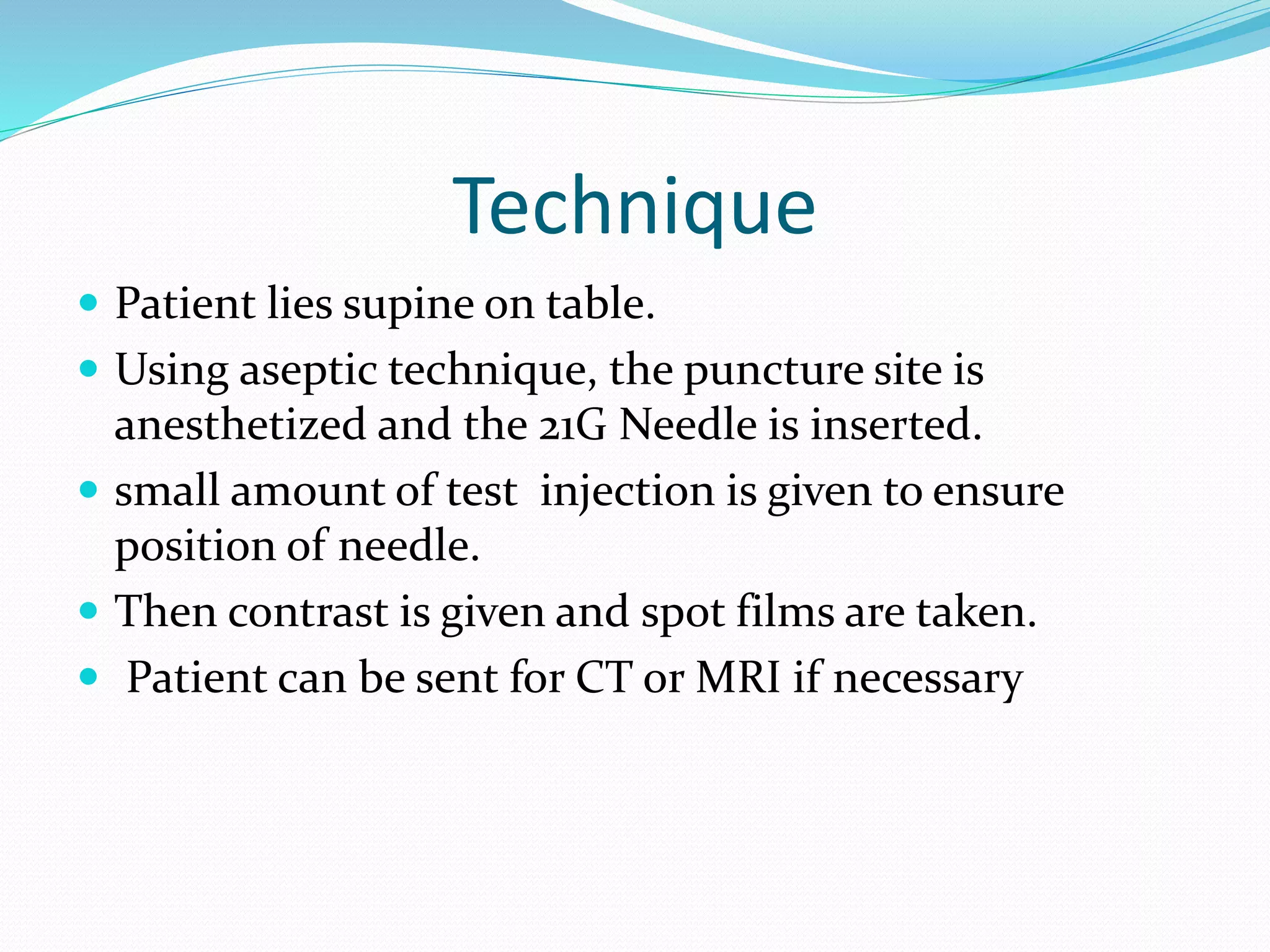
 It is the radiological examination that involves the
study of joint space and related soft tissue structures
by introduction of iodinated as well as negative
contrast media.
Indications
 To access the status of cartilage, labrum and tendons
 To access for presence, type, extent, gap, and edges of
torn capsular and pericapsular stuctures
 Assessment of the prosthesis
 Lesions or cyst
Contraindication:
• Hypersensitivity to contrast media
• Infection
Contrast media
 LOCM , 5-15ml depending on joint
Preliminary film
 Ap and lateral
Technique
 Patient lies supine on table.
 Using aseptic technique, the puncture site is
anesthetized and the 21G Needle is inserted.
 small amount of test injection is given to ensure
position of needle.
 Then contrast is given and spot films are taken.
 Patient can be sent for CT or MRI if necessary
Puncture sites for joints:
 For knee- puncture site 1-2cm posterior to patella
 For hip- puncture site below junction of femoral artery
 For shoulder- puncture site into glenohumeral joint
 For elbow- puncture site is proximal to radial head
 For ankle- puncture site is between 2 malleoli
 For wrist- puncture site is over midpoint of radiocarpal
joint
Aftercare:
 Patient might feel discomfort for few days in the joint.
 Patient should be asked not to do hard works.
Complications:
 Trauma, pain
 Infection
 Bleeding, redness, swelling at the site of puncture.

Arthrography.pptx

  • 2.
     It isthe radiological examination that involves the study of joint space and related soft tissue structures by introduction of iodinated as well as negative contrast media.
  • 3.
    Indications  To accessthe status of cartilage, labrum and tendons  To access for presence, type, extent, gap, and edges of torn capsular and pericapsular stuctures  Assessment of the prosthesis  Lesions or cyst
  • 4.
    Contraindication: • Hypersensitivity tocontrast media • Infection
  • 5.
    Contrast media  LOCM, 5-15ml depending on joint
  • 6.
  • 7.
    Technique  Patient liessupine on table.  Using aseptic technique, the puncture site is anesthetized and the 21G Needle is inserted.  small amount of test injection is given to ensure position of needle.  Then contrast is given and spot films are taken.  Patient can be sent for CT or MRI if necessary
  • 8.
    Puncture sites forjoints:  For knee- puncture site 1-2cm posterior to patella  For hip- puncture site below junction of femoral artery  For shoulder- puncture site into glenohumeral joint  For elbow- puncture site is proximal to radial head  For ankle- puncture site is between 2 malleoli  For wrist- puncture site is over midpoint of radiocarpal joint
  • 9.
    Aftercare:  Patient mightfeel discomfort for few days in the joint.  Patient should be asked not to do hard works.
  • 10.
    Complications:  Trauma, pain Infection  Bleeding, redness, swelling at the site of puncture.