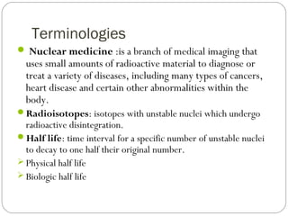
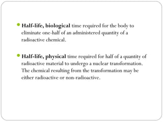
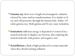
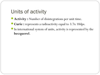
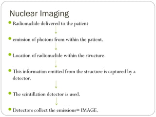
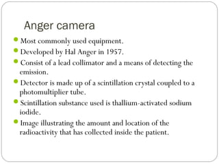
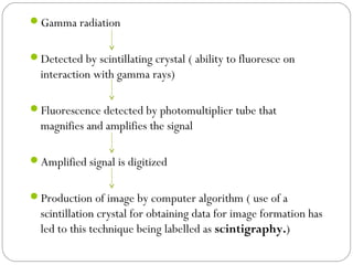
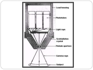
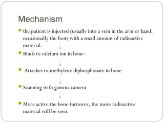
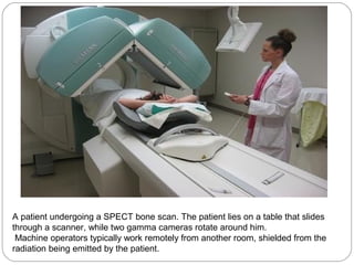
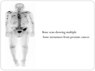
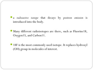
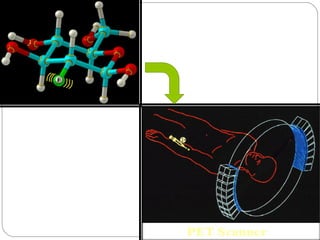
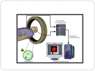
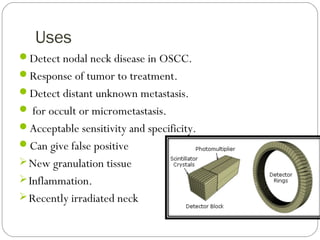
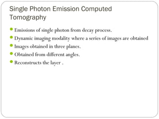
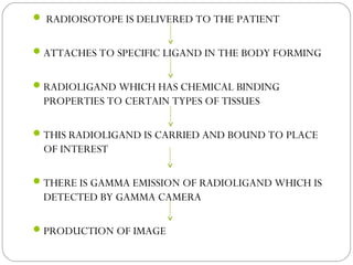
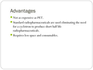
RADIONUCLIDE IMAGING
SEMINAR
Presented by:-
Aarti Dubey
Guided by:-
Dr Suvarna Dangore Dr R.R. Bhowate Dr S.S. Degwekar
“In this era of diagnostic radiology, progress lies not in
enhancing what is, but in advancing towards what will be”
PURPOSE STATEMENT
At the end of the presentation learners should be able to
explain about radionuclide imaging, history,indications,
contraindications, advantages ,disadvantages , describe
mechanism of radionuclide imaging & various newer
radionuclide imaging procedures.
S
no.
Learning objectives domain level criteria condition
1. Explain what is
radionuclide imaging.
Cognitive Must know All -
2 Describe the history of
radionuclide imaging.
Cognitive Must know All -
3 Enumerate indications
and contraindications of
radionuclide imaging.
Cognitive Must know All -
4 Enumerate its advantages
and disadvantages
Cognitive Must know All -
5 Compare Conventional
VS nuclear imaging
Cognitive Must know All -
6 Explain Mechanism of
radionuclide production
Cognitive Must know All -
7 Explain about PET,
SPECT and fusion
imaging
Cognitive Must know All -
Contents
Introduction
History
Terminology
Indications & Contraindications.
Advantages
Disadvantages
Conventional VS nuclear radiology
Radionuclides
Basics of radionuclide production
Radiopharmaceuticals
Newer techniques (SPECT,PET, PET-CT )
Applications
Conclusion
Introduction
Diagnostic tool that utilizes a radioactive substance to help
diagnose a disease process from inside the body.
Plain radiographs, CT & MRI – Structural/ morphological
alterations
Radionuclide imaging-Early physiologic changes/ metabolic
alterations
Allows the function of target tissue to be examined under
both static and dynamic conditions.
MILESTONES IN RADIONUCLIDE IMAGING
One of the earliest instances of nuclear medicine occurred in
1946 when radioactive iodine, via “atomic- cocktail”, was
first used to treat thyroid cancer.
Gamma ray detection (1947-1948)- Coltman and Marshall
Scintillation camera- Anger in 1957
Scintillation detector (1950’s)- Macintyre, Cassen et
al.,Widespread use of radionuclide imaging began in early
1950’s.
SPECT- (1963)- Kuhl and Edwards
In the 1960’s and the years to follow, the growth of
radionuclide imaging as a speciality discipline was
phenomenal.
Initially techniques were developed to measure blood flow to
lungs and to identify cancer “hot spots”.
By the 1970’s most organs of the body could be visualized
with nuclear medicine procedures including liver and spleen
scanning, brain tumour localisation and studies of GIT tract.
In 1971 the American Medical Association officially
recognised Radionuclide Imaging (nuclear imaging) as a
medical speciality.
1980’s- radiopharmaceuticals were designed, FDG was
developed.
1989- FDA approved rubidium-82 for myocardial perfusion
imaging.
1990’s- PET was becoming an important diagnostic tool.
2000- PET-CT( fusion imaging). The ability to detect the
exact location of the metabolic spot “hot spot” by overlaying
the PET and CT images provided priceless information.
Terminologies
 Nuclear medicine :is a branch of medical imaging that
uses small amounts of radioactive material to diagnose or
treat a variety of diseases, including many types of cancers,
heart disease and certain other abnormalities within the
body.
Radioisotopes: isotopes with unstable nuclei which undergo
radioactive disintegration.
Half life: time interval for a specific number of unstable nuclei
to decay to one half their original number.
 Physical half life
 Biologic half life
Half-life, biological time required for the body to
eliminate one-half of an administered quantity of a
radioactive chemical. 
Half-life, physical time required for half of a quantity of
radioactive material to undergo a nuclear transformation.
The chemical resulting from the transformation may be
either radioactive or non-radioactive. 
Gamma ray short wave-length electromagnetic radiation
released by some nuclear transformations. It is similar to X-
ray and will penetrate through the human body. Iodine 131
emits gamma rays. Both gamma and X-rays cause ionisation.
Ionisation sufficient energy is deposited or removed in a
neutral molecule to displace an electron, thus replacing the
neutral molecule with positive and negative ions.
Scintillation a flash of light produced in certain materials
when they absorb ionizing radiation
Units of activity
Activity : Number of disintegrations per unit time.
Curie : represents a radioactivity equal to 3.7x 10dps.
In international system of units, activity is represented by the
becquerel.
Indications
Assessment of site and extent of bone metastases.
Investigation of salivary gland function particularly in
Sjogren’s syndrome
Evaluation of bone grafts.
Assessment of continued growth in condylar hyperplasia.
Investigation of thyroid function.
Brain scans and assessment of a breakdown of blood brain
barrier.
Contraindications
Pregnancy
Allergic reactions
Previous surgical, radiologic procedures.
Prior medications
Advantages
Target tissue function is investigated.
All similar target tissue can be examined during one
investigation.
Computer analysis and enhancements of results are available.
Disadvantages
Poor image resolution.
High radiation dose.
Images are not disease specific.
Difficult to localize exact anatomical site of source of
emission.
Facilities are not widely available.
Nuclear Imaging vs Conventional
radiology
The patient, rather than the machine, is source of radiation.
The detection instrument is different.
The sensitivities of nuclear medicine are very great.
The specificities are very low.
Radioisotopes
RADIOISOTOPES TARGET TISSUE
Technetium(99m Tc-
pertechnetate)
SG,Thyroid,bone,blood,liver,l
ung and heart
Gallium(67 Ga) Tumours and inflammation.
Iodine(131I) Thyroid
Krypton(81 Kr) Lung
Radiopharmaceuticals
Radionuclide that has been modified by chemically
combining it with various biochemicals that may have
physiologic or metabolic properties that would be
beneficial in a particular study.
Incorporated into diverse and biologically important
compounds
Glucose
Amino acids
Ammonia
Why Technetium(99m Tc-pertechnetate) ???
Single 141ev gamma emissions which are ideal.
Short half life=6.5 hrs
Readily attached to variety of different substances that are
concentrated in different organs.
Safe, Easily produced , as and when required, on site.
Nuclear Imaging
Radionuclide delivered to the patient
emission of photons from within the patient.
Location of radionuclide within the structure.
This information emitted from the structure is captured by a
detector.
The scintillation detector is used.
Detectors collect the emissions= IMAGE.
Anger camera
Most commonly used equipment.
Developed by Hal Anger in 1957.
Consist of a lead collimator and a means of detecting the
emission.
Detector is made up of a scintillation crystal coupled to a
photomultiplier tube.
Scintillation substance used is thallium-activated sodium
iodide.
Image illustrating the amount and location of the
radioactivity that has collected inside the patient.
Gamma radiation
Detected by scintillating crystal ( ability to fluoresce on
interaction with gamma rays)
Fluorescence detected by photomultiplier tube that
magnifies and amplifies the signal
Amplified signal is digitized
Production of image by computer algorithm ( use of a
scintillation crystal for obtaining data for image formation has
led to this technique being labelled as scintigraphy.)
Imaging devices
Planar nuclear imaging
Single-photon-emission computed tomography
Positron emission tomography
Fusion imaging
Planar imaging
Scintillation cameras convert detected gamma radiation into
light emissions.
Image display &analysis – Photomultiplier tube and
computer systems.
Commonly used to examine:
Primary and secondary malignancies
Inflammatory conditions
Metabolic disease
Trauma
BONE SCAN
In contrast to a radiograph, bone scan gives no information
on morphology of lesion, either internally or in areas of bone
adjacent to the lesion.
The scan does demonstrate, however areas of altered bone
metabolism within and around the lesion, thus allowing a
reasonably accurate assessment of the growth of a lesion and
extent of its borders.
Bone scan also allows to view the entire skeleton with no
additional radiation burden to the patient.
Positive findings usually lead to conventional radiographs of
the suspicious areas, allowing morphologic study of regions
with altered metabolism.
Types of Bone scan
Standard whole body scan.
Three-phase bone scan.
Three-phase bone scan
Dynamic vascular flow phase- imaging every 2-3
seconds after injection of intravascular contrast medium.
Blood pool phase: images are taken after every 5 mins.
Osseous delayed static phase: occurs after 2-4 hours.
Agents used
Technetium bone scan
Gallium bone scan (half life=78 hrs)
 If a technetium bone scan is being contemplated, it should be performed
first.
 Gallium scan is mainly used in cases of osteomyelitis.
 Adjunct with technetium scan.
Mechanism
the patient is injected (usually into a vein in the arm or hand,
occasionally the foot) with a small amount of radioactive
material.
Binds to calcium ion in bone-
 Attaches to methylene diphosphonate in bone
Scanning with gamma camera
More active the bone turnover, the more radioactive
material will be seen.
A patient undergoing a SPECT bone scan. The patient lies on a table that slides
through a scanner, while two gamma cameras rotate around him.
Machine operators typically work remotely from another room, shielded from the
radiation being emitted by the patient.
Bone scan showing multiple
bone metastases from prostate cancer.
Positron Emission Tomography
A nuclear medicine imaging technique which provides high resolution
tomographic images of the bio-distribution of a radiopharmaceutical
or radiotracers in the body.
 A PET scan measures important body functions, such as blood flow,
oxygen use, and sugar (glucose) metabolism, to help doctors evaluate
how well organs and tissues are functioning.
a radioactive isotope that decays by positron emission is
introduced into the body.
Many different radioisotopes are there, such as Fluorine18,
Oxygen15, and Carbon11.
18F is the most commonly used isotope. It replaces hydroxyl
(OH) group in molecules of interest.
Within the cell, FDG is phosphorylated by enzyme
“hexokinase” to 2-deoxyglucose-6-phosphate
Increased proliferation of tumor cells manifest as increased
uptake of FDG in cancer cells compared to the surrounding
normal tissue
 Positron emitting radioisotopes are prepared by bombarding
stable atomic nuclei by protons.
 Protons are speeded up in a particle accelerator called
cyclotron which then impinge upon the stable nuclei, and
knocks out one neutron from its nucleus.
ANNIHILATION
 The Feynman diagram below is one of the most common occurrences
when an electron e- and a positron e+ collide and it is called electron-
positron annihilation.
 The result of this collision is the transformation of the electrons
into gamma ray photons.
TRACER
 EMITTS POSITRON
 POSITRON THEN ANNILATES WITH ELECTRON
TWO GAMMA PROTONS EMITTED IN OPPOSITE
DIRECTION
 PET SCANNER DETECTS THESE EMISSION COINCIDENT
IN TIME (PROVIDES RADIATION EVENT LOCALIZATION,
THUS INCREASING RESOLUTION OF IMAGES THAN
SPECT)
ADVANTAGES
Non-invasive
Low-risk infection compared to surgery
Identifying active diseases after therapy completion
Outcome of chemotherapy
E.g. In non-Hodgkin’s lymphoma, FDG uptake was found to
decrease as early as 1 d after the initiation of chemotherapy
early assessment of response during the first treatment cycles is
important to appreciate chemosensitivity and may potentially
guide further risk-adapted therapeutic strategies in aggressive
lymphoma
DISADVANTAGES
Radiation risk: although minimal (equivalent to 2 chest
radiograph)
Not indicated in pregnancy & lactation
Short t1/2, limited time for completing scan on patient
(~120 mins)
Expensive
Non-availability of cyclotron (expensive)
Uses
Detect nodal neck disease in OSCC.
Response of tumor to treatment.
Detect distant unknown metastasis.
 for occult or micrometastasis.
Acceptable sensitivity and specificity.
Can give false positive
New granulation tissue
Inflammation.
Recently irradiated neck
Single Photon Emission Computed
Tomography
Emissions of single photon from decay process.
Dynamic imaging modality where a series of images are obtained
Images obtained in three planes.
Obtained from different angles.
Reconstructs the layer .
 RADIOISOTOPE IS DELIVERED TO THE PATIENT
ATTACHES TO SPECIFIC LIGAND IN THE BODY FORMING
RADIOLIGAND WHICH HAS CHEMICAL BINDING
PROPERTIES TO CERTAIN TYPES OF TISSUES
THIS RADIOLIGAND IS CARRIED AND BOUND TO PLACE
OF INTEREST
THERE IS GAMMA EMISSION OF RADIOLIGAND WHICH IS
DETECTED BY GAMMA CAMERA
PRODUCTION OF IMAGE
Advantages
Not as expensive as PET.
Standard radiopharmaceuticals are used eliminating the need
for a cyclotron to produce short half life
radiopharmaceuticals.
Requires less space and consumables.
Fusion imaging
Combinations of two different modalities to produce final image.
CT and SPECT.
CT and PET.
PET and MRI
Structure and function.
Eliminate the mismatching of images.
Imaging times are reduced.
Accurate localization.
Coregistered FDG-PET and low-resolution MRI images and
image fusion
 Sublingual glands
 Tonsils
 Spinal cord
 Submandibular glands
Applications of radionuclide imaging
Lymphoscintigraphy
Salivary gland scintigraphy
Oral and maxillofacial inflammation
Tumors
Trauma
Bone healing
Tmj
Lymphoscintigraphy
Simple and non-invasive functional test for demonstrating
lymphatic pathways.
Technetium 99m sulphur colloid is injected in 4-6
subcutaneous sites around neoplastic sites.
Colloid will be carried in lymphatic channels to first lymph
node draining that area, so called sentinel node.
Best predictor of nodal spread of tumor.
Imaged using gamma camera.
Sentinel node is free, remaining nodes are free.
Sentinel nodes are positive- remove remaining nodes.
If the node is disease free, patient is spared an elective neck
dissection.
If node is positive, the patient goes on to a more formal neck
dissection.
Salivary Gland Studies
Used for functional evaluation and evaluating mass lesions.
Te99 mimics chloride influx into the acinar cells.
Involves administrating a radioactive tracer with affinity for
tissue of interest.
Recorded with scintillation camera.
Study is rarely diagnostic, but is useful adjunct.
Mass lesions in a gland present as areas of decreased uptake.
Patients with Sjogren’s syndrome may have poor uptake and
poor response to stimulation.
Agent used Tc99m Iodinated contrast
media
Administration Intravenous Retrograde
pressure of
contrast media
Detector Gamma camera Fluoroscope
Structures Glandular
parenchyma
Duct system
Advantages Function, sensitive High resolution of
ducts.
Disadvantages Non specific Contraindications
Salivary gland
scan Sialography
Inflammatory diseases
Bone scintigraphy can be used to disclose periapical lesions .
Used to detect inflammatory responses of TMJ.
SPECT is used to detect arthritic changes.
Osteomyelitis of jaws.
Bone-graft viability
Means of predicting graft failure before clinical or radiographic
changes are apparent.
Positive scan is correlated with a viable bone graft.
Negative bone scan corresponds to non-viable bone graft.
Periodontal disease
In various studies, it has been reported that the uptake of
radionuclide Tc99m was elevated in alveolar bone with chronic
destructive periodontal disease.
More sensitive method to detect bone loss than were standard
intraoral radiograph.
Tumors
Bone scanning at three and 24 hours following
radiopharmaceutical administration has been shown to
enhance differentiation of benign from malignant tumors.
Uptake increases in malignant tissue.
Uptake in degenerative lesions decreases.
FDG PET test has a good predictive value for identifying
recurrent malignancies in head and neck.
Trauma
Initial detection of subtle bone fractures , not apparent on
standard radiographs.
Evidence of stress fracture on radiographs appear late in the
healing process i.e. up to two to 12 weeks after nuclear
imaging.
An interesting case report described a stress fracture detected by
nuclear imaging in an ice cream scooper’s hand.
Temporomandibular joint
Evaluation of bone metabolism in bony components of
TMJ.
For assessment of skeletal facial growth.
Presence of active hyperplastic activity in these joints.
Effectiveness of SPECT for quantitative skeletal scintigraphy
of mandibular condyle.
Usefulness of Tc 99m uptake in correlation of mandibular
growth with chronological and skeletal age.
Radiation dose
CT Scan 7.6 Sv
Bone scan 4 Sv
F-18FDG PET 5.9 Sv
PET 6 Sv
If an injection of 3.7 X 10 (8) Bq of 99m Tc delivered = a whole body
radiation dose of 1 mGy which is about 1/3rd
the average annual
effective dose resulting from natural radiation.
CONCLUSION
Nuclear medicine procedures are costeffective.
 There are nearly 100 different nuclear medicine
imaging procedures available today.
 Unlike other tests/procedures, etc., nuclear
medicine provides information about the function of
virtually every major organ system within the body.
Nuclear medicine procedures are painless and do
not require anesthesia.
Nuclear medicine is an integral part of patient care
and contributes to the well being of patients
worldwide.
REFERENCES
Freny R Karjodkar; Dental & Maxillofacial Radiology; 2nd
ed
White & Pharoah; Oral Radiology principles and
interpretation; 5th
ed
Ghom’s ; Oral Radiology
Henry N. Wagner.What is nuclear medicine; Siemens
Medical Solutions, USA,Inc.; Philips Medical Systems; and
Digirad.
Johan Nuyts. Nuclear Medicine Technology and Techniques;

Radionuclide imaging- Aarti Dubey

  • 1.
    RADIONUCLIDE IMAGING SEMINAR Presented by:- AartiDubey Guided by:- Dr Suvarna Dangore Dr R.R. Bhowate Dr S.S. Degwekar “In this era of diagnostic radiology, progress lies not in enhancing what is, but in advancing towards what will be”
  • 2.
    PURPOSE STATEMENT At theend of the presentation learners should be able to explain about radionuclide imaging, history,indications, contraindications, advantages ,disadvantages , describe mechanism of radionuclide imaging & various newer radionuclide imaging procedures.
  • 3.
    S no. Learning objectives domainlevel criteria condition 1. Explain what is radionuclide imaging. Cognitive Must know All - 2 Describe the history of radionuclide imaging. Cognitive Must know All - 3 Enumerate indications and contraindications of radionuclide imaging. Cognitive Must know All - 4 Enumerate its advantages and disadvantages Cognitive Must know All - 5 Compare Conventional VS nuclear imaging Cognitive Must know All - 6 Explain Mechanism of radionuclide production Cognitive Must know All - 7 Explain about PET, SPECT and fusion imaging Cognitive Must know All -
  • 4.
    Contents Introduction History Terminology Indications & Contraindications. Advantages Disadvantages ConventionalVS nuclear radiology Radionuclides Basics of radionuclide production Radiopharmaceuticals Newer techniques (SPECT,PET, PET-CT ) Applications Conclusion
  • 5.
    Introduction Diagnostic tool thatutilizes a radioactive substance to help diagnose a disease process from inside the body. Plain radiographs, CT & MRI – Structural/ morphological alterations Radionuclide imaging-Early physiologic changes/ metabolic alterations Allows the function of target tissue to be examined under both static and dynamic conditions.
  • 6.
    MILESTONES IN RADIONUCLIDEIMAGING One of the earliest instances of nuclear medicine occurred in 1946 when radioactive iodine, via “atomic- cocktail”, was first used to treat thyroid cancer. Gamma ray detection (1947-1948)- Coltman and Marshall Scintillation camera- Anger in 1957 Scintillation detector (1950’s)- Macintyre, Cassen et al.,Widespread use of radionuclide imaging began in early 1950’s. SPECT- (1963)- Kuhl and Edwards In the 1960’s and the years to follow, the growth of radionuclide imaging as a speciality discipline was phenomenal. Initially techniques were developed to measure blood flow to lungs and to identify cancer “hot spots”.
  • 7.
    By the 1970’smost organs of the body could be visualized with nuclear medicine procedures including liver and spleen scanning, brain tumour localisation and studies of GIT tract. In 1971 the American Medical Association officially recognised Radionuclide Imaging (nuclear imaging) as a medical speciality. 1980’s- radiopharmaceuticals were designed, FDG was developed. 1989- FDA approved rubidium-82 for myocardial perfusion imaging. 1990’s- PET was becoming an important diagnostic tool. 2000- PET-CT( fusion imaging). The ability to detect the exact location of the metabolic spot “hot spot” by overlaying the PET and CT images provided priceless information.
  • 9.
    Terminologies  Nuclear medicine:is a branch of medical imaging that uses small amounts of radioactive material to diagnose or treat a variety of diseases, including many types of cancers, heart disease and certain other abnormalities within the body. Radioisotopes: isotopes with unstable nuclei which undergo radioactive disintegration. Half life: time interval for a specific number of unstable nuclei to decay to one half their original number.  Physical half life  Biologic half life
  • 10.
    Half-life, biological time requiredfor the body to eliminate one-half of an administered quantity of a radioactive chemical.  Half-life, physical time required for half of a quantity of radioactive material to undergo a nuclear transformation. The chemical resulting from the transformation may be either radioactive or non-radioactive. 
  • 11.
    Gamma ray short wave-lengthelectromagnetic radiation released by some nuclear transformations. It is similar to X- ray and will penetrate through the human body. Iodine 131 emits gamma rays. Both gamma and X-rays cause ionisation. Ionisation sufficient energy is deposited or removed in a neutral molecule to displace an electron, thus replacing the neutral molecule with positive and negative ions. Scintillation a flash of light produced in certain materials when they absorb ionizing radiation
  • 12.
    Units of activity Activity: Number of disintegrations per unit time. Curie : represents a radioactivity equal to 3.7x 10dps. In international system of units, activity is represented by the becquerel.
  • 13.
    Indications Assessment of siteand extent of bone metastases. Investigation of salivary gland function particularly in Sjogren’s syndrome Evaluation of bone grafts. Assessment of continued growth in condylar hyperplasia. Investigation of thyroid function. Brain scans and assessment of a breakdown of blood brain barrier.
  • 14.
  • 15.
    Advantages Target tissue functionis investigated. All similar target tissue can be examined during one investigation. Computer analysis and enhancements of results are available.
  • 16.
    Disadvantages Poor image resolution. Highradiation dose. Images are not disease specific. Difficult to localize exact anatomical site of source of emission. Facilities are not widely available.
  • 17.
    Nuclear Imaging vsConventional radiology The patient, rather than the machine, is source of radiation. The detection instrument is different. The sensitivities of nuclear medicine are very great. The specificities are very low.
  • 18.
    Radioisotopes RADIOISOTOPES TARGET TISSUE Technetium(99mTc- pertechnetate) SG,Thyroid,bone,blood,liver,l ung and heart Gallium(67 Ga) Tumours and inflammation. Iodine(131I) Thyroid Krypton(81 Kr) Lung
  • 19.
    Radiopharmaceuticals Radionuclide that hasbeen modified by chemically combining it with various biochemicals that may have physiologic or metabolic properties that would be beneficial in a particular study. Incorporated into diverse and biologically important compounds Glucose Amino acids Ammonia
  • 20.
    Why Technetium(99m Tc-pertechnetate)??? Single 141ev gamma emissions which are ideal. Short half life=6.5 hrs Readily attached to variety of different substances that are concentrated in different organs. Safe, Easily produced , as and when required, on site.
  • 21.
    Nuclear Imaging Radionuclide deliveredto the patient emission of photons from within the patient. Location of radionuclide within the structure. This information emitted from the structure is captured by a detector. The scintillation detector is used. Detectors collect the emissions= IMAGE.
  • 22.
    Anger camera Most commonlyused equipment. Developed by Hal Anger in 1957. Consist of a lead collimator and a means of detecting the emission. Detector is made up of a scintillation crystal coupled to a photomultiplier tube. Scintillation substance used is thallium-activated sodium iodide. Image illustrating the amount and location of the radioactivity that has collected inside the patient.
  • 23.
    Gamma radiation Detected byscintillating crystal ( ability to fluoresce on interaction with gamma rays) Fluorescence detected by photomultiplier tube that magnifies and amplifies the signal Amplified signal is digitized Production of image by computer algorithm ( use of a scintillation crystal for obtaining data for image formation has led to this technique being labelled as scintigraphy.)
  • 25.
    Imaging devices Planar nuclearimaging Single-photon-emission computed tomography Positron emission tomography Fusion imaging
  • 26.
    Planar imaging Scintillation camerasconvert detected gamma radiation into light emissions. Image display &analysis – Photomultiplier tube and computer systems. Commonly used to examine: Primary and secondary malignancies Inflammatory conditions Metabolic disease Trauma
  • 27.
    BONE SCAN In contrastto a radiograph, bone scan gives no information on morphology of lesion, either internally or in areas of bone adjacent to the lesion. The scan does demonstrate, however areas of altered bone metabolism within and around the lesion, thus allowing a reasonably accurate assessment of the growth of a lesion and extent of its borders.
  • 28.
    Bone scan alsoallows to view the entire skeleton with no additional radiation burden to the patient. Positive findings usually lead to conventional radiographs of the suspicious areas, allowing morphologic study of regions with altered metabolism.
  • 29.
    Types of Bonescan Standard whole body scan. Three-phase bone scan.
  • 30.
    Three-phase bone scan Dynamicvascular flow phase- imaging every 2-3 seconds after injection of intravascular contrast medium. Blood pool phase: images are taken after every 5 mins. Osseous delayed static phase: occurs after 2-4 hours.
  • 32.
    Agents used Technetium bonescan Gallium bone scan (half life=78 hrs)  If a technetium bone scan is being contemplated, it should be performed first.  Gallium scan is mainly used in cases of osteomyelitis.  Adjunct with technetium scan.
  • 33.
    Mechanism the patient isinjected (usually into a vein in the arm or hand, occasionally the foot) with a small amount of radioactive material. Binds to calcium ion in bone-  Attaches to methylene diphosphonate in bone Scanning with gamma camera More active the bone turnover, the more radioactive material will be seen.
  • 34.
    A patient undergoinga SPECT bone scan. The patient lies on a table that slides through a scanner, while two gamma cameras rotate around him. Machine operators typically work remotely from another room, shielded from the radiation being emitted by the patient.
  • 35.
    Bone scan showingmultiple bone metastases from prostate cancer.
  • 36.
    Positron Emission Tomography Anuclear medicine imaging technique which provides high resolution tomographic images of the bio-distribution of a radiopharmaceutical or radiotracers in the body.  A PET scan measures important body functions, such as blood flow, oxygen use, and sugar (glucose) metabolism, to help doctors evaluate how well organs and tissues are functioning.
  • 37.
    a radioactive isotopethat decays by positron emission is introduced into the body. Many different radioisotopes are there, such as Fluorine18, Oxygen15, and Carbon11. 18F is the most commonly used isotope. It replaces hydroxyl (OH) group in molecules of interest.
  • 38.
    Within the cell,FDG is phosphorylated by enzyme “hexokinase” to 2-deoxyglucose-6-phosphate Increased proliferation of tumor cells manifest as increased uptake of FDG in cancer cells compared to the surrounding normal tissue  Positron emitting radioisotopes are prepared by bombarding stable atomic nuclei by protons.  Protons are speeded up in a particle accelerator called cyclotron which then impinge upon the stable nuclei, and knocks out one neutron from its nucleus.
  • 39.
    ANNIHILATION  The Feynmandiagram below is one of the most common occurrences when an electron e- and a positron e+ collide and it is called electron- positron annihilation.  The result of this collision is the transformation of the electrons into gamma ray photons.
  • 40.
    TRACER  EMITTS POSITRON POSITRON THEN ANNILATES WITH ELECTRON TWO GAMMA PROTONS EMITTED IN OPPOSITE DIRECTION  PET SCANNER DETECTS THESE EMISSION COINCIDENT IN TIME (PROVIDES RADIATION EVENT LOCALIZATION, THUS INCREASING RESOLUTION OF IMAGES THAN SPECT)
  • 43.
    ADVANTAGES Non-invasive Low-risk infection comparedto surgery Identifying active diseases after therapy completion Outcome of chemotherapy E.g. In non-Hodgkin’s lymphoma, FDG uptake was found to decrease as early as 1 d after the initiation of chemotherapy early assessment of response during the first treatment cycles is important to appreciate chemosensitivity and may potentially guide further risk-adapted therapeutic strategies in aggressive lymphoma
  • 44.
    DISADVANTAGES Radiation risk: althoughminimal (equivalent to 2 chest radiograph) Not indicated in pregnancy & lactation Short t1/2, limited time for completing scan on patient (~120 mins) Expensive Non-availability of cyclotron (expensive)
  • 45.
    Uses Detect nodal neckdisease in OSCC. Response of tumor to treatment. Detect distant unknown metastasis.  for occult or micrometastasis. Acceptable sensitivity and specificity. Can give false positive New granulation tissue Inflammation. Recently irradiated neck
  • 46.
    Single Photon EmissionComputed Tomography Emissions of single photon from decay process. Dynamic imaging modality where a series of images are obtained Images obtained in three planes. Obtained from different angles. Reconstructs the layer .
  • 47.
     RADIOISOTOPE ISDELIVERED TO THE PATIENT ATTACHES TO SPECIFIC LIGAND IN THE BODY FORMING RADIOLIGAND WHICH HAS CHEMICAL BINDING PROPERTIES TO CERTAIN TYPES OF TISSUES THIS RADIOLIGAND IS CARRIED AND BOUND TO PLACE OF INTEREST THERE IS GAMMA EMISSION OF RADIOLIGAND WHICH IS DETECTED BY GAMMA CAMERA PRODUCTION OF IMAGE
  • 48.
    Advantages Not as expensiveas PET. Standard radiopharmaceuticals are used eliminating the need for a cyclotron to produce short half life radiopharmaceuticals. Requires less space and consumables.
  • 49.
    Fusion imaging Combinations oftwo different modalities to produce final image. CT and SPECT. CT and PET. PET and MRI Structure and function. Eliminate the mismatching of images. Imaging times are reduced. Accurate localization.
  • 50.
    Coregistered FDG-PET andlow-resolution MRI images and image fusion  Sublingual glands  Tonsils  Spinal cord  Submandibular glands
  • 51.
    Applications of radionuclideimaging Lymphoscintigraphy Salivary gland scintigraphy Oral and maxillofacial inflammation Tumors Trauma Bone healing Tmj
  • 52.
    Lymphoscintigraphy Simple and non-invasivefunctional test for demonstrating lymphatic pathways. Technetium 99m sulphur colloid is injected in 4-6 subcutaneous sites around neoplastic sites. Colloid will be carried in lymphatic channels to first lymph node draining that area, so called sentinel node. Best predictor of nodal spread of tumor. Imaged using gamma camera. Sentinel node is free, remaining nodes are free. Sentinel nodes are positive- remove remaining nodes.
  • 53.
    If the nodeis disease free, patient is spared an elective neck dissection. If node is positive, the patient goes on to a more formal neck dissection.
  • 54.
    Salivary Gland Studies Usedfor functional evaluation and evaluating mass lesions. Te99 mimics chloride influx into the acinar cells. Involves administrating a radioactive tracer with affinity for tissue of interest. Recorded with scintillation camera. Study is rarely diagnostic, but is useful adjunct. Mass lesions in a gland present as areas of decreased uptake. Patients with Sjogren’s syndrome may have poor uptake and poor response to stimulation.
  • 55.
    Agent used Tc99mIodinated contrast media Administration Intravenous Retrograde pressure of contrast media Detector Gamma camera Fluoroscope Structures Glandular parenchyma Duct system Advantages Function, sensitive High resolution of ducts. Disadvantages Non specific Contraindications Salivary gland scan Sialography
  • 56.
    Inflammatory diseases Bone scintigraphycan be used to disclose periapical lesions . Used to detect inflammatory responses of TMJ. SPECT is used to detect arthritic changes. Osteomyelitis of jaws.
  • 57.
    Bone-graft viability Means ofpredicting graft failure before clinical or radiographic changes are apparent. Positive scan is correlated with a viable bone graft. Negative bone scan corresponds to non-viable bone graft.
  • 58.
    Periodontal disease In variousstudies, it has been reported that the uptake of radionuclide Tc99m was elevated in alveolar bone with chronic destructive periodontal disease. More sensitive method to detect bone loss than were standard intraoral radiograph.
  • 59.
    Tumors Bone scanning atthree and 24 hours following radiopharmaceutical administration has been shown to enhance differentiation of benign from malignant tumors. Uptake increases in malignant tissue. Uptake in degenerative lesions decreases. FDG PET test has a good predictive value for identifying recurrent malignancies in head and neck.
  • 60.
    Trauma Initial detection ofsubtle bone fractures , not apparent on standard radiographs. Evidence of stress fracture on radiographs appear late in the healing process i.e. up to two to 12 weeks after nuclear imaging. An interesting case report described a stress fracture detected by nuclear imaging in an ice cream scooper’s hand.
  • 61.
    Temporomandibular joint Evaluation ofbone metabolism in bony components of TMJ. For assessment of skeletal facial growth. Presence of active hyperplastic activity in these joints. Effectiveness of SPECT for quantitative skeletal scintigraphy of mandibular condyle. Usefulness of Tc 99m uptake in correlation of mandibular growth with chronological and skeletal age.
  • 62.
    Radiation dose CT Scan7.6 Sv Bone scan 4 Sv F-18FDG PET 5.9 Sv PET 6 Sv If an injection of 3.7 X 10 (8) Bq of 99m Tc delivered = a whole body radiation dose of 1 mGy which is about 1/3rd the average annual effective dose resulting from natural radiation.
  • 63.
    CONCLUSION Nuclear medicine proceduresare costeffective.  There are nearly 100 different nuclear medicine imaging procedures available today.  Unlike other tests/procedures, etc., nuclear medicine provides information about the function of virtually every major organ system within the body. Nuclear medicine procedures are painless and do not require anesthesia. Nuclear medicine is an integral part of patient care and contributes to the well being of patients worldwide.
  • 64.
    REFERENCES Freny R Karjodkar;Dental & Maxillofacial Radiology; 2nd ed White & Pharoah; Oral Radiology principles and interpretation; 5th ed Ghom’s ; Oral Radiology Henry N. Wagner.What is nuclear medicine; Siemens Medical Solutions, USA,Inc.; Philips Medical Systems; and Digirad. Johan Nuyts. Nuclear Medicine Technology and Techniques;