1. The document discusses bone scintigraphy (bone scan), providing information on its uses, procedures, interpretations, and applications.
2. A bone scan uses radiopharmaceuticals like technetium-99m MDP to detect areas of abnormal bone metabolism that could indicate conditions like fractures, infections, tumors and metastases.
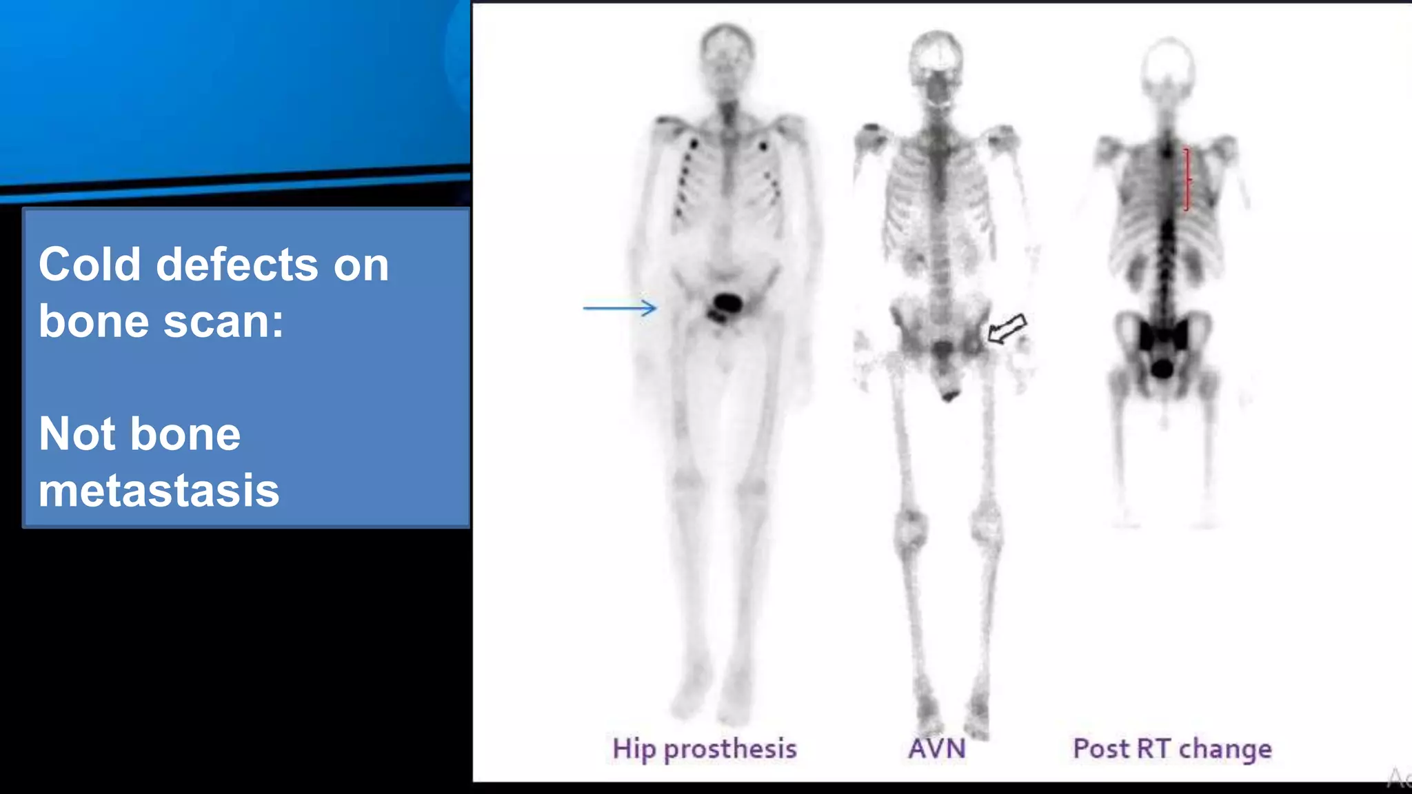
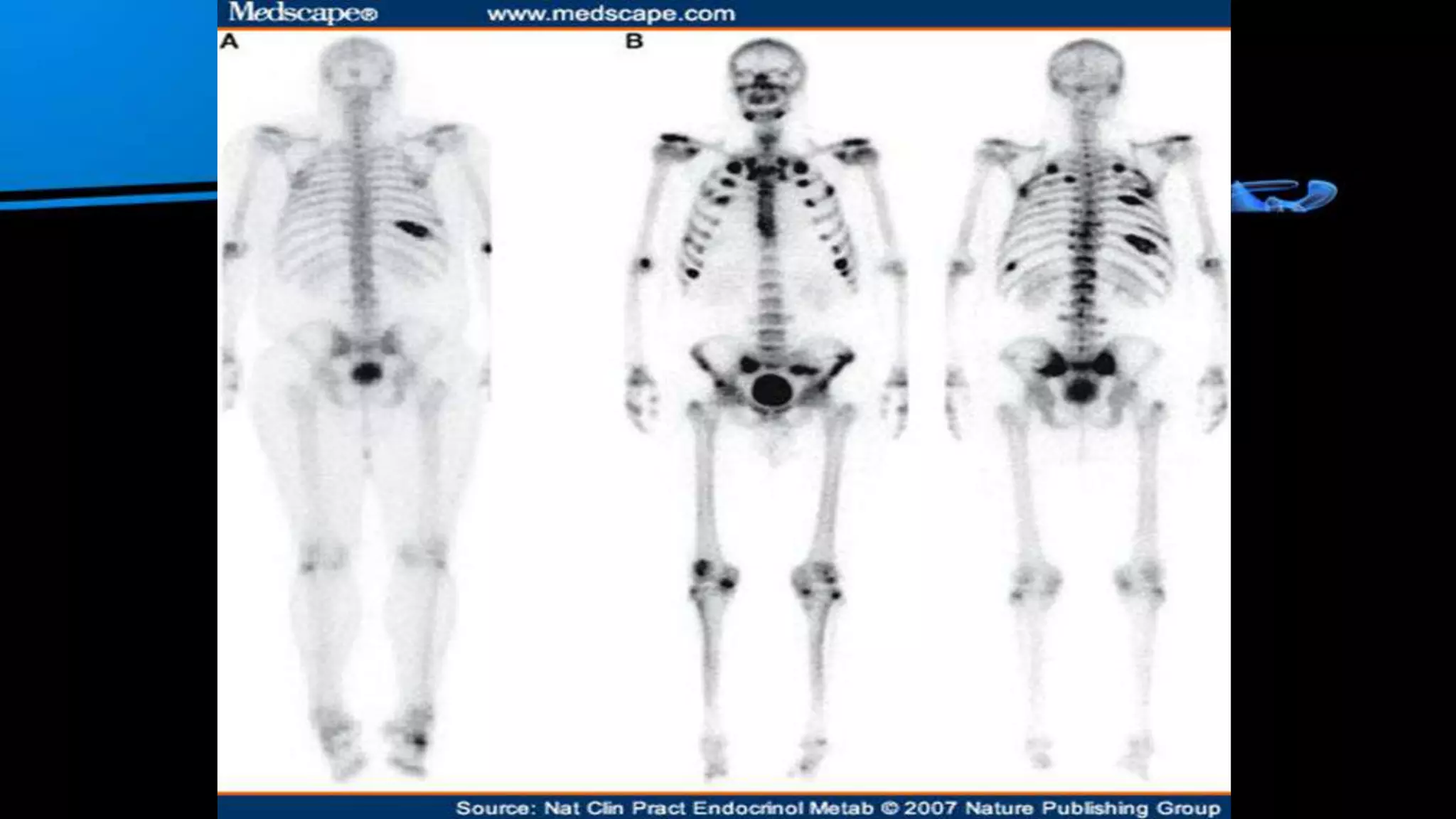
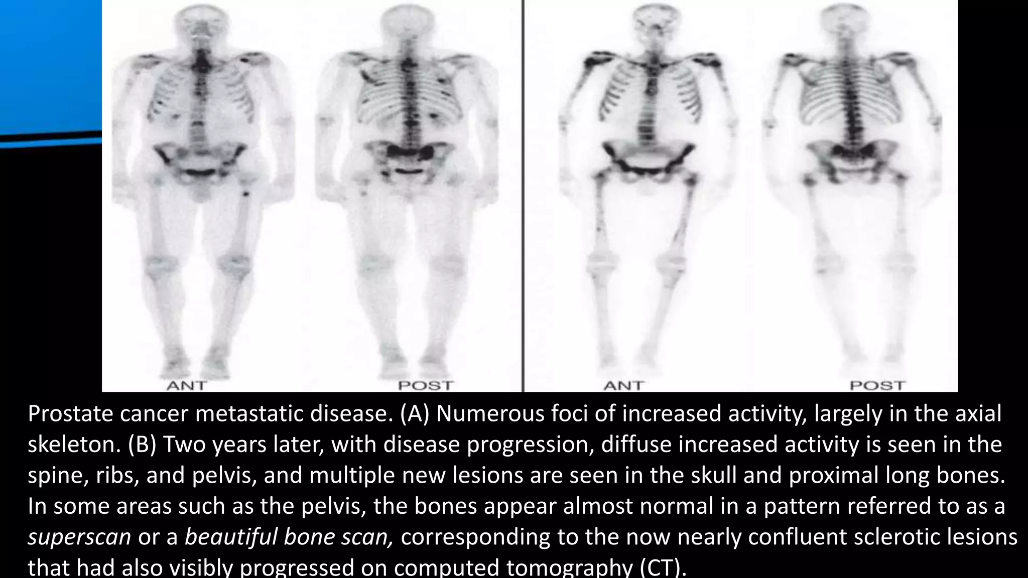
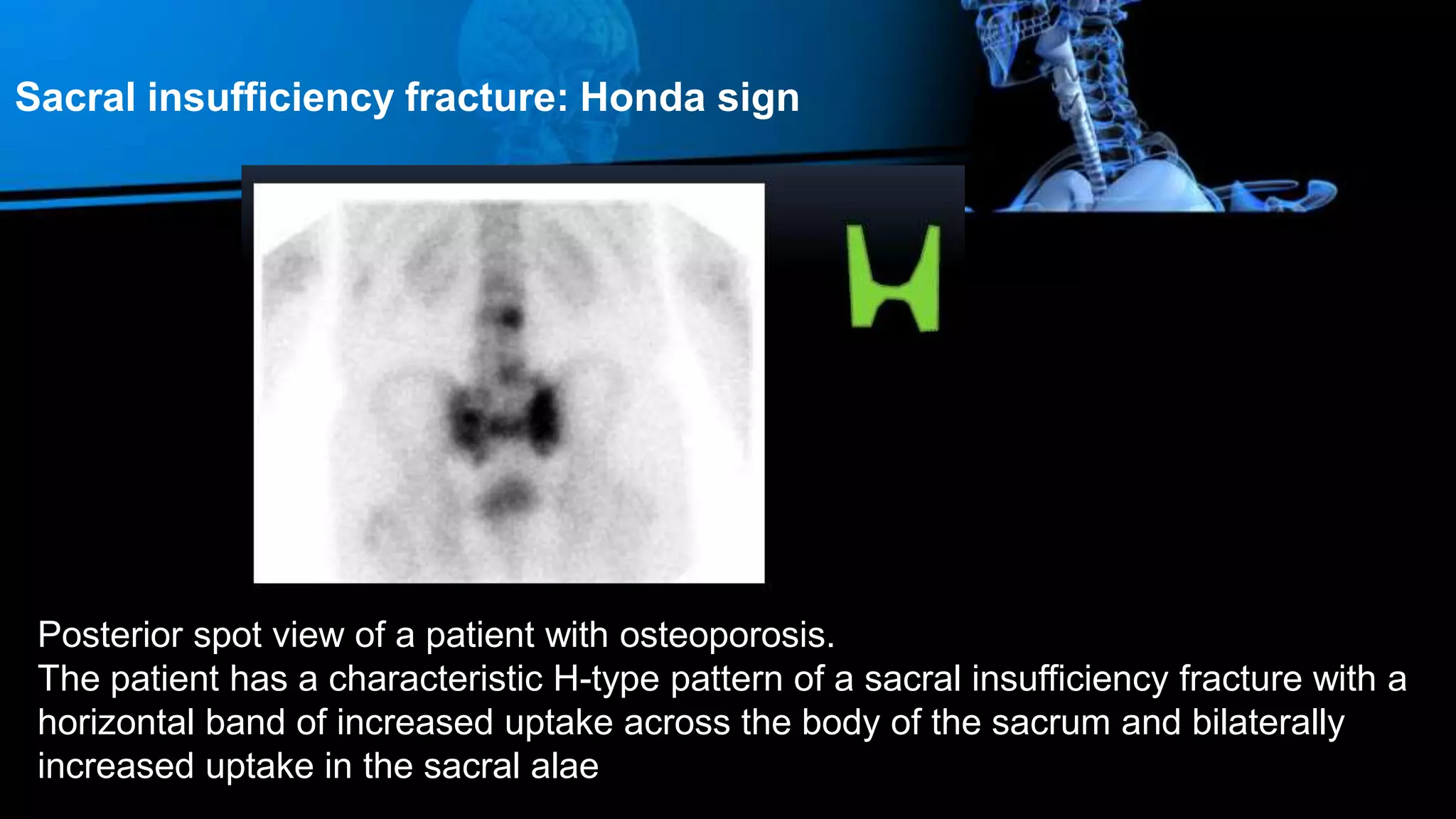
3. It is a sensitive test but not specific, so findings must be interpreted in the full clinical context. The document outlines patterns for various bone diseases and cancers.
The bone scan is a nuclear medicine procedure that uses radiopharmaceuticals like technetium-99m to generate images of the skeletal system. It is useful for evaluating bone disorders and can detect abnormalities earlier than other imaging modalities. The scan involves injection of the radiotracer and acquisition of blood flow, blood pool, and bone scan phase images over hours. It is helpful for detecting fractures, metastases, infections and other bone abnormalities. Precise diagnosis may require correlation with clinical history and other imaging tests.
Introduction and indications of BONE SCANAmir Bahadur
Bone scintigraphy, or a bone scan, is a nuclear medicine imaging technique used to evaluate bone abnormalities. It involves injecting technetium-99m labeled diphosphonate which is absorbed by areas of high bone turnover and then imaging its distribution throughout the skeleton. A bone scan has high sensitivity but low specificity for detecting bone abnormalities. It is commonly used to detect bone metastases from cancer, evaluate orthopedic conditions like fractures and infections, and monitor treatment response. The presentation provides an overview of bone scanning techniques and its clinical indications.
Introduction to Bone Scan: Techniques and Diagnosis Waseem M.Nizamani
This document provides an overview of nuclear medicine imaging techniques like bone scans. It discusses how nuclear medicine shows physiological functioning rather than anatomy. Bone scans specifically involve injecting radioactive tracers that are absorbed by bone and then imaging their distribution to identify abnormalities. The document reviews normal scan findings, uses of bone scans to detect conditions like metastases and fractures, and presents several case examples to illustrate bone scan interpretations for conditions like osteomyelitis, bone metastases, and trauma.
This document discusses fusion imaging, which combines images from different modalities to create a hybrid image. It describes fusion imaging techniques like PET-CT and SPECT-CT that merge functional imaging data with anatomical images. The primary advantage of fusion imaging is that it allows correlation of findings from two concurrent imaging modalities, providing both anatomical and functional/metabolic information in a single exam. Specifically, PET-CT fusion improves diagnostic accuracy and lesion localization by overcoming the limitations of each individual modality. In conclusion, combined PET-CT exams are more effective than PET alone for localizing lesions and differentiating normal variants from tumors.
Multiplanar reformatted images in CT scans of the chest allow radiologists to view the scans in different planes and orientations beyond just axial sections. This helps identify patterns of diffuse lung disease, distributions of lesions, and other findings more quickly. Techniques like longitudinal reformation, minimum intensity projection (mIP), maximum intensity projection (MIP), multiplanar virtual reality (VR) averaging, and three-dimensional VR and volume intensity projection (VIP) generate reformatted images that provide additional clinical information about characteristics and locations of abnormalities in a more efficient manner compared to just reviewing numerous axial images.
Dual energy CT utilizes two different x-ray spectra to characterize tissues. It can help address challenges with single energy CT like lesion detection and image noise. Dual energy CT works by analyzing how materials attenuate x-rays differently at various energies, allowing differentiation of substances like iodine and calcium. There are several technical approaches to dual energy CT, including sequential acquisition with two scans, rapid voltage switching between two voltages, and dual-source CT with two tube-detector pairs. Post-processing involves material decomposition and differentiation using image-domain or projection-domain algorithms.
This document discusses breast MRI protocols, techniques, and the interpretation of findings. It provides details on coil and patient positioning, recommended MRI field strength, and standard breast MRI protocols. It discusses recognizing normal enhancing structures like vessels, nipples, and lymph nodes. Guidelines are presented for analyzing lesion enhancement and characterizing benign masses based on criteria like smooth margins, shape, homogeneous enhancement, fat content, T2 signal, and rim enhancement. Examples of benign findings like fibroadenomas and fat-containing lesions are also described.
This document provides details on the technique of CT enterography for evaluating diseases of the small bowel. It discusses the history and advantages of CT enterography over other imaging methods. It describes the optimal protocol for CT enterography, including the use of oral contrast agents to distend the bowel as well as intravenous contrast. It also discusses variations such as multiphase scanning and methods for reducing radiation dose. Additional imaging techniques for evaluating the small bowel like MRI enterography are also summarized.
MR spectroscopy is a non-invasive technique that uses MRI to measure brain chemistry. It provides information about metabolites like NAA, creatine, and choline to help characterize lesions and diseases. Single-voxel MRS is less advanced but faster, while multi-voxel MRS examines more areas but takes longer. MRS is an additive test that is interpreted along with conventional MRI images to aid diagnosis.
The bone scan shows normal uptake in kidneys, joints, and growing bones in children. Increased uptake elsewhere can indicate metastatic bone disease, hyperparathyroidism, or renal failure. Soft tissue uptake may be seen in tumors, necrosis, breast tissue in young women, and radiotracer impurities collecting in other organs. While bone scans can detect abnormalities, further testing is needed to differentiate between benign and malignant causes. Interpretation requires correlating scan findings with clinical history and other imaging results.
Bone scintigraphy uses radiolabeled phosphonates injected intravenously to evaluate bone formation. It produces whole body images of tracer distribution in the skeleton. Increased uptake indicates elevated osteoblastic activity such as might occur with fractures, tumors, or metastases. The scan has high sensitivity but low specificity for bone abnormalities, so findings must be interpreted in clinical context. It is useful for detecting skeletal involvement by cancer or other bone diseases.
Ultrasound contrast agents rely on the different ways sound waves are reflected at interfaces between substances. Commercially available contrast media are gas-filled microbubbles administered intravenously, which have a high echogenicity compared to soft tissues. Contrast-enhanced ultrasound can image blood perfusion in organs and measure blood flow. Microbubbles are around 1-4 μm, similar to red blood cell size, and consist of a gas core surrounded by a lipid shell. Non-targeted contrast agents remain in circulation temporarily, while targeted agents are designed to bind specific molecules expressed in tissues of interest. Contrast imaging techniques include linear and nonlinear methods.
Perfusion MRI (DSC and DCE perfusion techniques) for radiology residentsRiham Dessouky
This document provides an overview of perfusion weighted MR imaging techniques. It discusses three main types: dynamic susceptibility contrast (DSC) MR perfusion, dynamic contrast enhanced (DCE) MR perfusion, and arterial spin labeling (ASL) MR perfusion. DSC relies on signal loss from gadolinium contrast to measure parameters like relative cerebral blood volume (rCBV) and flow (rCBF). DCE uses T1 shortening effects of contrast to calculate permeability and perfusion. Both techniques are used to evaluate brain tumors and strokes by analyzing signal intensity curves. DCE is also used in breast MRI to classify enhancement curves and measure permeability with the Ktrans parameter.
Physicians have used palpation to detect differences in tissue stiffness as an aid to diagnosis based on the fact that the mechanical properties of tissues are often dramatically affected by the presence of disease processes such as cancer, inflammation, and fibrosis. Elastography depends on the same differences in mechanical properties between healthy and abnormal tissues using imaging to detect these differences at depths not reachable by manual palpation and presents data in color-coded display, can be performed with ultrasound, using manual pressure or low frequency sonic waves, or by MR Elastography imaging.
Magnetic Resonance Elastography is an advanced imaging technique in MRI. This method is a method of "virtual palpation" of internal organs with the help of MRI.
This document provides an overview of CT procedures for imaging the abdomen and pelvis. It describes the gross anatomy visualized, common indications for CT, patient preparation including use of oral, IV and rectal contrast agents, and techniques for routine and specialized abdominal CT exams of various organs. Modifications to the routine technique are outlined for exams of the stomach, liver, pancreas, small intestine, colon and other structures.
This document discusses various MRI sequences. It describes spin echo sequences, inversion recovery sequences, gradient echo sequences, and echo planar imaging. Free induction decay is discussed as a short-lived signal appearing after a 90 degree RF pulse that does not contribute to image formation. Parameters, modifications, and uses of different sequences are outlined.
MDCT AN INTRODUCTION TO CLINICAL APPLICATIONShazem youssef
This document provides an introduction to multi-slice CT (MSCT) and describes some of its clinical applications. MSCT allows for faster scanning, improved resolution, and the ability to acquire multiple slices simultaneously. This enables more detailed 3D imaging and angiography of various organs and body systems. Some examples given include MSCT angiography of the brain, thoracic aorta, abdomen, and lower limbs. MSCT is also used to image the liver, biliary system, kidneys, ureters, and bladder. It can detect abnormalities such as cysts, masses, stones, and fractures. In summary, MSCT provides improved imaging capabilities for evaluating many anatomical structures and pathologies.
This document discusses brain tumor imaging modalities. It covers the types of primary and secondary brain tumors, as well as treatment and outcomes. Imaging modalities like MRI, CT, and nuclear medicine techniques are described. MRI is the clinical gold standard but has limitations. PET tracers like FDG and radiolabeled amino acids can help distinguish tumor recurrence from treatment effects. Amino acid tracers show promise as they have high tumor uptake and low normal brain uptake, aiding in detection of low-grade tumors.
A comprehensive study about new and upcoming modalities in imaging and screening of breast lesions with description about every new modalities with their advantages and pitfalls.
MRI is useful for evaluating various liver conditions. It is superior to CT for detecting small liver lesions and characterizing lesions. MRI can identify diffuse liver diseases affecting hepatocytes or reticuloendothelial cells, causing homogeneous or segmental changes. Cirrhosis appears as numerous low signal regenerative nodules on T2-weighted images. Hemangiomas are intensely hyperintense on T2-weighted images and enhance peripherally on contrast images. Dysplastic nodules are generally hypointense on T1-weighted images and do not enhance with contrast. MRI utilizes multiple sequences and techniques to comprehensively evaluate liver tumors, diffuse diseases, and incidental findings.
MRI uses strong magnetic fields and radio waves to generate images of the inside of the body. It works by aligning hydrogen atoms in water molecules and fat in tissues when placed in a magnetic field. Radio waves are then used to stimulate the hydrogen atoms, which emit signals as they relax back to their original positions. These signals can be used to construct detailed images of tissues and organs inside the body. The document discusses key concepts in MRI physics including precession, relaxation times T1 and T2, spin echo and gradient echo sequences, and how varying pulse sequence parameters affects contrast in the resulting images.
presentation about bone scintigraphy or bone scan study on nuclear medicine field done by students of nuclear medicine course in deparment of radiological techniques, Qassim univeristy
This document discusses various techniques for reducing radiation dose in computed tomography (CT) scans. It outlines strategies such as using automatic exposure control, adjusting scan parameters based on patient size, employing noise-tolerant images when possible, limiting scan lengths and phases, and utilizing newer reconstruction techniques. The goal is to lower radiation dose without compromising diagnostic image quality.
MR imaging is useful for staging prostate cancer once diagnosis is established through biopsy. It allows for identification of extracapsular extension, seminal vesicle invasion, and lymph node involvement. The departmental cases demonstrated various MRI findings of prostate cancer, including low T2 signal in the peripheral zone, restricted diffusion, and increased choline on MR spectroscopy. MRI is more sensitive and specific than other imaging modalities for local staging of prostate cancer when combined with MR spectroscopy.
Highly malignant tumor of mesenchymal origin.Spindle shaped cells that produce osteoid.2nd most common primary malignant bone tumor after MM.Incidence – 1 to 3 per million per year
Treated by chemo,amputation or rotationplasty
Skeletal radionuclide imaging plays an important role in understanding bone metabolism and evaluating bone abnormalities. Various radiotracers are used including 99mTc-MDP, 18F-NaF, and 18F-FDG. SPECT and PET are employed to produce images showing radiotracer distribution. Normal scans show clear, symmetric uptake throughout the skeleton with increased uptake in joints, while abnormal scans can detect changes in blood flow, osteoblastic activity, or bone destruction.
Nuclear medicine in musculoskeletal disordersfatmahoceny
This document discusses nuclear medicine techniques for evaluating musculoskeletal disorders like rheumatoid arthritis. It provides an overview of normal bone scintigraphy patterns and discusses how nuclear medicine can detect functional changes in RA before structural damage occurs. Specifically, it notes that three-phase bone scintigraphy and SPECT are sensitive for detecting early RA abnormalities and monitoring treatment response. Positron emission tomography using FDG can also quantify joint inflammation and correlate with disease activity measures. Nuclear medicine thus provides functional imaging that complements anatomical imaging for evaluating musculoskeletal diseases.
Bone isotope scans are a sensitive method for detecting bone abnormalities. They involve injecting radioactive tracers that are drawn to bone tissue, allowing areas of abnormal bone metabolism to be visualized. Common clinical uses include detecting bone cancer metastases, stress fractures, and infections like osteomyelitis. The scan may reveal single or multiple problematic areas and provides diagnostic information beyond plain X-rays.
This document provides an overview of osteosarcoma, including its definition, epidemiology, pathogenesis, clinical presentation, evaluation, classification, investigations, treatment, and prognosis. Some key points are:
- Osteosarcoma is a highly malignant bone tumor arising from bone-forming cells. It produces malignant bone and most commonly affects adolescents and young adults. The most common sites are the distal femur, proximal tibia, and proximal humerus.
- Evaluation involves imaging like X-rays, CT, MRI and bone scans. Biopsy is needed to confirm diagnosis. Staging uses the Enneking system. Treatment typically involves neoadjuvant chemotherapy, surgical resection with wide margins, and reconstruction with
This document discusses various medical applications of radioisotopes and isotope scans, including salivary gland scanning, Meckel's diverticulum scans, carotid body tumor scans, bone scans, osteoporosis scans using gadolinium or x-rays, venous thrombosis scans using fibrinogen or albumin isotopes, and mediastinal scans. It provides technical details on the tracers and principles used in each type of scan.
This document discusses patterns, variants, artifacts, and pitfalls seen on conventional radionuclide bone imaging and SPECT/CT scans. It notes that bone scans have high sensitivity but variable specificity. It describes common normal variants seen in the skull, sternum, vertebrae and other bones that can mimic pathology. Knowing these variants is important to avoid misinterpretation of scans. The document also discusses how hybrid SPECT/CT imaging can help characterize indeterminate lesions seen on bone scans.
The IOSR Journal of Pharmacy (IOSRPHR) is an open access online & offline peer reviewed international journal, which publishes innovative research papers, reviews, mini-reviews, short communications and notes dealing with Pharmaceutical Sciences( Pharmaceutical Technology, Pharmaceutics, Biopharmaceutics, Pharmacokinetics, Pharmaceutical/Medicinal Chemistry, Computational Chemistry and Molecular Drug Design, Pharmacognosy & Phytochemistry, Pharmacology, Pharmaceutical Analysis, Pharmacy Practice, Clinical and Hospital Pharmacy, Cell Biology, Genomics and Proteomics, Pharmacogenomics, Bioinformatics and Biotechnology of Pharmaceutical Interest........more details on Aim & Scope).
All manuscripts are subject to rapid peer review. Those of high quality (not previously published and not under consideration for publication in another journal) will be published without delay
Ewing's sarcoma is a rare cancer that affects bones or soft tissue. It is characterized by small, round cancer cells and commonly affects children and young adults. Treatment involves induction chemotherapy, followed by local therapy with surgery or radiation if possible, along with additional maintenance chemotherapy. The multimodal approach has improved survival rates in recent decades, though long-term outcomes remain challenging due to the risk of recurrence or secondary cancers.
The document summarizes skeletal scintigraphy (bone scan) imaging. It describes how bone scan works by using radiotracers that localize to areas of bone formation/turnover. Technetium-99m methylene diphosphonate (Tc-99m MDP) is commonly used as it rapidly distributes to bone and is cleared from soft tissues. Imaging involves whole body scans 2-4 hours later to visualize bone abnormalities indicative of tumors, fractures or infections. Single photon emission computed tomography (SPECT) provides three-dimensional localization of lesions and improved specificity when fused to CT images. The distribution of uptake in normal bone changes with age and osteoarthritic changes are common benign findings.
This document discusses the management of bone metastases. It begins by explaining how tumor cells interact with bone cells, disrupting normal bone metabolism and increasing osteoclast activity. This leads to skeletal complications over several years for cancers like myeloma, breast, and prostate. Common sites of bone metastases are then outlined. Treatment options discussed include systemic therapies like bisphosphonates and denosumab which target osteoclasts and RANKL, as well as local therapies like surgery, radiation, vertebroplasty, and kyphoplasty. Denosumab is positioned as an alternative to zoledronic acid, with potential advantages of subcutaneous dosing and reduced risks of osteonecrosis of the jaw and renal toxicity. Guidelines recommend
Metastatic Bone Disease & Role of Zoledronic AcidMRINMOY ROY
Metastatic Bone Disease is Cancer that begins in an organ, such as the lungs, breast, or prostate, and then spreads to bone.
More than 1.2 million new cancer cases are diagnosed each year. Approximately 50% of these tumours can spread (metastasize) to the skeleton.
With improved medical treatment of many cancers — especially breast, lung, and prostate — patients are living longer. However, the primary cancers in more of these patients are spreading to bone. The tumours that result are called bone metastases.
Here the role of Zoledronic Acid have been fall in place in treatment.
This document discusses metastatic carcinoma to bone. It notes that the spine is the most common site of metastatic disease. Breast cancer is the most common cause of bone metastases in women, while prostate cancer is most common in men. Metastases typically appear lytic on x-ray but may have a sclerotic component. Treatment involves radiation, chemotherapy, surgery such as fixation of fractures, or vertebroplasty for spinal metastases. Case studies demonstrate various presentations of bone metastases from primary cancers such as breast, prostate, lung, kidney and thyroid.
This document discusses osteosarcoma, including its classification, clinical presentation, investigations, and treatment techniques. It notes that osteosarcoma is the most common primary bone cancer and often occurs in teenagers. The main investigations discussed are plain X-rays, MRI, CT scan, bone scan, and biopsy. Treatment involves preoperative chemotherapy, surgical resection with wide margins (either amputation or limb-sparing surgery), and postoperative chemotherapy. Limb-sparing techniques like rotationplasty are described. The role of chemotherapy in improving outcomes is also summarized.
The document discusses metastatic carcinoma to bone. Some key points:
- Metastatic carcinoma commonly spreads to bone, with breast and prostate cancers being most common. The spine is the most frequent site.
- Lesions from different cancers have varying appearances. Breast cancers tend to be lytic while prostate cancers are often blastic.
- Treatment involves radiation, chemotherapy, surgery like fixation or prostheses, and bisphosphonates to reduce osteoclastic activity.
- Prognosis depends on cancer type, with prostate having best survival and lung having worst. Pathological fractures worsen survival.
PPT.pptxfhj iytd jhjkj hgf uykjjn khgyfu iuf tyyf uyu v ctrc67 v ciytr57ikishansuyal
This document discusses the management of small renal masses (SRMs). Key points include:
1. SRMs are detected more frequently due to increased use of imaging and are usually less than 4cm.
2. Goals for managing early stage renal cell carcinoma include cancer survival, preserving renal function, and avoiding treatment morbidity.
3. Treatment options for SRMs include radical nephrectomy, partial nephrectomy, thermal ablation, and active surveillance. Partial nephrectomy is the gold standard.
4. Cryoablation and radiofrequency ablation are emerging minimally invasive techniques for treating SRMs but long term data on oncologic outcomes is still lacking.
Bone tumors introduction and general principlesBarun Patel
This document discusses bone tumors. It covers the initial evaluation, presenting symptoms, history taking, physical examination, laboratory tests, investigations such as x-rays and scans, biopsy procedures and principles, classification, staging, principles of surgery including amputation vs limb salvage and achieving appropriate surgical margins, and treatment techniques such as curettage.
A bone scan uses radioactive tracers to detect areas of increased or decreased bone activity. It can evaluate bone abnormalities throughout the entire skeleton. Some key points:
- Bone scans are useful for detecting cancer metastases, fractures, stress fractures, bone infections, and other bone diseases.
- They have advantages of being able to image the whole body and having relatively low radiation exposure.
- The most common tracer used is technetium-99m MDP, which concentrates in areas of increased bone formation.
- Abnormal findings on bone scans include multiple areas of abnormal uptake indicating cancer metastases, linear areas of uptake indicating fractures, and photopenic defects indicating bone infarcts.
Similar to Skeletal scintigraphy presenatation, dr.mustafa (20)
This document provides an anatomy tutorial on abdominal CT scans. It identifies and labels key abdominal structures visible on CT images, including the stomach, pancreas, inferior vena cava, celiac artery and its branches, superior mesenteric vein, abdominal aorta, ovaries, and rectum. The tutorial also notes the normal appearance and location of these structures to aid in anatomical identification on abdominal CT scans.
This document discusses head and neck PET/CT scans. It provides information on:
- The types of cancers that PET/CT scans are used for in the head and neck region.
- The superiority of PET/CT over CT and MRI for detecting lymph node involvement, distant metastases, and unknown primary cancers.
- The key applications of PET/CT including pretreatment staging, radiotherapy planning, monitoring treatment response, follow-up care, and detecting unknown primary cancers.
Benign breast conditions can mimic breast cancer on PET/CT imaging. The most common cause of benign FDG uptake in the breast is inflammatory lesions. PET/CT is used along with other imaging like mammography and MRI to make a diagnosis. Common benign causes of FDG uptake include dense breast tissue, fat necrosis, lactation, silicone implants or granulomas, infection, trauma, and benign breast tumors. While normal breast tissue FDG uptake depends on density and menopausal status, benign conditions like fat necrosis, lactation, silicone rupture, infection, and benign tumors can have increased FDG uptake. Poorly FDG-avid cancers, lobular carcinoma, DCIS, and small tumors are also important considerations in
A 48-year-old woman underwent a thyroidectomy for papillary thyroid cancer. A PET scan found uptake in her thyroid, and an ultrasound confirmed a nodule. A post-operative iodine scan found uptake in thyroid tissue, but no metastases. She was diagnosed with the tall cell variant of papillary thyroid cancer based on pathology. Papillary thyroid cancer is characterized by mutations affecting the MAPK pathway and is usually differentiated and radioiodine avid. Imaging with iodine scans, PET, and ultrasound can help identify the primary tumor and check for metastases following treatment.
This document discusses brain PET imaging for tumors, including normal brain uptake patterns, radiotracers like FDG and amino acids, factors affecting glioma uptake, and clinical indications for PET/CT and PET/MR imaging in gliomas. It provides details on the various radiotracers, their uses, and examples of images. Key points are that PET is useful for differentiating tumor recurrence from radiation necrosis, tumor grading, delineating edges for surgery/radiation, and provides prognostic information. Amino acids are best for recurrence differentiation while FDG is more advantageous for grading.
Adhd Medication Shortage Uk - trinexpharmacy.comreignlana06
The UK is currently facing a Adhd Medication Shortage Uk, which has left many patients and their families grappling with uncertainty and frustration. ADHD, or Attention Deficit Hyperactivity Disorder, is a chronic condition that requires consistent medication to manage effectively. This shortage has highlighted the critical role these medications play in the daily lives of those affected by ADHD. Contact : +1 (747) 209 – 3649 E-mail : sales@trinexpharmacy.com
TEST BANK For Community Health Nursing A Canadian Perspective, 5th Edition by...Donc Test
TEST BANK For Community Health Nursing A Canadian Perspective, 5th Edition by Stamler, Verified Chapters 1 - 33, Complete Newest Version Community Health Nursing A Canadian Perspective, 5th Edition by Stamler, Verified Chapters 1 - 33, Complete Newest Version Community Health Nursing A Canadian Perspective, 5th Edition by Stamler Community Health Nursing A Canadian Perspective, 5th Edition TEST BANK by Stamler Test Bank For Community Health Nursing A Canadian Perspective, 5th Edition Pdf Chapters Download Test Bank For Community Health Nursing A Canadian Perspective, 5th Edition Pdf Download Stuvia Test Bank For Community Health Nursing A Canadian Perspective, 5th Edition Study Guide Test Bank For Community Health Nursing A Canadian Perspective, 5th Edition Ebook Download Stuvia Test Bank For Community Health Nursing A Canadian Perspective, 5th Edition Questions and Answers Quizlet Test Bank For Community Health Nursing A Canadian Perspective, 5th Edition Studocu Test Bank For Community Health Nursing A Canadian Perspective, 5th Edition Quizlet Test Bank For Community Health Nursing A Canadian Perspective, 5th Edition Stuvia Community Health Nursing A Canadian Perspective, 5th Edition Pdf Chapters Download Community Health Nursing A Canadian Perspective, 5th Edition Pdf Download Course Hero Community Health Nursing A Canadian Perspective, 5th Edition Answers Quizlet Community Health Nursing A Canadian Perspective, 5th Edition Ebook Download Course hero Community Health Nursing A Canadian Perspective, 5th Edition Questions and Answers Community Health Nursing A Canadian Perspective, 5th Edition Studocu Community Health Nursing A Canadian Perspective, 5th Edition Quizlet Community Health Nursing A Canadian Perspective, 5th Edition Stuvia Community Health Nursing A Canadian Perspective, 5th Edition Test Bank Pdf Chapters Download Community Health Nursing A Canadian Perspective, 5th Edition Test Bank Pdf Download Stuvia Community Health Nursing A Canadian Perspective, 5th Edition Test Bank Study Guide Questions and Answers Community Health Nursing A Canadian Perspective, 5th Edition Test Bank Ebook Download Stuvia Community Health Nursing A Canadian Perspective, 5th Edition Test Bank Questions Quizlet Community Health Nursing A Canadian Perspective, 5th Edition Test Bank Studocu Community Health Nursing A Canadian Perspective, 5th Edition Test Bank Quizlet Community Health Nursing A Canadian Perspective, 5th Edition Test Bank Stuvia
ABDOMINAL TRAUMA in pediatrics part one.drhasanrajab
Abdominal trauma in pediatrics refers to injuries or damage to the abdominal organs in children. It can occur due to various causes such as falls, motor vehicle accidents, sports-related injuries, and physical abuse. Children are more vulnerable to abdominal trauma due to their unique anatomical and physiological characteristics. Signs and symptoms include abdominal pain, tenderness, distension, vomiting, and signs of shock. Diagnosis involves physical examination, imaging studies, and laboratory tests. Management depends on the severity and may involve conservative treatment or surgical intervention. Prevention is crucial in reducing the incidence of abdominal trauma in children.
Rasamanikya is a excellent preparation in the field of Rasashastra, it is used in various Kushtha Roga, Shwasa, Vicharchika, Bhagandara, Vatarakta, and Phiranga Roga. In this article Preparation& Comparative analytical profile for both Formulationon i.e Rasamanikya prepared by Kushmanda swarasa & Churnodhaka Shodita Haratala. The study aims to provide insights into the comparative efficacy and analytical aspects of these formulations for enhanced therapeutic outcomes.
Vestibulocochlear Nerve by Dr. Rabia Inam Gandapore.pptx
Skeletal scintigraphy presenatation, dr.mustafa
1. Dr. Mustafa Ahmed Alazam
Tehran university of medical sciences
Research center for Nuclear medicine
National center of excellence
Shariati hospital
Skeletal scintigraphy
3. Topics:
Part one: Introduction and imaging procedure:
What is Bone Scintigraphy?
Sensitivity and Specificity.
Radiopharmaceuticals.
Uptake and Pharmacokinetics
Normal Tc-99m MDP WBBS
Imaging Protocol of Tc-99m MDP
Preparation.
The protocol.
Bone-Scan Applications.
Part Two: Metastatic diseases.
Part Three: Traumatic, metabolic diseases, inflammation and benign conditions.
4.
5. What is Bone Scintigraphy?
Bone scan is one of the most commonly performed procedures in nuclear
medicine.
Bone scan often provides an earlier diagnosis and demonstrates more lesions than
are found by radiographic procedures. Scintigraphic image quality has improved
dramatically over time, due to advances in camera detector technology and processing
software. Accuracy is also improved through the use of three-dimensional (3-D) single-
photon emission computed tomography (SPECT). In addition, as uptake may be the result of
many different processes, CT correlation can be used to explain the etiology of nonspecific
abnormal activity. Fusing the CT to the SPECT is especially helpful in correcting the low
specificity of the bone scan. This fusion will very often be better when images are acquired
on a hybrid SPECT/CT scanner.
6. Sensitivity and Specificity
• Bone scan is very sensitive study but it is not specific.
• Although findings on bone scan are non-specific, its monostotic or polyostotic
status and anatomical distribution can provide important clues to the differential
diagnosis.
• Because of its ability to image the entire skeleton with high sensitivity at a
reasonable cost, it remains widely used decades after its introduction, despite
technological advances in CT and magnetic resonance (MR) imaging.
7. They are bone seeking agents. They are labeled with Tc99m. They are phosphate
analogs.
Most commonly used one is MDP ( Methylene Diphosphonates) and the HDP
(Hydroxy methylene diphosphonates).
It is a highly versatile examination, able to assess the effects of tumor, infection,
trauma, arthritis, and metabolic bone disease.
Radiopharmaceuticals
8. Technetium-99m MDP
The combination of Tc-99m with a phosphate analog carrier molecule creates an agent that can
demonstrate skeletal turnover. Initially, pyrophosphates (Tc-99m PYP) were used, characterized
by their P–O–P bond. However, agents containing a diphosphonate structure were ultimately
found superior: Their P–C–P bond is more stable and allows faster background clearance
by renal excretion. Tc-99m hydroxymethylene diphosphonate (Tc-99m HMDP or HDP) and Tc-
99m methylene diphosphonate (Tc-99m MDP) are both able to demonstrate a high level of
detail, although Tc-99m MDP is more commonly used.
9. The injected Tc-99m MDP rapidly distributes into the extracellular fluid and is quickly taken up
into bone.
Although accumulation relates to the amount of blood flow to a region, uptake is primarily the
result of osteogenic activity, being much higher in areas of active bone formation and repair
than in mature bone.
Tc-99m MDP binding occurs by chemisorption in the hydroxyapatite mineral component of the
osseous matrix. Accumulation in areas of amorphous calcium phosphate may account for the
Tc-99m MDP uptake sometimes seen in sites outside the bone, such as dystrophic soft tissue
ossification.
Decreased activity is seen in areas of reduced or absent blood flow or infarction. Diminished
uptake or cold areas are also often seen in lytic metastases.
Uptake and Pharmacokinetics
10. Approximately 50% of the dose is localized to the bone, with the remainder excreted
by the kidneys.
Although peak bone uptake occurs approximately 1 hour after injection, the highest
target-to-background ratios are seen after 6 to 12 hours.
Images are typically taken at approximately 3 hours to balance the need for
background clearance with the relatively short 6-hour half-life of Tc-99m and patient
convenience.
Also, the radiotracer half-life limits imaging to a maximum of 24 hours after injection.
11.
12.
13. Routine bone scan (single phase at 2-4 hrs.)
Usually performed in cancer patients for osseous metastasis.
Three-phase or triple-phase bone scan
Recommended for evaluation of bone pain of unknown causes or diseases
that may affect blood flow eg. Inflammatory conditions.
Bone Scan: Techniques
14.
15. PREPARATION
Position gamma camera immediately over area of concern.
RADIOPHARMACEUTICAL ADMINISTRATION
Bolus intravenous injection of Tc-99m MDP
VASCULAR PHASE
Obtain dynamic 2- to 5-second images for 60 seconds
BLOOD POOL or SOFT-TISSUE PHASE
Obtain immediate static images for time (5 minutes) or counts (300k).
SKELETAL PHASE (Delayed 3-hr)
Delayed 300k-1000k images at 2 to 4 hours.
Three-phase bone scan (3PBS)
22. Typical findings:
Multiple randomly distributed areas of
abnormal increased uptake, varying
in size, shape, and intensity.
Axial skeleton*
Multiple bone metastases
23. DDx Cold defects on bone scan
Bone metastases
Metal artifact (pacemaker, prosthesis)
Radiation changes
Early avascular necrosis
Early infarct
Benign tumors, cysts
28. Post Rx bone scan shows increase in intensity and looks like progressive metastasis.
but it actually represents increased reparative process due to therapeutic
response*.
•This phenomenon may last upto 3-6 months post systemic treatment eg. CMT,
hormonal Rx.
•Early change on bone scintigraphy a marker for a successful cancer treatment.
•F/U bone scan 6 months after treatment more accurate.
Flare Phenomena
29.
30. Super scan
-“Super scan” is intense symmetric activity in
the bones with diminished renal and soft
tissue activity.
-This findings may be called as “beautiful
bone scan”
Common cancers: Prostate, lung, breast
32. Axial skeleton
Expansile bony lesions
Presence of photopenic areas due to osteolytic lesion
Patterns that are suggestive for bone metastases
33.
34. Up to 50% of patients dying from a primary lung cancer have osseous metastasis at autopsy.
Bone is a common site of metastatic cancer spread in NSCLC patients (20–40%), comparable in
frequency to liver (25–30%) and the contralateral lung (40–50%)
Also, increased cortical activity, prominent in the extremities, can be seen in lung cancer as a
result of hypertrophic osteoarthropathy.
Thus, bone scans are not generally performed in patients with stage I or II disease. However,
initial bone scan is helpful for subsequent bone scan evaluation.
Skeletal scintigraphy is highly sensitive in breast cancer.
Abnormal soft tissue activity can be seen from tumor in the breast, metastatic disease in the
liver, and in malignant pleural effusions.
Cancer Types:
35. PSA < 10 ng/ml < 2 % bone metastasis
PSA < 20 ng/ml 2-10% bone metastasis
Very low risk to have bone metastasis from prostate cancer.
Patients with normal alkaline phosphatase levels, PSA level <10, and a Gleason
score <6 represent
Patients with a PSA <10, Gleason score ≤6, and T2 prostate cancer:
Low risk
Patients with elevated alkaline phosphatise level, PSA <10, and a Gleason score
<6.
Bone imaging may be appropriate.
Patients with PSA = 10 - 20 or a Gleason score =7 with a predominantly Gleason 4
pattern.
Patients with PSA < 10, Gleason score =7, and T2 prostate cancer, especially with
a dominant Gleason 4 pattern.
Prostate cancer
36. 80% of the cases with sternal lesions
sternal metastasis
Breast Cancer with Sternal metastasis
Sternal metastasis & left malignant pleural effusion
37. (A) Anterior and posterior whole-body images of a
patient with breast carcinoma have the advantage
of depicting the entire skeleton in a single view. Note the
abnormal activity in one of the lower left ribs.
(B) High-count-density left posterior oblique spot view of
the same patient. The location and appearance of
lesions are often clearer on the spot view. In this case,
the lesion tracking along the rib is classic for a metastatic
lesion.
38. Abnormal mild uptake in the distended
abdomen is characteristic
of malignant ascites on bone scan.
39. Prostate cancer metastatic disease. (A) Numerous foci of increased activity, largely in the axial
skeleton. (B) Two years later, with disease progression, diffuse increased activity is seen in the
spine, ribs, and pelvis, and multiple new lesions are seen in the skull and proximal long bones.
In some areas such as the pelvis, the bones appear almost normal in a pattern referred to as a
superscan or a beautiful bone scan, corresponding to the now nearly confluent sclerotic lesions
that had also visibly progressed on computed tomography (CT).
40.
41. Traumatic fractures
Occult fractures
Vertebral compression fractures: traumatic vs osteoporotic fractures.
Stress fractures, hardly seen on early plain radiograph, bone scan is more sensitive.
Fatigue fractures: sport injury
Insufficiency fractures: sacrum
Trauma
42. Rib Fractures
Most fractures show early increase in activity as a result of hyperemia and inflammation.
Repair begins within a few hours and reaches a maximum in 2 to 3 weeks.
Rib fractures arranging as a linear pattern in consecutive ribs.
Time after Fracture at which Bone Scan Becomes Abnormal
43.
44.
45. This athlete had bilateral lower leg pain.
A, Vascular phase is normal
B, Blood pool image is also normal. (normally a lot of blood flow to the calf muscles.)
C and D, On delayed 3-hour images, there is increased activity (arrows) in a long
linear distribution of the osteromedial tibial shafts.
3PBS: Shin Splints
46. Sacral insufficiency fracture: Honda sign
Posterior spot view of a patient with osteoporosis.
The patient has a characteristic H-type pattern of a sacral insufficiency fracture with a
horizontal band of increased uptake across the body of the sacrum and bilaterally
increased uptake in the sacral alae
47.
48.
49. Osteomyelitis of the right clavicle.
Anterior scintigram in a
child showing that the uptake on the
right is markedly greater than in
the left clavicle.
50. Three-phase bone scan may be highly useful when magnetic resonance (MR) is limited, as in this
case of a 34-year-old with prior reduction of a talar and fifth metatarsal fracture 2 years prior, with new
pain and swelling. (A) T1-weighted sagittal ankle MR is limited by metal susceptibility artifact (arrows).
(B) Blood flow and (C) soft tissue activity are increased in the areas of prior surgery, as is the delayed image
(D). Subsequent positive white blood cell and normal sulfur colloid marrow studies of the region confirmed
the presence of osteomyelitis.
51.
52.
53.
54.
55. Hypertrophic Osteoarthropathy (HOA)
Symmetric linear increase in tracer accumulation along
diaphyseal and metaphyseal surfaces of long bones
"tram-track" appearance
- Periostitis: metaphyseal and
diaphyseal regions of long
bones show smooth
periosteal reaction
- With disease progression
periostitis extend to epiphysis