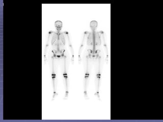
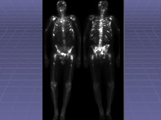
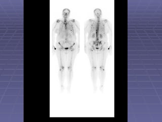
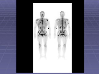
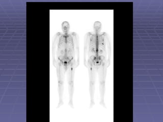
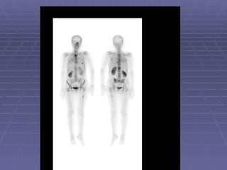
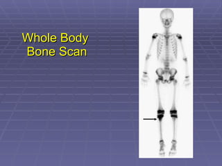
BONE ISOTOPE SCAN الدكتور قصي المقبل أستاذ مشارك -  قسم الأشعة والطب النووي كلية الطب - جامعة العلوم والتكنولوجيا الأردنية
Introduction Bone scan is one of the most commonly performed procedures in nuclear medicine. Bone scan often provides an earlier diagnosis and demonstrates more lesions than are found by radiographic procedures.
Sensitivity and Specificity Bone scan is very sensitive study but it is not specific.  Although findings on bone scan are non-specific, its monostotic or polyostotic status and anatomical distribution can provide important clues to the differential diagnosis.
Radiopharmaceuticals They are bone seeking agents. They are labeled with Tc99m. They are phosphate analogs. Most commonly used one is HDP (Hydroxy Methylene Diphosphonates). They are given intravenously.
Mechanism of Localization Phosphate groups bind to the hydroxyapatite [Ca3(Po4)2] structure of bone tissue by a mechanism called chemisorption. The hydroxyapatite structure of the bone is exposed during bone remodeling. So, more radiopharmaceutical will deposit in that region giving “hot” area. 50-60% of injected dose localized on bone, remain dose is cleared by kidneys.
Whole body bone scan This is the bony phase of bone scan. Inject radiopharmaceutical and image in 2-4 hours. When we say bone scan, we usually mean whole body bone scan.


Three Phase Bone Scan It is done to see if there is soft tissue hyperemia. First phase is the perfusion phase or vascular phase. Second phase is the blood pooling phase or soft tissue phase. Third phase is the bony phase.
First Phase   30-60 dynamic images are usually obtained over 1 minute immediately after injection. This is radionuclide angiography and gives an idea about the local vasculature. During the first minute after injection, injected dose is still intravascular.
Perfusion
Second Phase   Static image is obtained in 5 minutes after dose injection. Within 5 minutes post injection, radiopharmaceutical moves from intravascular space to extravascular space (soft tissue). It gives idea about soft tissue edema.
Blood Pool
Hyperemia If there is focal increased activity in the first and second phases, hyperemia or acute inflammatory process is present .
Third Phase   It is the bony phase image obtained in 2-4 hours post injection. It is the same as whole body bone scan.
Clinical Applications Malignancy  1- Primary bone cancer 2- Secondary metastatic bone disease Osteomyelitis  Stress fractures
Primary Bone Malignancy (Sarcomas)   MRI provides more exact information regarding tumor extent, particularly in soft tissue. So, bone scan is not a diagnosis image in primary bone tumors.
Bone sarcomas-cont.. Bone scan is a staging and restaging image in bone sarcomas. It is performed to see if there is bone metastatic disease (bone to bone). 40 – 50% of patients with either Ewing’s sarcoma or osteosarcoma develop osseous metastases within 2 years of presentation.
 
 
Metastatic Bone Disease Bone scan is an extremely important tool in decision making during management of cancer patients. Any cancer potentially could cause bone metastatic disease.  However, prostate, breast and lung cancers have propensity to metastasize to bone.
Metastatic Bone Disease and Bone Pain About 80% of patients with known cancer and bone pain have metastases documented by bone scan. 30 – 50% of patients with metastatic bone disease do not have bone pain.
Metastatic Bone Disease and Image Findings The hallmark of metastatic bone disease is multiple foci of increased osteoblastic activity in bony skeleton. However, single lesion could be also metastatic.


Stress Fractures   It is often difficult to visualized on a plain radiograph. Fractures may be identified by bone scan as early as 24 hours after occurrence. 3-phase bone scan is usually done.  There is hyperemia and osteoblastic process (three phases are positive).
Stress Fracture Blood Pool Anterior Posterior Anterior Posterior Left Lat Right Lat Left Lat Right Lat 2 hour delay
Stress Fracture
Acute Osteomyelitis   Early plain radiography signs of osteomyelitis are non-specific. 3-phase bone scan is usually the procedure of choice to differentiate between osteomyelitis and cellulitis.
Acute Osteomyelitis-cont.. If first and 2nd phases are positive (hyperemia) with normal third phase, diagnosis would be cellulitis. In acute osteomyelitis all 3-phases are positive (hyperemia and osteoblastic process in the bone).
Perfusion
Blood Pool
Whole Body  Bone Scan

2. bone scan (2010)

  • 1.
    BONE ISOTOPE SCANالدكتور قصي المقبل أستاذ مشارك - قسم الأشعة والطب النووي كلية الطب - جامعة العلوم والتكنولوجيا الأردنية
  • 2.
    Introduction Bone scanis one of the most commonly performed procedures in nuclear medicine. Bone scan often provides an earlier diagnosis and demonstrates more lesions than are found by radiographic procedures.
  • 3.
    Sensitivity and SpecificityBone scan is very sensitive study but it is not specific. Although findings on bone scan are non-specific, its monostotic or polyostotic status and anatomical distribution can provide important clues to the differential diagnosis.
  • 4.
    Radiopharmaceuticals They arebone seeking agents. They are labeled with Tc99m. They are phosphate analogs. Most commonly used one is HDP (Hydroxy Methylene Diphosphonates). They are given intravenously.
  • 5.
    Mechanism of LocalizationPhosphate groups bind to the hydroxyapatite [Ca3(Po4)2] structure of bone tissue by a mechanism called chemisorption. The hydroxyapatite structure of the bone is exposed during bone remodeling. So, more radiopharmaceutical will deposit in that region giving “hot” area. 50-60% of injected dose localized on bone, remain dose is cleared by kidneys.
  • 6.
    Whole body bonescan This is the bony phase of bone scan. Inject radiopharmaceutical and image in 2-4 hours. When we say bone scan, we usually mean whole body bone scan.
  • 7.
  • 8.
  • 9.
  • 10.
  • 11.
    Three Phase BoneScan It is done to see if there is soft tissue hyperemia. First phase is the perfusion phase or vascular phase. Second phase is the blood pooling phase or soft tissue phase. Third phase is the bony phase.
  • 12.
    First Phase 30-60 dynamic images are usually obtained over 1 minute immediately after injection. This is radionuclide angiography and gives an idea about the local vasculature. During the first minute after injection, injected dose is still intravascular.
  • 13.
  • 14.
    Second Phase Static image is obtained in 5 minutes after dose injection. Within 5 minutes post injection, radiopharmaceutical moves from intravascular space to extravascular space (soft tissue). It gives idea about soft tissue edema.
  • 15.
  • 16.
    Hyperemia If thereis focal increased activity in the first and second phases, hyperemia or acute inflammatory process is present .
  • 17.
    Third Phase It is the bony phase image obtained in 2-4 hours post injection. It is the same as whole body bone scan.
  • 18.
    Clinical Applications Malignancy 1- Primary bone cancer 2- Secondary metastatic bone disease Osteomyelitis Stress fractures
  • 19.
    Primary Bone Malignancy(Sarcomas) MRI provides more exact information regarding tumor extent, particularly in soft tissue. So, bone scan is not a diagnosis image in primary bone tumors.
  • 20.
    Bone sarcomas-cont.. Bonescan is a staging and restaging image in bone sarcomas. It is performed to see if there is bone metastatic disease (bone to bone). 40 – 50% of patients with either Ewing’s sarcoma or osteosarcoma develop osseous metastases within 2 years of presentation.
  • 21.
  • 22.
  • 23.
    Metastatic Bone DiseaseBone scan is an extremely important tool in decision making during management of cancer patients. Any cancer potentially could cause bone metastatic disease. However, prostate, breast and lung cancers have propensity to metastasize to bone.
  • 24.
    Metastatic Bone Diseaseand Bone Pain About 80% of patients with known cancer and bone pain have metastases documented by bone scan. 30 – 50% of patients with metastatic bone disease do not have bone pain.
  • 25.
    Metastatic Bone Diseaseand Image Findings The hallmark of metastatic bone disease is multiple foci of increased osteoblastic activity in bony skeleton. However, single lesion could be also metastatic.
  • 26.
  • 27.
  • 28.
  • 29.
  • 30.
  • 31.
    Stress Fractures It is often difficult to visualized on a plain radiograph. Fractures may be identified by bone scan as early as 24 hours after occurrence. 3-phase bone scan is usually done. There is hyperemia and osteoblastic process (three phases are positive).
  • 32.
    Stress Fracture BloodPool Anterior Posterior Anterior Posterior Left Lat Right Lat Left Lat Right Lat 2 hour delay
  • 33.
  • 34.
    Acute Osteomyelitis Early plain radiography signs of osteomyelitis are non-specific. 3-phase bone scan is usually the procedure of choice to differentiate between osteomyelitis and cellulitis.
  • 35.
    Acute Osteomyelitis-cont.. Iffirst and 2nd phases are positive (hyperemia) with normal third phase, diagnosis would be cellulitis. In acute osteomyelitis all 3-phases are positive (hyperemia and osteoblastic process in the bone).
  • 36.
  • 37.
  • 38.
    Whole Body Bone Scan

Editor's Notes

  • #33 57 year old man with painful left ankle. Plain films showed an area of sclerosis at the distal left tibia. Pertinent Findings: Blood flow images demonstrate an increase in activity in the distal left tibia. The bone scan shows increased activity along the anterior cortex and at the site of sclerosis. Diagnosis: Insufficiency fracture of the distal left tibia. Discussion : Stress fractures are either insufficiency or fatigue fractures. Insufficiency fractures occur in the setting of normal stress applied to abnormal bone. Abnormality of bone includes diminshed elasticity, deficient mineralization. Fatigue fractures result when abnormal stress is applied to normal bone.
  • #34 Clinical History: Pain in the right foot for three weeks. Findings: Nuclear Medicine Bone Scan: A single focus of increased radiopharmaceutical activity is identified on all three phases within the distal right fourth metatarsal. Diagnosis: Stress fracture. Discussion: A stress fracture is the result of repetitive, prolonged muscular action on a bone that has not accommodated itself to that action. In most instances, persons who have stress fractures are engaged in vigorous activity to which they have not yet become conditioned. Under normal circumstances, the muscle tone takes up at a faster rate than bones which result in a mechanical imbalance. Stress fractures are divided into two categories, fatigue fractures and insufficiency fractures. A fatigue fracture is caused by the application of abnormal stress or torque on a bone that has normal elastic resistance. On the other hand, an insufficiency fracture occurs when normal muscular activity stresses a bone that is deficient in mineral or elastic resistance. The term pathologic fracture should be reserved for fractures that occur in bone already weakened by neoplasm or infection. The stress placed on bone causes resorption and microfractures. If the patient rests and the stress decreases then repair may continue with resultant relief of pain. However, if the repair process can no longer keep up with the resorptive process, then cortical disruption and a radiographically visible fracture may ensue. Findings develop earlier on bone scan and MRI than on plain film roentgenograms. A hot spot on all three phases of a bone scan is a bone that has received repetitive trauma is indicative of a stress fracture. Type sites for athletic stress fractures include:Tibial and fibular shaft ;Medial aspect of the femoral neck ;Inferior pubic ramus ;Metatarsals and os calcis ;Navicular and tarsal sesamoids