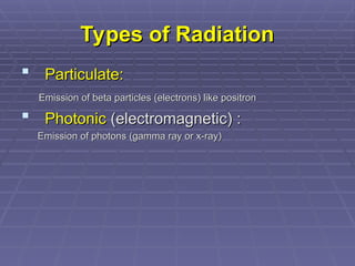
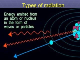
Types of Radiation
Typesof Radiation
Particulate:
Particulate:
Emission of beta particles (electrons) like positron
Emission of beta particles (electrons) like positron
Photonic
Photonic (electromagnetic) :
(electromagnetic) :
Emission of photons (gamma ray or x-ray)
Emission of photons (gamma ray or x-ray)
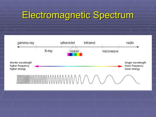
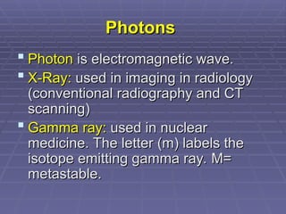
Photons
Photons
Photon
Photon iselectromagnetic wave.
is electromagnetic wave.
X-Ray:
X-Ray: used in imaging in radiology
used in imaging in radiology
(conventional radiography and CT
(conventional radiography and CT
scanning)
scanning)
Gamma ray:
Gamma ray: used in nuclear
used in nuclear
medicine. The letter (m) labels the
medicine. The letter (m) labels the
isotope emitting gamma ray. M=
isotope emitting gamma ray. M=
metastable.
metastable.
6.
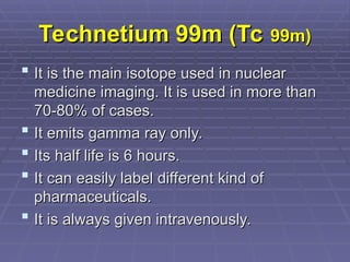
Technetium 99m (Tc
Technetium99m (Tc 99m)
99m)
It is the main isotope used in nuclear
It is the main isotope used in nuclear
medicine imaging. It is used in more than
medicine imaging. It is used in more than
70-80% of cases.
70-80% of cases.
It emits gamma ray only.
It emits gamma ray only.
Its half life is 6 hours.
Its half life is 6 hours.
It can easily label different kind of
It can easily label different kind of
pharmaceuticals.
pharmaceuticals.
It is always given intravenously.
It is always given intravenously.
7.
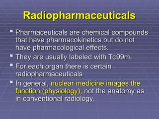
Radiopharmaceuticals
Radiopharmaceuticals
Pharmaceuticals arechemical compounds
Pharmaceuticals are chemical compounds
that have pharmacokinetics but do not
that have pharmacokinetics but do not
have pharmacological effects.
have pharmacological effects.
They are usually labeled with Tc99m.
They are usually labeled with Tc99m.
For each organ there is certain
For each organ there is certain
radiopharmaceuticals
radiopharmaceuticals
In general,
In general, nuclear medicine images the
nuclear medicine images the
function (physiology),
function (physiology), not the anatomy as
not the anatomy as
in conventional radiology.
in conventional radiology.
Introduction
Introduction
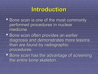
Bone scanis one of the most commonly
Bone scan is one of the most commonly
performed procedures in nuclear
performed procedures in nuclear
medicine.
medicine.
Bone scan often provides an earlier
Bone scan often provides an earlier
diagnosis and demonstrates more lesions
diagnosis and demonstrates more lesions
than are found by radiographic
than are found by radiographic
procedures.
procedures.
Bone scan has the advantage of screening
Bone scan has the advantage of screening
the entire bone skeleton.
the entire bone skeleton.
11.
High Sensitivity
High Sensitivity
Bone scan is very sensitive in lesion detection even
Bone scan is very sensitive in lesion detection even
better than radiographic images (plain x-ray and CT
better than radiographic images (plain x-ray and CT
scan). Even a
scan). Even a 5% bone turnover
5% bone turnover can be detected by
can be detected by
bone scan, whereas radiographs require a minimum
bone scan, whereas radiographs require a minimum
mineral loss of 50% before a lesion is visualized.
mineral loss of 50% before a lesion is visualized.
Higher sensitivity means earlier diagnosis before
Higher sensitivity means earlier diagnosis before
structural changes appear on radiographic images
structural changes appear on radiographic images
(plain x-ray and CT scan).
(plain x-ray and CT scan).
Higher sensitive because bone scan allows screening
Higher sensitive because bone scan allows screening
of entire bone skeleton.
of entire bone skeleton.
Higher sensitive because of high contrast of the lesion
Higher sensitive because of high contrast of the lesion
(easy detectability of the lesions).
(easy detectability of the lesions).
12.
Low Specificity
Low Specificity
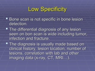
Bone scan is not specific in bone lesion
Bone scan is not specific in bone lesion
detection.
detection.
The differential diagnosis of any lesion
The differential diagnosis of any lesion
seen on bon scan is wide including tumor,
seen on bon scan is wide including tumor,
infection and fracture.
infection and fracture.
The diagnosis is usually made based on
The diagnosis is usually made based on
clinical history, lesion location, number of
clinical history, lesion location, number of
lesions, correlation with lab and other
lesions, correlation with lab and other
imaging data (x-ray, CT, MRI…).
imaging data (x-ray, CT, MRI…).
13.
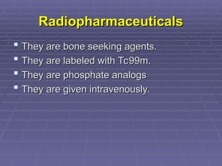
Radiopharmaceuticals
Radiopharmaceuticals
They arebone seeking agents.
They are bone seeking agents.
They are labeled with Tc99m.
They are labeled with Tc99m.
They are phosphate analogs
They are phosphate analogs
They are given intravenously.
They are given intravenously.
14.
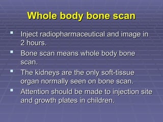
Whole body bonescan
Whole body bone scan
Inject radiopharmaceutical and image in
Inject radiopharmaceutical and image in
2 hours.
2 hours.
Bone scan means whole body bone
Bone scan means whole body bone
scan.
scan.
The kidneys are the only soft-tissue
The kidneys are the only soft-tissue
organ normally seen on bone scan.
organ normally seen on bone scan.
Attention should be made to injection site
Attention should be made to injection site
and growth plates in children.
and growth plates in children.
15.
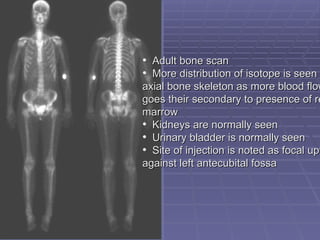
• Adult bonescan
Adult bone scan
• More distribution of isotope is seen
More distribution of isotope is seen i
axial bone skeleton as more blood flow
axial bone skeleton as more blood flow
goes their secondary to presence of re
goes their secondary to presence of re
marrow
marrow
• Kidneys are normally seen
Kidneys are normally seen
• Urinary bladder is normally seen
Urinary bladder is normally seen
• Site of injection is noted as focal upt
Site of injection is noted as focal upt
against left antecubital fossa
against left antecubital fossa
16.
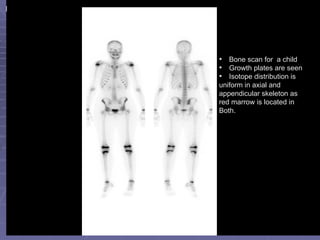
• Bone scanfor a child
Bone scan for a child
• Growth plates are seen
Growth plates are seen
• Isotope distribution is
Isotope distribution is
uniform in axial and
uniform in axial and
appendicular skeleton as
appendicular skeleton as
red marrow is located in
red marrow is located in
Both.
Both.
17.
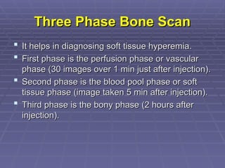
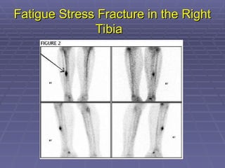
Three Phase BoneScan
Three Phase Bone Scan
It helps in diagnosing soft tissue hyperemia.
It helps in diagnosing soft tissue hyperemia.
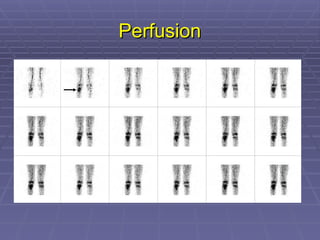
First phase is the perfusion phase or vascular
First phase is the perfusion phase or vascular
phase (30 images over 1 min just after injection).
phase (30 images over 1 min just after injection).
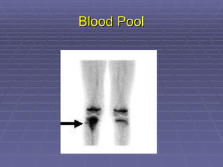
Second phase is the blood pool phase or soft
Second phase is the blood pool phase or soft
tissue phase (image taken 5 min after injection).
tissue phase (image taken 5 min after injection).
Third phase is the bony phase (2 hours after
Third phase is the bony phase (2 hours after
injection).
injection).
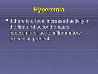
Hyperemia
Hyperemia
If thereis a focal increased activity in
If there is a focal increased activity in
the first and second phases,
the first and second phases,
hyperemia or acute inflammatory
hyperemia or acute inflammatory
process is present
process is present.
.
21.
Third Phase
Third Phase
It is the bony phase image obtained
It is the bony phase image obtained
in 2 hours post injection.
in 2 hours post injection.
It is the same as whole body bone
It is the same as whole body bone
scan.
scan.
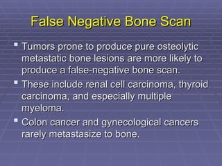
False Negative BoneScan
False Negative Bone Scan
Tumors prone to produce pure osteolytic
Tumors prone to produce pure osteolytic
metastatic bone lesions are more likely to
metastatic bone lesions are more likely to
produce a false-negative bone scan.
produce a false-negative bone scan.
These include renal cell carcinoma, thyroid
These include renal cell carcinoma, thyroid
carcinoma, and especially multiple
carcinoma, and especially multiple
myeloma.
myeloma.
Colon cancer and gynecological cancers
Colon cancer and gynecological cancers
rarely metastasize to bone.
rarely metastasize to bone.
24.
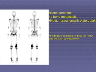
•Bone sarcoma:
Bone sarcoma:
nobone metastasis
no bone metastasis
•Note: normal growth plate uptake
Note: normal growth plate uptake
•Findings: focal uptake in distal left femur
Findings: focal uptake in distal left femur
due to known osteosarcoma
due to known osteosarcoma
25.
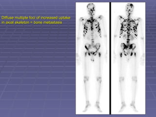
Diffuse multiple fociof increased uptake
Diffuse multiple foci of increased uptake
in axial skeleton = bone metastasis
in axial skeleton = bone metastasis
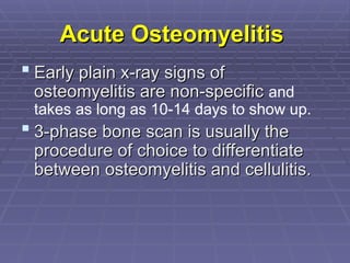
Acute Osteomyelitis
Acute Osteomyelitis
Early plain x-ray signs of
Early plain x-ray signs of
osteomyelitis are non-specific
osteomyelitis are non-specific and
takes as long as 10-14 days to show up.
3-phase bone scan is usually the
3-phase bone scan is usually the
procedure of choice to differentiate
procedure of choice to differentiate
between osteomyelitis and cellulitis.
between osteomyelitis and cellulitis.
28.
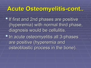
Acute Osteomyelitis-cont..
Acute Osteomyelitis-cont..
If first and 2nd phases are positive
If first and 2nd phases are positive
(hyperemia) with normal third phase,
(hyperemia) with normal third phase,
diagnosis would be cellulitis.
diagnosis would be cellulitis.
In acute osteomyelitis all 3-phases
In acute osteomyelitis all 3-phases
are positive (hyperemia and
are positive (hyperemia and
osteoblastic process in the bone).
osteoblastic process in the bone).
Editor's Notes
#4 Ionizing radiation is radiation with enough energy so that during an interaction with an atom, it can remove tightly bound electrons from their orbits, causing the atom to become charged or ionized.
Here we are concerned with only one type of radiation, ionizing radiation, which occurs in two forms - waves or particles.


![[5]Isotope_Scan_Surgical_Diseases](https://cdn.slidesharecdn.com/ss_thumbnails/1664464-thumbnail.jpg?width=640&height=640&fit=bounds)


