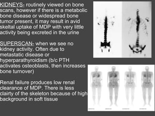
The Bone Scan You have to know what is normal before you determine what is ABNORMAL!
KIDNEYS-  routinely viewed on bone scans, however if there is a metabolic bone disease or widespread bone tumor present, it may result in avid skeltal uptake of MDP with very little activity being excreted in the urine   SUPERSCAN-  when we see no kidney activity. Often due to metastatic disease or hyperparathyroidism (b/c PTH activates osteoblasts, then increases bone turnover)   Renal failure produces low renal clearance of MDP. There is less clairty of the skeleton because of high background in soft tissue  
NORMAL "HOT" AREAS...  SI Joints   Acromioclavicular joints   Hip joints    Sternoclavicular joints   Ends of long bones   Growing epiphysis (in pediatric patients)   Fractures and orthopedic surgery produce uptake for 1 or more years   Variations in spine uptake are casued by normal curvature of the spine
CAUSES OF SOFT TISSUE UPTAKE Soft tissue necrosis or calcification   Tumors   Breast uptake can be normal in young females (beastfeeding), however it can also be seen in Breast CA patients   Malignant pleural effusions   Radiochemical impurity will have uptake in thyroid, salivary glands, gastric mucosa 
MAKING A DIAGNOSIS... Increased activity is not diagnositc of nepplastic process   Positive bone scan findings does NOT differentiate between malignant or benign   Final diagnosis is made by correlating bone scan images with clinical data (remember how important it is to take a good history!!!) and other imaging modalities (think SPECT CT) 
PITFALLS IN BONE SCANS  Epiphyeal plates in kids will appear hot due to increased blood flow and metabolism in growing bones   Bone images may be negative when bony healing is completed (healed fx's, cured osteo, treated bone mets). However, this may take 1+ years.   When bone images are positive but nonspecific, radiographic correlation is important   Ga-67 or In-111 used with Tc-99m MDP for osteo. They help define active osteo and may detect deep soft tissue infection
MDP may concentrate at sites of soft tissue pathologic conditions such as breast CA, malignant pleural effusions, ascites, soft tissue malignancies, myositis ossificans, MI, electrical burns, ectopic calcification, splenic infarction, IM injections   Bone grafts show increased activity at the ends of the bone graft, with little to no uptake in the middle for 6 months post sx.    http://www.skeletalscintigraphy.com/fp/index.htm   A good website to view interesting bone scan cases INTERESTING FINDINGS
Myositis Ossificans A reparative process that causes benign heterotropic ossifications in soft tissue. Usually a result of trauma.      Myositis ossificans.   A bone scintigram (A-B) demonstrates marked accumulation of radiotracer in the soft tissues of both distal thighs and knees left greater than right . A radiograph (C) of the left knee shows soft tissue ossification consistent with myositis ossificans. Myositis ossificans.   A bone scintigram (A) demonstrates marked accumulation of radiotracer in the region of the right hip. Plain radiograph (B) of the right hipshows ossification in the soft tissues consistent with myositis ossificans.
Myocardial Findings in a Bone Scan Ischemic cardiomyopathy. This whole-body bone scintigram shows diffuse radionuclide uptake in the region of the heart of a patient with two previous myocardial infarctions and ischemic cardiomyopathy. The ejection fraction was 14%.       Scintigraphy with Tc-99m pyrophosphate is commonly used to detect acute myocardial infarction. Similar findings are present with Tc-99m diphosphonates, when there is an excess of tissue calcium, following local tissue necrosis or damage. Focal uptake is seen with transmural myocardial infarction. Patchy uptake has been noted in unstable angina pectoris, and diffuse uptake has been observed with  cardiomyopathies   (ref)  , pericardial tumors, and diffuse pericarditis. Persistent uptake following infarction correlates with a higher rate of complications and with left ventricular aneurysm. Other causes of myocardial uptake include myocardial contusion, electro-cardioversion, and  amyloidosis  (ref) Amyloidosis  Anterior view of the whole-body bone scan show intense radiotracer uptake in the myocardium.
Electrical Burns Electrical burn.   The bone scan shows increased radionuclide uptake in the muscle of the forearm caused by rhabdomyolysis as a result of electrical burn.
Lymph node uptake due to extravasation A bone scintigram (A) demonstrates focal accumulation of radiotracer in the axilla due to extravasation at injection site. A repeat bone scan (B) 3 months later shows resolution of the previous finding.  * Note RIGHT axilla ---> Injection site  (AKA extravasation) --->
SCAR Whole-body bone scan in anterior (A) and lateral (B) projections show linear radiotracer accumulation along the midepigastric region, where prior laparotomy scar is evident.
Urine contamination Anterior image (A) of the bone scan shows 2 hot spots projecting over the right femur; repeat image (B) after decontamination shows disappearance of both hot spots.
Malignant Fibrohistiocytoma Malignant fibrous histiocytoma  (MFH) is a type of  cancer  that is the most common  soft tissue sarcoma  of late adult life, most commonly occurring between age 50-70. In rare cases MFH does occur in children, but it is usually in a less aggressive form.                    The bone scintigram (A) shows radionuclide accumulation in the soft tissue of the right thigh. Axial MRI (B) demonstrates an intramuscular soft tissue mass, that was biopsy-proven malignatn fibrohistiocytoma.
Breast CA The bone scintigram shows intense soft­ issue uptake in a large carcinoma of the left breast. Calcifications in the mass were present on xeromammogram.
More FYI's  In avascular processes, there is absence of delivery of tracer to the area distal to the occluded vessels. Above the avascular area, there is usually generalized increased uptake.   Fractures - within 72 hours from injury, most people will have increased uptake at the site of the fx. However, geriatric patients sometimes won't show increased uptake unitl 5 days. Use 3-5 days post injury event as a rule of thumb when imaging to r/o fx.   Post partum females - increase activity in SI and pubic symphysis can be seen approx 18 months post delivery.

The Bone Scan

  • 1.
    The Bone ScanYou have to know what is normal before you determine what is ABNORMAL!
  • 2.
    KIDNEYS- routinelyviewed on bone scans, however if there is a metabolic bone disease or widespread bone tumor present, it may result in avid skeltal uptake of MDP with very little activity being excreted in the urine   SUPERSCAN- when we see no kidney activity. Often due to metastatic disease or hyperparathyroidism (b/c PTH activates osteoblasts, then increases bone turnover)   Renal failure produces low renal clearance of MDP. There is less clairty of the skeleton because of high background in soft tissue  
  • 3.
    NORMAL "HOT" AREAS... SI Joints   Acromioclavicular joints   Hip joints    Sternoclavicular joints   Ends of long bones   Growing epiphysis (in pediatric patients)   Fractures and orthopedic surgery produce uptake for 1 or more years   Variations in spine uptake are casued by normal curvature of the spine
  • 4.
    CAUSES OF SOFTTISSUE UPTAKE Soft tissue necrosis or calcification   Tumors   Breast uptake can be normal in young females (beastfeeding), however it can also be seen in Breast CA patients   Malignant pleural effusions   Radiochemical impurity will have uptake in thyroid, salivary glands, gastric mucosa 
  • 5.
    MAKING A DIAGNOSIS...Increased activity is not diagnositc of nepplastic process   Positive bone scan findings does NOT differentiate between malignant or benign   Final diagnosis is made by correlating bone scan images with clinical data (remember how important it is to take a good history!!!) and other imaging modalities (think SPECT CT) 
  • 6.
    PITFALLS IN BONESCANS  Epiphyeal plates in kids will appear hot due to increased blood flow and metabolism in growing bones   Bone images may be negative when bony healing is completed (healed fx's, cured osteo, treated bone mets). However, this may take 1+ years.   When bone images are positive but nonspecific, radiographic correlation is important   Ga-67 or In-111 used with Tc-99m MDP for osteo. They help define active osteo and may detect deep soft tissue infection
  • 7.
    MDP may concentrateat sites of soft tissue pathologic conditions such as breast CA, malignant pleural effusions, ascites, soft tissue malignancies, myositis ossificans, MI, electrical burns, ectopic calcification, splenic infarction, IM injections   Bone grafts show increased activity at the ends of the bone graft, with little to no uptake in the middle for 6 months post sx.    http://www.skeletalscintigraphy.com/fp/index.htm   A good website to view interesting bone scan cases INTERESTING FINDINGS
  • 8.
    Myositis Ossificans Areparative process that causes benign heterotropic ossifications in soft tissue. Usually a result of trauma.     Myositis ossificans. A bone scintigram (A-B) demonstrates marked accumulation of radiotracer in the soft tissues of both distal thighs and knees left greater than right . A radiograph (C) of the left knee shows soft tissue ossification consistent with myositis ossificans. Myositis ossificans. A bone scintigram (A) demonstrates marked accumulation of radiotracer in the region of the right hip. Plain radiograph (B) of the right hipshows ossification in the soft tissues consistent with myositis ossificans.
  • 9.
    Myocardial Findings ina Bone Scan Ischemic cardiomyopathy. This whole-body bone scintigram shows diffuse radionuclide uptake in the region of the heart of a patient with two previous myocardial infarctions and ischemic cardiomyopathy. The ejection fraction was 14%.     Scintigraphy with Tc-99m pyrophosphate is commonly used to detect acute myocardial infarction. Similar findings are present with Tc-99m diphosphonates, when there is an excess of tissue calcium, following local tissue necrosis or damage. Focal uptake is seen with transmural myocardial infarction. Patchy uptake has been noted in unstable angina pectoris, and diffuse uptake has been observed with cardiomyopathies (ref) , pericardial tumors, and diffuse pericarditis. Persistent uptake following infarction correlates with a higher rate of complications and with left ventricular aneurysm. Other causes of myocardial uptake include myocardial contusion, electro-cardioversion, and amyloidosis (ref) Amyloidosis Anterior view of the whole-body bone scan show intense radiotracer uptake in the myocardium.
  • 10.
    Electrical Burns Electricalburn. The bone scan shows increased radionuclide uptake in the muscle of the forearm caused by rhabdomyolysis as a result of electrical burn.
  • 11.
    Lymph node uptakedue to extravasation A bone scintigram (A) demonstrates focal accumulation of radiotracer in the axilla due to extravasation at injection site. A repeat bone scan (B) 3 months later shows resolution of the previous finding. * Note RIGHT axilla ---> Injection site (AKA extravasation) --->
  • 12.
    SCAR Whole-body bonescan in anterior (A) and lateral (B) projections show linear radiotracer accumulation along the midepigastric region, where prior laparotomy scar is evident.
  • 13.
    Urine contamination Anteriorimage (A) of the bone scan shows 2 hot spots projecting over the right femur; repeat image (B) after decontamination shows disappearance of both hot spots.
  • 14.
    Malignant Fibrohistiocytoma Malignantfibrous histiocytoma (MFH) is a type of cancer that is the most common soft tissue sarcoma of late adult life, most commonly occurring between age 50-70. In rare cases MFH does occur in children, but it is usually in a less aggressive form.                   The bone scintigram (A) shows radionuclide accumulation in the soft tissue of the right thigh. Axial MRI (B) demonstrates an intramuscular soft tissue mass, that was biopsy-proven malignatn fibrohistiocytoma.
  • 15.
    Breast CA Thebone scintigram shows intense soft­ issue uptake in a large carcinoma of the left breast. Calcifications in the mass were present on xeromammogram.
  • 16.
    More FYI's  Inavascular processes, there is absence of delivery of tracer to the area distal to the occluded vessels. Above the avascular area, there is usually generalized increased uptake.   Fractures - within 72 hours from injury, most people will have increased uptake at the site of the fx. However, geriatric patients sometimes won't show increased uptake unitl 5 days. Use 3-5 days post injury event as a rule of thumb when imaging to r/o fx.   Post partum females - increase activity in SI and pubic symphysis can be seen approx 18 months post delivery.