Downloaded 642 times
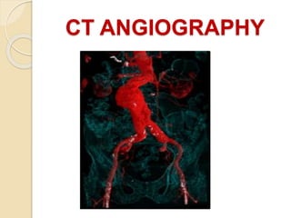

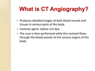


CT angiography uses x-rays and iodine contrast dye to produce detailed images of blood vessels and tissues. A CT scan is performed after the contrast dye is injected into the bloodstream. CT angiography can be used to diagnose and evaluate diseases of the blood vessels like injuries, aneurysms, and blockages. It provides more precise anatomical detail than MRI for small blood vessels. Potential risks include radiation exposure and allergic reaction to the contrast dye.


![ct angio group c ,omer drazbsrit ]]].pptx](https://cdn.slidesharecdn.com/ss_thumbnails/ctangiogroupcomerdraz-240927172646-77eeb8b5-thumbnail.jpg?width=640&height=640&fit=bounds)