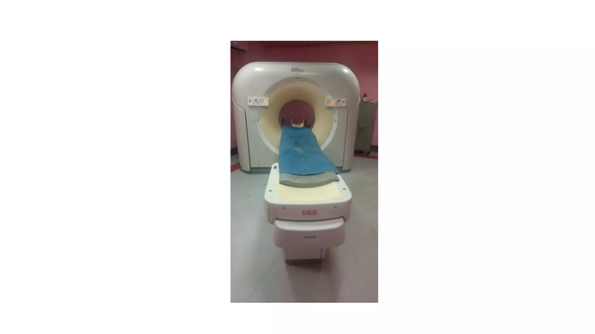
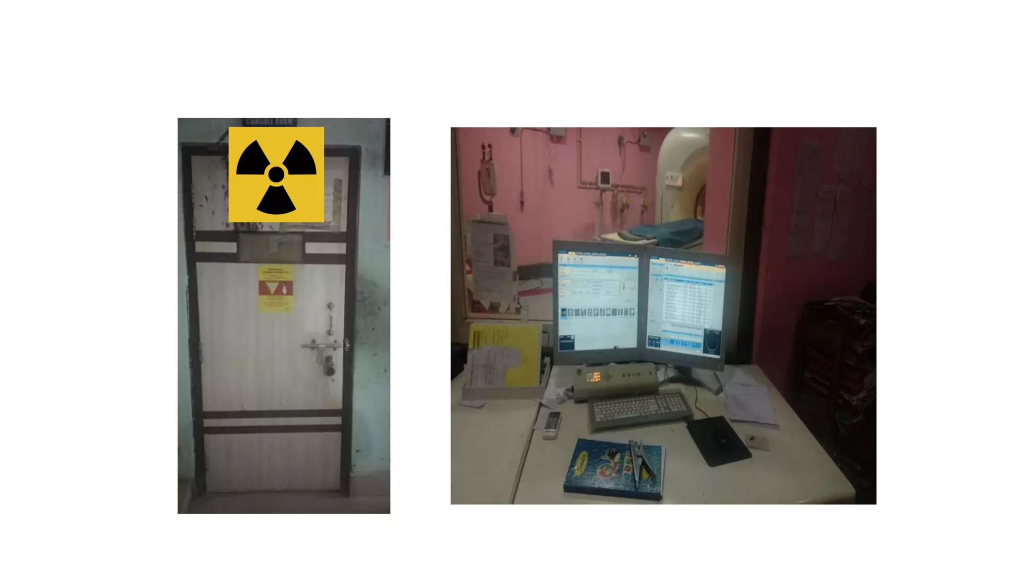
BASICS OF
COMPUTED TOMOGRAPHY
PRESENTED BY : DR.B.BORTHAKUR
PROFRSSOR AND HEAD, DEPT. OF ORTHOPAEDICS,SMCH
• Computed Tomography (CT) scanning, also known as,
Computed Axial Tomography (CAT) scanning, is a
diagnostic imaging procedure that uses x-rays to
build cross-sectional images of the body.
• CT is based on the fundamental principle that the
density of the tissue passed by the x-ray beam can be
measured
 The patient lies on the table within the CT gantry,
 During each slice acquisition, an X-ray tube circling the
patient produces an X-ray beam that passes through the
patient and is absorbed by a ring of detectors surrounding
the patient.
 The intensity of the X-ray beam that reaches the detector is
dependent on the absorption characteristics of the tissues
it passes through.
 Since the beam is moving around the patient, each
tissue will be exposed from multiple directions. The
basic principle of CT lies in the reconstruction of the
internal structure of an object from multiple
projections of the object.
 Using a process called Fourier analysis, the computer
ultimately forms the image.
Components of CT scanner:-
1. Gantry assembly & patient table/couch
2. Computer
3. Operating console
Gantry assembly & patient table/couch
Console Room and Computer
Operating console
Image reconstruction
 The computer receives a signal in analog form and converts it to
a binary digit. The digital signal is stored and the image is
reconstructed after the scan is over.
 In CT, a cross-sectional layer of the body is divided into many
tiny blocks, called as voxels. Voxel is a 3D tissue element.
 Pixel is the 2D projection of a voxel on the computer screen
 Therefore, a voxel is a volume element and a pixel is a picture
element. Each is displayed on a matrix.
CT number and Hounsfield units:-
o Each pixel is assigned a numerical value (CT number), which is
the average of all the attenuation values contained within the
corresponding voxel.
o This number is compared to the attenuation value of water and
displayed on a scale of arbitrary units named Hounsfield units
(HU) after Sir Godfrey Hounsfield.
Evolution of CT scanners
First generation
• detectors: one
• type of beam: pencil-like x-ray beam
• tube-detector movements: translate-rotate
• duration of scan (average): 25-30 mins
• single X-ray source and single X-ray detector cell to collect all the data
for a single slice.
• Process is repeated once for each projection angle until 180
projections , across a 24 cm FOV
First Generation CT
Second generation
• detectors: multiple (up to 30)
• type of beam: fan-shaped x-ray beam
• tube-detector movements: translate-rotate
• duration of scan (average): less than 90 sec
• By adding detectors angularly displaced , several projections could be
obtained in a single translation
Third generation
• detectors: multiple, originally 288; newer ones use over 700 arranged
in an arc
• type of beam: fan-shaped x-ray beam
• tube-detector movements: rotate-rotate
• duration of scan (average): approximately 5 sec
• Sampling considerations required scanning an additional arc of one
fan angle beyond 180°, although most scanners rotate 360°for each
scan.
Fourth generation
• detectors: multiple (more than 2000) arranged in an outer ring which
is fixed
• type of beam: fan-shaped x-ray beam
• tube-detector movements: rotate-fixed
• duration of scan (average): few seconds
• Design: stationary detector ring & rotating X-ray tube
Spiral/Helical CT
• Design: x-ray tube rotates as patient is moved smoothly into x-ray
scan field
• Simultaneous source rotation, table translation and data acquisition
• Produces one continuous volume set of data for entire region
• Data for multiple slices from patient acquired at 1sec/slice
SSCT VS MSCT
 MSCT Detectors The primary difference
between single-slice CT (SSCT) and MSCT
hardware is in the design of the detector
arrays.
 SSCT detector arrays are one
dimensional, that is, they consist of a
large number (typically 750 or more) of
detector elements in a single row across
the irradiated slice to intercept the x-ray
fanbeam.
MDCT or MSCT
 MDCT is a commonly used acronym that stands for Multiple-
row Detector Computed Tomography.
 The other commonly used term MSCT stands for Multi Slice
Computed Tomography.
 It is the latest breakthrough in CT technology.
 It has transformed CT from trans-axial cross sectional
technique into a true three dimensional imaging modality.
Advantages of MDCT
1. Shorter scan time: Reduces movement artifacts. Useful in
children, trauma patients & acutely ill patients.
2. Improved scanning of parenchymal organs:- different phases of
contrast enhancement can be elicited
3. Minimization of partial volume averaging effects.
4. Thinner sections:- excellent multiplaner reformations can be
performed.
5. Reduction of radiation exposure.
Disadvantages:-
 Radiation exposure.
 Relatively poor tissue contrast when compared to MRI is a
problem,
 There can be contrast media-related complications,
including allergic reactions and renal toxicity.
THANK YOU

Basics of CT

  • 1.
    BASICS OF COMPUTED TOMOGRAPHY PRESENTEDBY : DR.B.BORTHAKUR PROFRSSOR AND HEAD, DEPT. OF ORTHOPAEDICS,SMCH
  • 2.
    • Computed Tomography(CT) scanning, also known as, Computed Axial Tomography (CAT) scanning, is a diagnostic imaging procedure that uses x-rays to build cross-sectional images of the body. • CT is based on the fundamental principle that the density of the tissue passed by the x-ray beam can be measured
  • 3.
     The patientlies on the table within the CT gantry,  During each slice acquisition, an X-ray tube circling the patient produces an X-ray beam that passes through the patient and is absorbed by a ring of detectors surrounding the patient.  The intensity of the X-ray beam that reaches the detector is dependent on the absorption characteristics of the tissues it passes through.
  • 4.
     Since thebeam is moving around the patient, each tissue will be exposed from multiple directions. The basic principle of CT lies in the reconstruction of the internal structure of an object from multiple projections of the object.  Using a process called Fourier analysis, the computer ultimately forms the image.
  • 5.
    Components of CTscanner:- 1. Gantry assembly & patient table/couch 2. Computer 3. Operating console
  • 6.
    Gantry assembly &patient table/couch
  • 7.
    Console Room andComputer Operating console
  • 8.
    Image reconstruction  Thecomputer receives a signal in analog form and converts it to a binary digit. The digital signal is stored and the image is reconstructed after the scan is over.  In CT, a cross-sectional layer of the body is divided into many tiny blocks, called as voxels. Voxel is a 3D tissue element.  Pixel is the 2D projection of a voxel on the computer screen  Therefore, a voxel is a volume element and a pixel is a picture element. Each is displayed on a matrix.
  • 10.
    CT number andHounsfield units:- o Each pixel is assigned a numerical value (CT number), which is the average of all the attenuation values contained within the corresponding voxel. o This number is compared to the attenuation value of water and displayed on a scale of arbitrary units named Hounsfield units (HU) after Sir Godfrey Hounsfield.
  • 11.
    Evolution of CTscanners First generation • detectors: one • type of beam: pencil-like x-ray beam • tube-detector movements: translate-rotate • duration of scan (average): 25-30 mins • single X-ray source and single X-ray detector cell to collect all the data for a single slice. • Process is repeated once for each projection angle until 180 projections , across a 24 cm FOV
  • 12.
  • 14.
    Second generation • detectors:multiple (up to 30) • type of beam: fan-shaped x-ray beam • tube-detector movements: translate-rotate • duration of scan (average): less than 90 sec • By adding detectors angularly displaced , several projections could be obtained in a single translation
  • 15.
    Third generation • detectors:multiple, originally 288; newer ones use over 700 arranged in an arc • type of beam: fan-shaped x-ray beam • tube-detector movements: rotate-rotate • duration of scan (average): approximately 5 sec • Sampling considerations required scanning an additional arc of one fan angle beyond 180°, although most scanners rotate 360°for each scan.
  • 17.
    Fourth generation • detectors:multiple (more than 2000) arranged in an outer ring which is fixed • type of beam: fan-shaped x-ray beam • tube-detector movements: rotate-fixed • duration of scan (average): few seconds • Design: stationary detector ring & rotating X-ray tube
  • 20.
    Spiral/Helical CT • Design:x-ray tube rotates as patient is moved smoothly into x-ray scan field • Simultaneous source rotation, table translation and data acquisition • Produces one continuous volume set of data for entire region • Data for multiple slices from patient acquired at 1sec/slice
  • 23.
    SSCT VS MSCT MSCT Detectors The primary difference between single-slice CT (SSCT) and MSCT hardware is in the design of the detector arrays.  SSCT detector arrays are one dimensional, that is, they consist of a large number (typically 750 or more) of detector elements in a single row across the irradiated slice to intercept the x-ray fanbeam.
  • 24.
    MDCT or MSCT MDCT is a commonly used acronym that stands for Multiple- row Detector Computed Tomography.  The other commonly used term MSCT stands for Multi Slice Computed Tomography.  It is the latest breakthrough in CT technology.  It has transformed CT from trans-axial cross sectional technique into a true three dimensional imaging modality.
  • 25.
    Advantages of MDCT 1.Shorter scan time: Reduces movement artifacts. Useful in children, trauma patients & acutely ill patients. 2. Improved scanning of parenchymal organs:- different phases of contrast enhancement can be elicited 3. Minimization of partial volume averaging effects. 4. Thinner sections:- excellent multiplaner reformations can be performed. 5. Reduction of radiation exposure.
  • 26.
    Disadvantages:-  Radiation exposure. Relatively poor tissue contrast when compared to MRI is a problem,  There can be contrast media-related complications, including allergic reactions and renal toxicity.
  • 27.