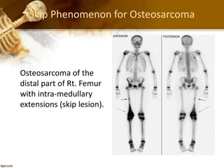
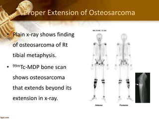
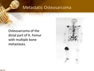
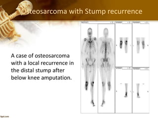
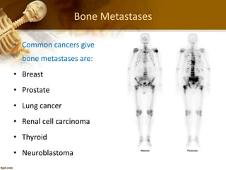
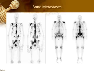
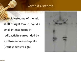
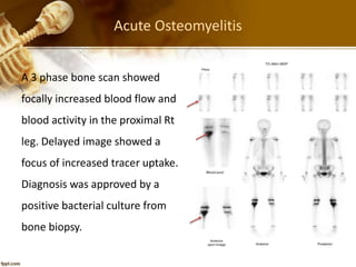
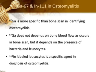
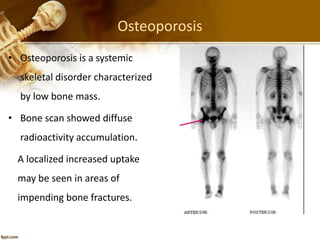
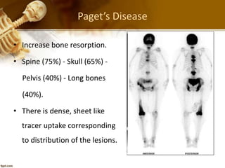
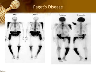
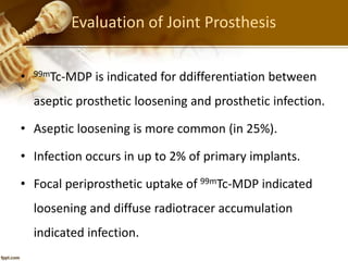
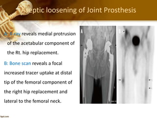
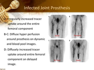
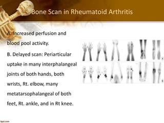
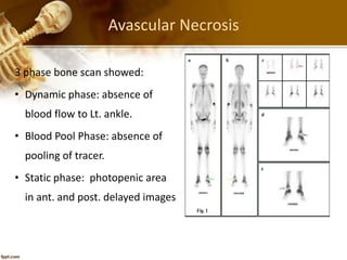
2. Normal Bone Scan
Normal increased uptake:
• Inferior angle of scapula.
• Ends of long bones.
• Sacroiliac joints.
• Growing epiphysis.
• Both kidneys.
• Urinary bladder.
4. Skip Phenomenon for Osteosarcoma
Osteosarcoma of the
distal part of Rt. Femur
with intra-medullary
extensions (skip lesion).
5. Proper Extension of Osteosarcoma
• Plain x-ray shows finding
of osteosarcoma of Rt
tibial metaphysis.
• 99mTc-MDP bone scan
shows osteosarcoma
that extends beyond its
extension in x-ray.
10. Osteoid Osteoma
Osteoid osteoma of the mid
shaft of right femur should a
small intense focus of
radioactivity surrounded by
a diffuse increased uptake
(Double density sign).
11. Osteomyelitis
• Occurs commonly in diaphyseal ends of long bones.
• Caused by pyogenic organisms, usually staphylococci.
• Bl. Culture is positive in about 50% of patients.
• X-ray may not positive until 10-14 days after clinical
onset
• Bone scan usually become positive as early as 24 hr.
after clinical onset.
12. Acute Osteomyelitis
A 3 phase bone scan showed
focally increased blood flow and
blood activity in the proximal Rt
leg. Delayed image showed a
focus of increased tracer uptake.
Diagnosis was approved by a
positive bacterial culture from
bone biopsy.
13. Ga-67 & In-111 in Osteomyelitis
• 67Ga is more specific than bone scan in identifying
osteomyelitis.
• 67Ga does not depends on bone blood flow as occurs
in bone scan, but it depends on the presence of
bacteria and leucocytes.
• 111In labeled leucocytes is a specific agent in
diagnosis of osteomyelitis.
14. Metabolic Bone Diseases
• Metabolic bone diseases are a group of disorders
affecting the whole skeleton.
• The disorders are mostly related to increased bone
turnover and increased uptake of radiolabelled
diphosphonate.
• Ex: Osteoporosis - Osteomalacia - Hyperparathyroidism
and Paget’s disease.
15. Osteoporosis
• Osteoporosis is a systemic
skeletal disorder characterized
by low bone mass.
• Bone scan showed diffuse
radioactivity accumulation.
A localized increased uptake
may be seen in areas of
impending bone fractures.
16. Paget’s Disease
• Increase bone resorption.
• Spine (75%) - Skull (65%) -
Pelvis (40%) - Long bones
(40%).
• There is dense, sheet like
tracer uptake corresponding
to distribution of the lesions.
18. Evaluation of Joint Prosthesis
• 99mTc-MDP is indicated for ddifferentiation between
aseptic prosthetic loosening and prosthetic infection.
• Aseptic loosening is more common (in 25%).
• Infection occurs in up to 2% of primary implants.
• Focal periprosthetic uptake of 99mTc-MDP indicated
loosening and diffuse radiotracer accumulation
indicated infection.
19. Aseptic loosening of Joint Prosthesis
A: X-ray reveals medial protrusion
of the acetabular component of
the Rt. hip replacement.
B: Bone scan reveals a focal
increased tracer uptake at distal
tip of the femoral component of
the right hip replacement and
lateral to the femoral neck.
20. Infected Joint Prosthesis
A: Irregularly increased tracer
uptake around the entire
femoral component
B-C: Diffuse hyper perfusion
around prosthesis on dynamic
and blood pool images.
D: Diffusely increased tracer
uptake around entire femoral
component on delayed
image.
Dynamic
Bl. Pool Static
Static
21. Bone Scan in Rheumatoid Arthritis
A. Increased perfusion and
blood pool activity.
B. Delayed scan: Periarticular
uptake in many interphalangeal
joints of both hands, both
wrists, Rt. elbow, many
metatarsophalangeal of both
feet, Rt. ankle, and in Rt knee.
22. Avascular Necrosis
3 phase bone scan showed:
• Dynamic phase: absence of
blood flow to Lt. ankle.
• Blood Pool Phase: absence of
pooling of tracer.
• Static phase: photopenic area
in ant. and post. delayed images
23. Advantages of Bone Scan
❶
• Early detection of bone lesions.
• Can detect proper extension of malignant lesions.
• Can detect skip phenomenon in osteosarcoma.
• Can detect asymptomatic bone secondaries.
• Three-phase bone scan assess vascular supply of
bone lesions.
24. Advantages of Bone Scan
❷
• Three-phase bone scan can assess soft tissue
extension.
• Follow-up of treatment of malignant bone lesions.
• Can assess renal morphology and function.
• Low radiation exposure to patients.
• Reasonable price.
25. 18F-NaF PET/CT
• Fluorine 18–Sodium Fluoride (18F–NaF) has been
introduced as a bone-seeking agent first in 1962 and
approved by FDA in 1972 for detection of osteogenic
activity.
• 18F-NaF PET/CT also contributes to the true evaluation of
bone marrow involvement.
• Similar to 99mTc-MDP, in which bone deposition occurs via
chemisorption, in which the OH- ions in hydroxyapatite
are exchanged for 18F - ions, converting hydroxyapatite
to fluorapatite.
26. 18F-NaF for PET/CT
• An 81-y-old woman with
history of metastatic breast
cancer. Sagittal 18F-NaF PET (A)
and CT (B) images and anterior
maximum-intensity projection
(C) show diffuse sclerotic
metastasis to entire spine and
sternum with abnormally
increased tracer uptake.
• Maximum-intensity projection
also demonstrates abnormal
uptake in ribs and pelvis.
27. 18F-NaF Vs 99mTc-MDP Bone Scan
• An 42-y-old woman with
history of Rt breast cancer.
• (A) posterior view of whole-body
99mTc-MDP bone scan shows
multiple bone lesions.
• (B) posterior maximum-
intensity projection of 18F-
NaF PET scan of same
patient performed 3 wk later
shows more metastatic
lesions than detected in
99mTc-MDP scan.