Highly malignant tumor of mesenchymal origin.Spindle shaped cells that produce osteoid.2nd most common primary malignant bone tumor after MM.Incidence – 1 to 3 per million per year
Treated by chemo,amputation or rotationplasty
Identified in 1921 by James Ewing
2nd most common bone tumor in children
Ewing’s Sarcoma Family of tumors:
Ewing’s sarcoma (Bone –87%)
Extraosseous Ewing’s sarcoma (8%)
Peripheral PNET(5%)
Askin’s tumor
Identified in 1921 by James Ewing
2nd most common bone tumor in children
Ewing’s Sarcoma Family of tumors:
Ewing’s sarcoma (Bone –87%)
Extraosseous Ewing’s sarcoma (8%)
Peripheral PNET(5%)
Askin’s tumor
Conformal Radiotherapy in Head and neck cancers is essential in terms of improving quality of life and local control in this era. This presentation aimed at giving an overview of conformal radiotherapy and its role in HNC to a 'general audience'.
When most normal cells grow
old or get damaged, they die, and new cells take their place. Sometimes, this
process goes wrong. New cells form when the body doesn't need them, and old or
damaged cells don't die as they should. The buildup of extra cells often forms
a mass of tissue called a growth or tumor.
Primary brain tumors can be benign or malignant:
Benign brain tumors do not contain cancer cells:
Usually, benign tumors can be removed, and
they seldom grow back.
Benign brain tumors usually have an obvious
border or edge. Cells from benign tumors rarely invade tissues around them.
They don't spread to other parts of the body. However, benign tumors can press
on sensitive areas of the brain and cause serious health problems.
Unlike benign tumors in most other parts of
the body, benign brain tumors are sometimes life threatening.
Benign brain tumors may become malignant.
Malignant brain tumors (also called brain
cancer) contain cancer cells:
Malignant brain tumors are generally more
serious and often are a threat to life.
They are likely to grow rapidly and crowd
or invade the nearby healthy brain tissue.
Cancer cells may break away from malignant
brain tumors and spread to other parts of the brain or to the spinal cord. They
rarely spread to other parts of the body.
Tumor Grade
Doctors group brain tumors by grade.
The grade of a tumor refers to the way the cells look under a microscope:
Grade I: The tissue is benign. The cells
look nearly like normal brain cells, and they grow slowly.
Grade II: The tissue is malignant. The
cells look less like normal cells than do the cells in a Grade I tumor.
Grade III: The malignant tissue has cells
that look very different from normal cells. The abnormal cells are actively
growing (anaplastic).
Grade IV: The malignant tissue has cells
that look most abnormal and tend to grow quickly.
Cells from low-grade tumors (grades I and
II) look more normal and generally grow more slowly than cells from high-grade
tumors (grades III and IV).
Over time, a low-grade tumor may become a
highgrade tumor. However, the change to a high-grade tumor happens more often
among adults than children.
You may want to read the NCI fact sheet Tumor
Grade.
Types of Primary Brain
Tumors
There are many types of primary brain
tumors. Primary brain tumors are named according to the type of cells or the
part of the brain in which they begin. For example, most primary brain tumors
begin in glial cells. This type of tumor is called a glioma.
Among adults, the most common types are:
Astrocytoma:
The tumor arises from star-shaped glial cells called astrocytes.
It can be any grade. In adults, an astrocytoma most often arises in the
cerebrum.
Grade I or II astrocytoma: It may be called
a low-grade glioma.
Grade III astrocytoma: It's sometimes
called a high-grade or an anaplastic astrocytoma.
Grade IV astrocytoma: It may be called a glioblastoma or
malignant astrocytic glioma.
Meningioma:
The tumor arises i
This PPT presentation talks about osteosarcoma from the clinical point of view, summarizing the recent guidelines in diagnosis and treatment of osteosarcoma.
Learn about the process of radiation therapy to treat soft tissue sarcoma, and how new radiation technology has improved treatment of the disease.
This presentation was given by Elizabeth H. Baldini, MD, MPH, radiation oncology director for the Center for Sarcoma and Bone Oncology at Dana-Farber Cancer Institute. It was originally presented as part of the "15 Years of GIST/Soft Tissue Sarcoma Symposium," held on Sept. 12, 2015 at Dana-Farber in Boston, Mass.
This is a powerpoint(case presentation) for radiology and imaging resident.There are many animations used inside this presentation so to see all the pictures which are placed layer by layer with the help of animations you simple need to download this presentation first.... Thanx.
It is about a disease called osteosarcoma which is a disease . It is about healthcare and health buisness . It was presented by Dr. Avinash gulati , a famous doctor in Norway.
micro teaching on communication m.sc nursing.pdfAnurag Sharma
Microteaching is a unique model of practice teaching. It is a viable instrument for the. desired change in the teaching behavior or the behavior potential which, in specified types of real. classroom situations, tends to facilitate the achievement of specified types of objectives.
These simplified slides by Dr. Sidra Arshad present an overview of the non-respiratory functions of the respiratory tract.
Learning objectives:
1. Enlist the non-respiratory functions of the respiratory tract
2. Briefly explain how these functions are carried out
3. Discuss the significance of dead space
4. Differentiate between minute ventilation and alveolar ventilation
5. Describe the cough and sneeze reflexes
Study Resources:
1. Chapter 39, Guyton and Hall Textbook of Medical Physiology, 14th edition
2. Chapter 34, Ganong’s Review of Medical Physiology, 26th edition
3. Chapter 17, Human Physiology by Lauralee Sherwood, 9th edition
4. Non-respiratory functions of the lungs https://academic.oup.com/bjaed/article/13/3/98/278874
- Video recording of this lecture in English language: https://youtu.be/lK81BzxMqdo
- Video recording of this lecture in Arabic language: https://youtu.be/Ve4P0COk9OI
- Link to download the book free: https://nephrotube.blogspot.com/p/nephrotube-nephrology-books.html
- Link to NephroTube website: www.NephroTube.com
- Link to NephroTube social media accounts: https://nephrotube.blogspot.com/p/join-nephrotube-on-social-media.html
Ozempic: Preoperative Management of Patients on GLP-1 Receptor Agonists Saeid Safari
Preoperative Management of Patients on GLP-1 Receptor Agonists like Ozempic and Semiglutide
ASA GUIDELINE
NYSORA Guideline
2 Case Reports of Gastric Ultrasound
Pulmonary Thromboembolism - etilogy, types, medical- Surgical and nursing man...VarunMahajani
Disruption of blood supply to lung alveoli due to blockage of one or more pulmonary blood vessels is called as Pulmonary thromboembolism. In this presentation we will discuss its causes, types and its management in depth.
Report Back from SGO 2024: What’s the Latest in Cervical Cancer?bkling
Are you curious about what’s new in cervical cancer research or unsure what the findings mean? Join Dr. Emily Ko, a gynecologic oncologist at Penn Medicine, to learn about the latest updates from the Society of Gynecologic Oncology (SGO) 2024 Annual Meeting on Women’s Cancer. Dr. Ko will discuss what the research presented at the conference means for you and answer your questions about the new developments.
Ethanol (CH3CH2OH), or beverage alcohol, is a two-carbon alcohol
that is rapidly distributed in the body and brain. Ethanol alters many
neurochemical systems and has rewarding and addictive properties. It
is the oldest recreational drug and likely contributes to more morbidity,
mortality, and public health costs than all illicit drugs combined. The
5th edition of the Diagnostic and Statistical Manual of Mental Disorders
(DSM-5) integrates alcohol abuse and alcohol dependence into a single
disorder called alcohol use disorder (AUD), with mild, moderate,
and severe subclassifications (American Psychiatric Association, 2013).
In the DSM-5, all types of substance abuse and dependence have been
combined into a single substance use disorder (SUD) on a continuum
from mild to severe. A diagnosis of AUD requires that at least two of
the 11 DSM-5 behaviors be present within a 12-month period (mild
AUD: 2–3 criteria; moderate AUD: 4–5 criteria; severe AUD: 6–11 criteria).
The four main behavioral effects of AUD are impaired control over
drinking, negative social consequences, risky use, and altered physiological
effects (tolerance, withdrawal). This chapter presents an overview
of the prevalence and harmful consequences of AUD in the U.S.,
the systemic nature of the disease, neurocircuitry and stages of AUD,
comorbidities, fetal alcohol spectrum disorders, genetic risk factors, and
pharmacotherapies for AUD.
Title: Sense of Taste
Presenter: Dr. Faiza, Assistant Professor of Physiology
Qualifications:
MBBS (Best Graduate, AIMC Lahore)
FCPS Physiology
ICMT, CHPE, DHPE (STMU)
MPH (GC University, Faisalabad)
MBA (Virtual University of Pakistan)
Learning Objectives:
Describe the structure and function of taste buds.
Describe the relationship between the taste threshold and taste index of common substances.
Explain the chemical basis and signal transduction of taste perception for each type of primary taste sensation.
Recognize different abnormalities of taste perception and their causes.
Key Topics:
Significance of Taste Sensation:
Differentiation between pleasant and harmful food
Influence on behavior
Selection of food based on metabolic needs
Receptors of Taste:
Taste buds on the tongue
Influence of sense of smell, texture of food, and pain stimulation (e.g., by pepper)
Primary and Secondary Taste Sensations:
Primary taste sensations: Sweet, Sour, Salty, Bitter, Umami
Chemical basis and signal transduction mechanisms for each taste
Taste Threshold and Index:
Taste threshold values for Sweet (sucrose), Salty (NaCl), Sour (HCl), and Bitter (Quinine)
Taste index relationship: Inversely proportional to taste threshold
Taste Blindness:
Inability to taste certain substances, particularly thiourea compounds
Example: Phenylthiocarbamide
Structure and Function of Taste Buds:
Composition: Epithelial cells, Sustentacular/Supporting cells, Taste cells, Basal cells
Features: Taste pores, Taste hairs/microvilli, and Taste nerve fibers
Location of Taste Buds:
Found in papillae of the tongue (Fungiform, Circumvallate, Foliate)
Also present on the palate, tonsillar pillars, epiglottis, and proximal esophagus
Mechanism of Taste Stimulation:
Interaction of taste substances with receptors on microvilli
Signal transduction pathways for Umami, Sweet, Bitter, Sour, and Salty tastes
Taste Sensitivity and Adaptation:
Decrease in sensitivity with age
Rapid adaptation of taste sensation
Role of Saliva in Taste:
Dissolution of tastants to reach receptors
Washing away the stimulus
Taste Preferences and Aversions:
Mechanisms behind taste preference and aversion
Influence of receptors and neural pathways
Impact of Sensory Nerve Damage:
Degeneration of taste buds if the sensory nerve fiber is cut
Abnormalities of Taste Detection:
Conditions: Ageusia, Hypogeusia, Dysgeusia (parageusia)
Causes: Nerve damage, neurological disorders, infections, poor oral hygiene, adverse drug effects, deficiencies, aging, tobacco use, altered neurotransmitter levels
Neurotransmitters and Taste Threshold:
Effects of serotonin (5-HT) and norepinephrine (NE) on taste sensitivity
Supertasters:
25% of the population with heightened sensitivity to taste, especially bitterness
Increased number of fungiform papillae
TEST BANK for Operations Management, 14th Edition by William J. Stevenson, Ve...kevinkariuki227
TEST BANK for Operations Management, 14th Edition by William J. Stevenson, Verified Chapters 1 - 19, Complete Newest Version.pdf
Surgical Site Infections, pathophysiology, and prevention.pptx
Osteosarcoma (1)
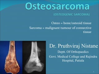
1. Osteo = bone/osteoid tissue Sarcoma = malignant tumour of connective tissue 02/04/12 Dr. Pruthviraj Nistane Deptt. Of Orthopaedics Govt. Medical College and Rajindra Hospital, Patiala