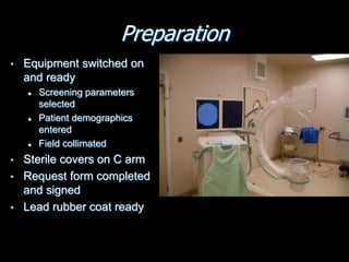
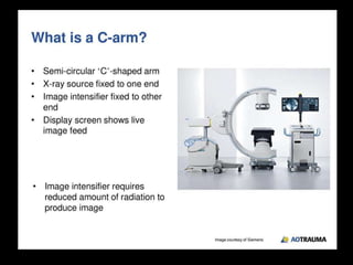
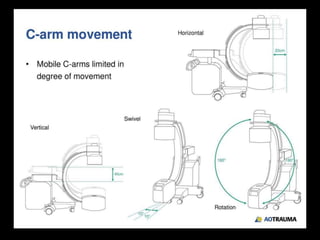
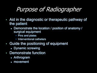
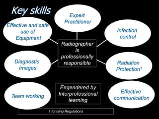
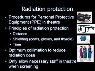
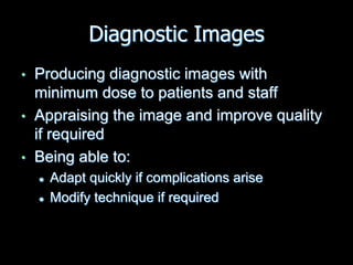
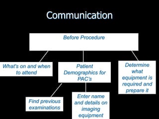
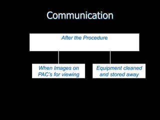
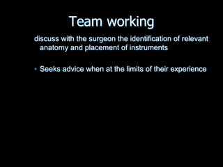
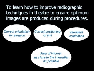
Radiography in the operating
theatre
Preparation
• Equipment switched on
and ready
 Screening parameters
selected
 Patient demographics
entered
 Field collimated
• Sterile covers on C arm
• Request form completed
and signed
• Lead rubber coat ready
Purpose of Radiographer
• Aid in the diagnostic or therapeutic pathway of
the patient
 Demonstrate the location / position of anatomy /
surgical equipment
• Pins and plates
• Interventional catheters
• Guide the positioning of equipment
 Dynamic screwing
• Demonstrate function
 Arthrogram
 movement
Radiographer
is
professionally
responsible
1 Ionising Regulations
Engendered by
Interprofessional
learning
Key skills Expert
Practitioner
Effective and safe
use of
Equipment
Diagnostic
Images
Team working
Infection
control
Effective
communication
Radiation
Protection1
• Procedures for Personal Protective
Equipment (PPE) in theatre
• Principles of radiation protection
• Distance
• Shielding (coats, gloves, and thyroid)
• Time
• Optimum collimation to reduce
radiation dose
• Only allow necessary staff in theatre
when screening
Radiation protection
Infection Control
• Procedures for Personal Protective
Equipment (PPE) in theatre
• Procedures to eliminate cross infection and
maintain a sterile environment
Diagnostic Images
• Producing diagnostic images with
minimum dose to patients and staff
• Appraising the image and improve quality
if required
• Being able to:
 Adapt quickly if complications arise
 Modify technique if required
Communication
Before Procedure
What's on and when
to attend
Patient
Demographics for
PAC’s
Find previous
examinations
Enter name
and details on
imaging
equipment
Determine
what
equipment is
required and
prepare it
Communication
During Procedure
All Staff
Radiation Protection
Surgeon / nurse
Image quality
Only relevant
staff present
Personal
Protective
Equipment
(PPE)
When to
arrive and
leave
Communication
After the Procedure
When Images on
PAC’s for viewing
Equipment cleaned
and stored away
Team working
discuss with the surgeon the identification of relevant
anatomy and placement of instruments
• Seeks advice when at the limits of their experience
To learn how to improve radiographic
techniques in theatre to ensure optimum
images are produced during procedures.
Intelligent
collimation
Correct orientation
for surgeon
Correct positioning
of unit
Area of interest
as close to the intensifier
as possible
Image quality
• Is this image
diagnostic?
• Is this an
optimum
image?
Image quality
Thank you

OT theatre radiography.ppt

  • 1.
    Radiography in theoperating theatre
  • 2.
    Preparation • Equipment switchedon and ready  Screening parameters selected  Patient demographics entered  Field collimated • Sterile covers on C arm • Request form completed and signed • Lead rubber coat ready
  • 18.
    Purpose of Radiographer •Aid in the diagnostic or therapeutic pathway of the patient  Demonstrate the location / position of anatomy / surgical equipment • Pins and plates • Interventional catheters • Guide the positioning of equipment  Dynamic screwing • Demonstrate function  Arthrogram  movement
  • 19.
    Radiographer is professionally responsible 1 Ionising Regulations Engenderedby Interprofessional learning Key skills Expert Practitioner Effective and safe use of Equipment Diagnostic Images Team working Infection control Effective communication Radiation Protection1
  • 20.
    • Procedures forPersonal Protective Equipment (PPE) in theatre • Principles of radiation protection • Distance • Shielding (coats, gloves, and thyroid) • Time • Optimum collimation to reduce radiation dose • Only allow necessary staff in theatre when screening Radiation protection
  • 21.
    Infection Control • Proceduresfor Personal Protective Equipment (PPE) in theatre • Procedures to eliminate cross infection and maintain a sterile environment
  • 22.
    Diagnostic Images • Producingdiagnostic images with minimum dose to patients and staff • Appraising the image and improve quality if required • Being able to:  Adapt quickly if complications arise  Modify technique if required
  • 23.
    Communication Before Procedure What's onand when to attend Patient Demographics for PAC’s Find previous examinations Enter name and details on imaging equipment Determine what equipment is required and prepare it
  • 24.
    Communication During Procedure All Staff RadiationProtection Surgeon / nurse Image quality Only relevant staff present Personal Protective Equipment (PPE) When to arrive and leave
  • 25.
    Communication After the Procedure WhenImages on PAC’s for viewing Equipment cleaned and stored away
  • 26.
    Team working discuss withthe surgeon the identification of relevant anatomy and placement of instruments • Seeks advice when at the limits of their experience
  • 27.
    To learn howto improve radiographic techniques in theatre to ensure optimum images are produced during procedures. Intelligent collimation Correct orientation for surgeon Correct positioning of unit Area of interest as close to the intensifier as possible
  • 28.
    Image quality • Isthis image diagnostic? • Is this an optimum image?
  • 29.
  • 30.

Editor's Notes