Downloaded 347 times
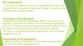
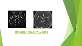
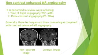


This document discusses magnetic resonance angiography (MRA) and its advantages and disadvantages compared to catheter angiography. It describes different MRA techniques including contrast enhanced MRA, time of flight angiography, phase contrast angiography, and non-contrast techniques. It also discusses artifacts that can appear on MRA such as metal artifacts and blooming artifacts. Key features and images of each technique are provided.


