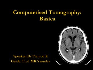
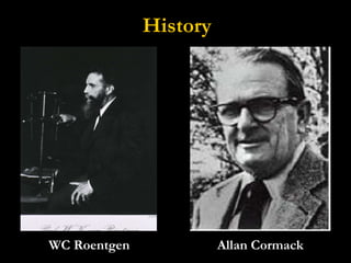
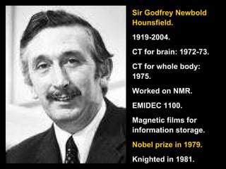
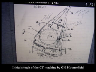
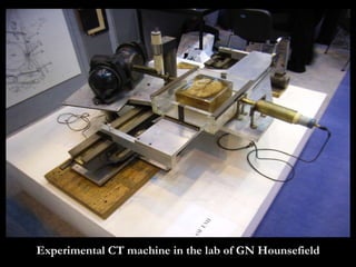
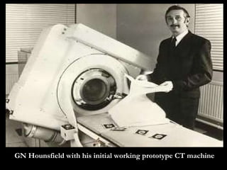
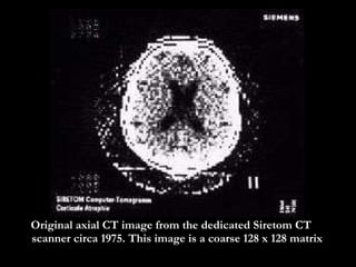
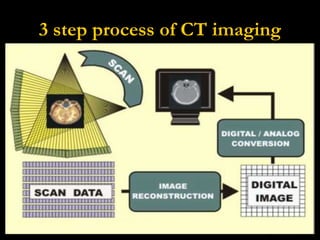
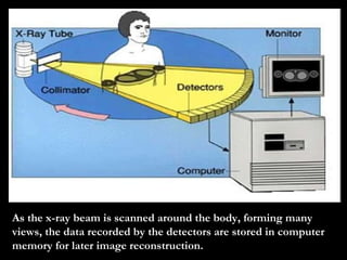
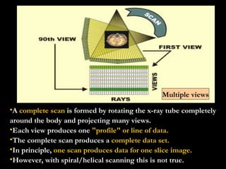
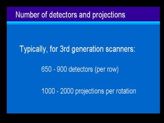
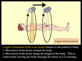
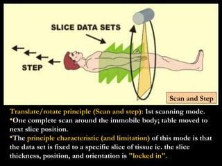
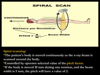
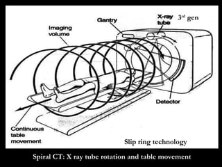
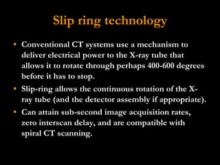
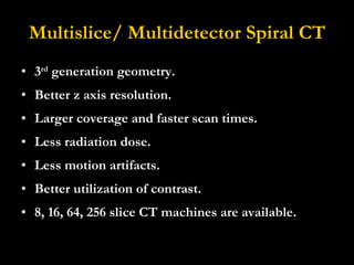
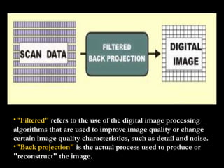
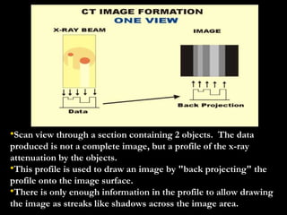
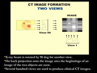
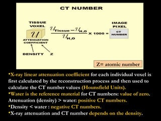
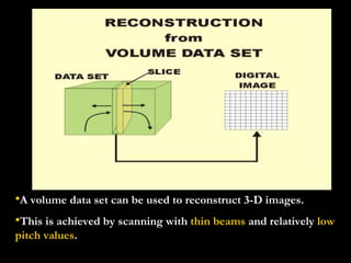
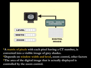
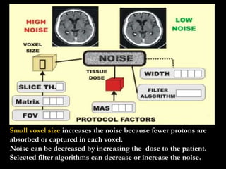
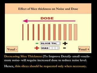
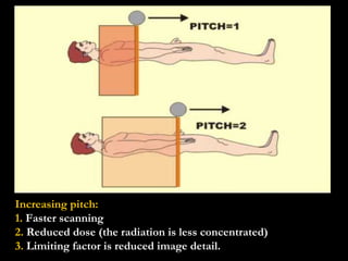
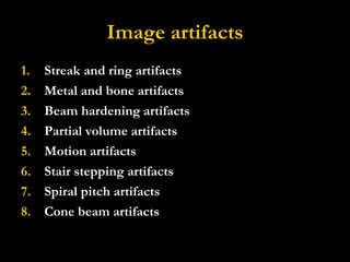
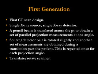
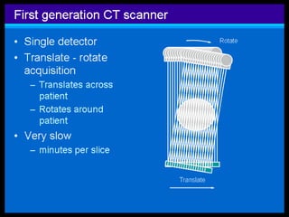
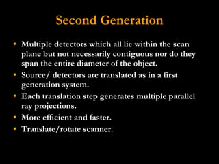
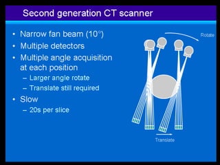
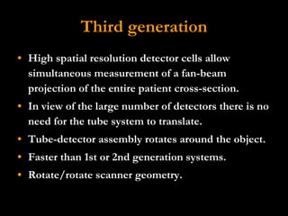
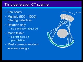
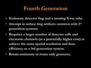
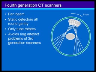
Computerized tomography (CT) was pioneered by Godfrey Hounsfield and Allan Cormack in the 1970s. CT uses X-rays and computer processing to create cross-sectional images of the body. The first CT scanners used a translate-rotate design, while later generations used multiple detectors and spiral scanning for faster, more detailed imaging. Image reconstruction uses back projection to convert attenuation measurements into pixel values and display slices. CT provides excellent anatomical detail and is widely used for diagnosing conditions of the brain, blood vessels, lungs and other organs.