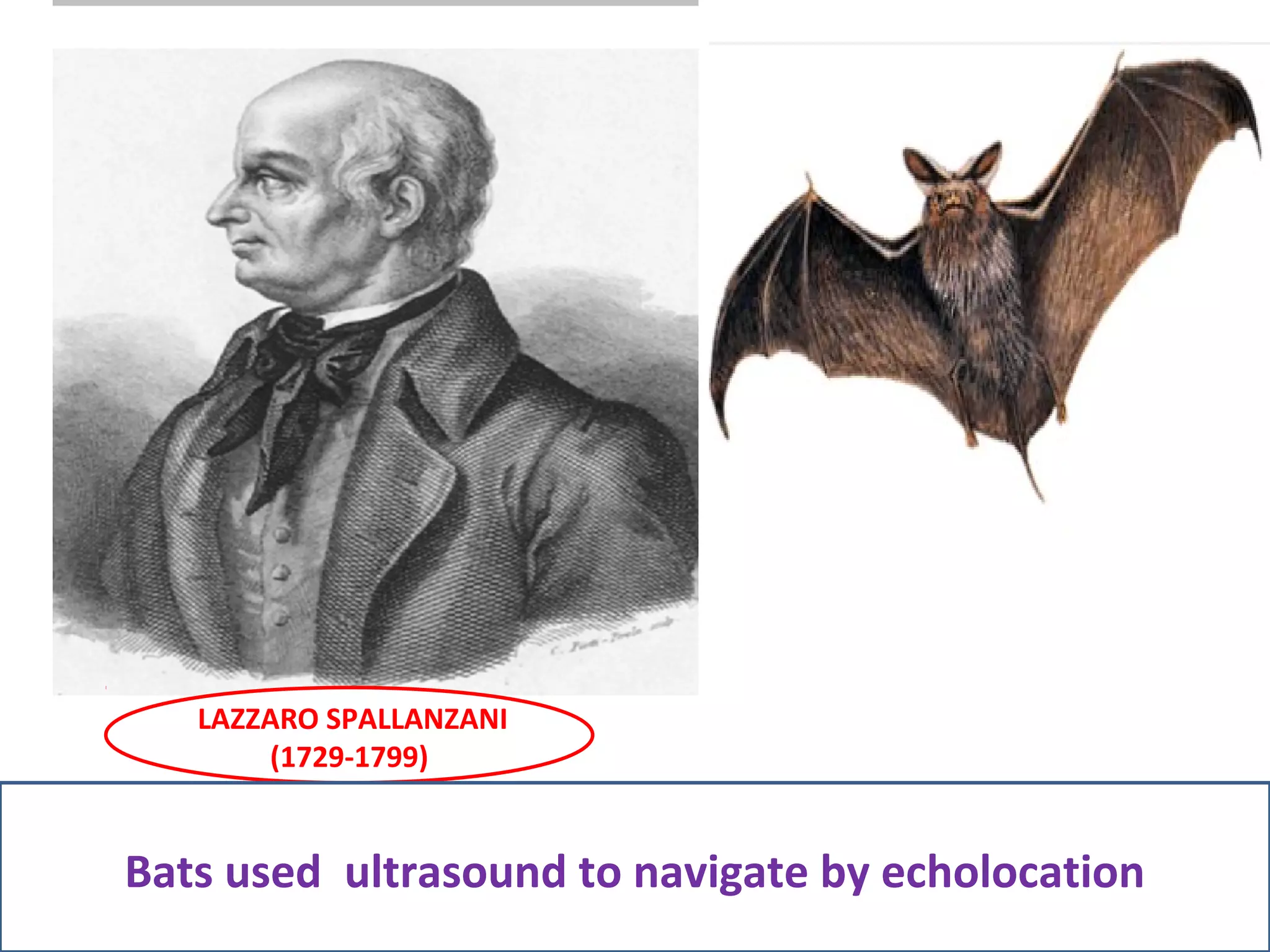
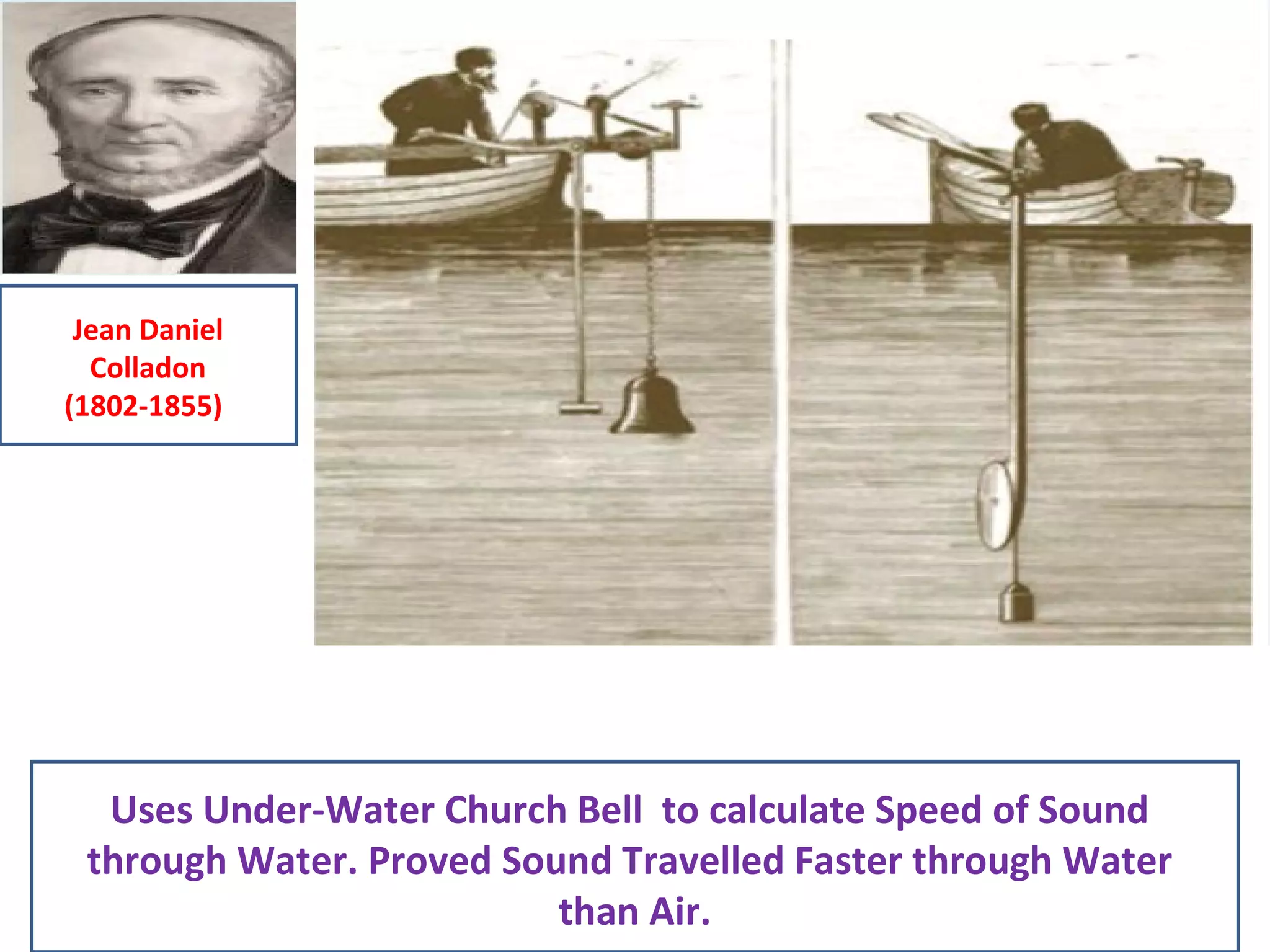
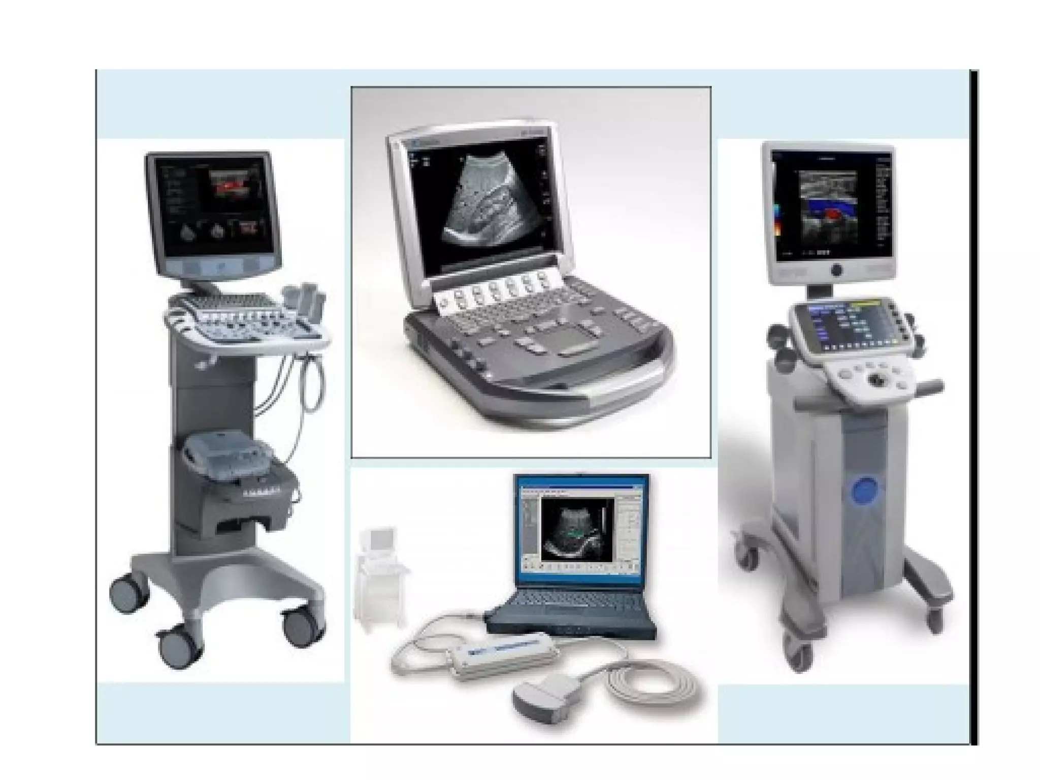
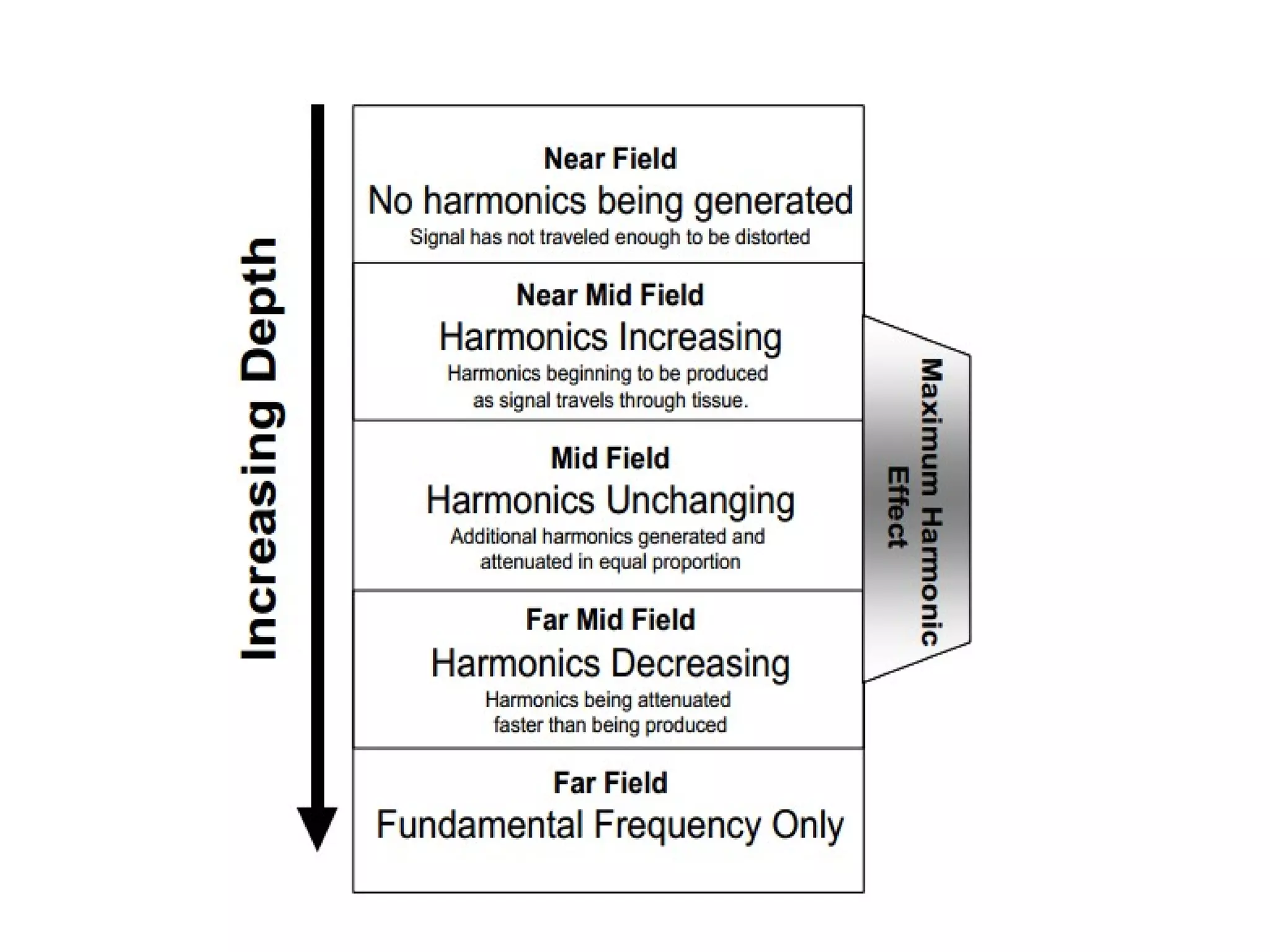
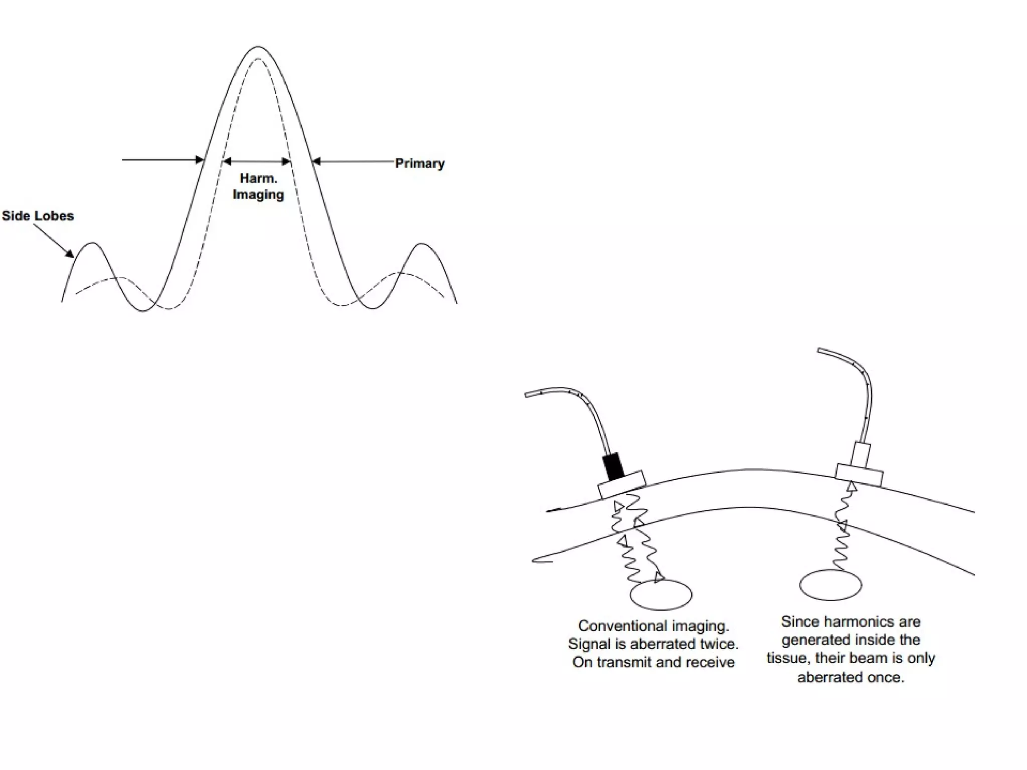
This document discusses various topics related to ultrasound imaging including goals, early pioneers, transducer types, Doppler instrumentation and physics, harmonic imaging, spatial compounding, extended field of view, fusion imaging, 3D and 4D ultrasound, and contrast enhanced ultrasound. It provides details on transducer selection, control settings, tissue harmonic imaging principles, spatial compounding benefits, fusion imaging steps, and contrast agent interactions.
Ultrasound elastography is a relatively advanced technique used to know the stiffness of the tissue. It is a non-invasive technique. Broadly classified into Quasistatic and Dynamic elastography.
Ultrasound Physics Made easy - By Dr Chandni WadhwaniChandni Wadhwani
History of ultrasound, Principle of Ultrasound.
Ultrasound wave and its interactions
Ultrasound machine and its parts, Image display, Artifacts and their clinical importance
what is Doppler ultrasound, Elastography and Recent advances in field of ultrasound.
Safety issues in ultrasound.
An overview of Doppler Effect in Ultrasonography - the medical imaging of the body using Ultrasound.
Includes Colour Doppler, Power Doppler, Spectral Doppler, Continuous Wave Doppler, Pulsed Wave Doppler, and comparisons with other Radiographic imaging modalities.
Usg transducer and basic principles of ultrasound Doppler, this slide describe the basic physics of ultrasound transducer and Doppler , must know thing is given in this presentaion. Good review for radiology resident. Thanks.
this power-point slide presentation includes lots of information like how MRI coil works. what is shimming, magnet, fringe, and design of mri coil and also magnet. this will help a lot for radiologist and technician radiographers.. thanks.
Do you know probe lens is repairable ? Are you experiencing extra cost for your ultrasound machine maintenance ?
Are you the people who expend unnecessarily ? Are you accustomed of purchasing a new one than repair ? Have you ever contacted Probelogic Pty Ltd ?
We know you will be amazed. We can reduce your cost surprisingly. Probe repair,probe replacements, probe testing, ultrasound transducer repair and lots more.
Contact Probelogic Pty Ltd to day - Get expert help - Let us reduce your cost.
Visit us www.probelogic.com.au
Virtual Navigator Real-Time Ultrasound Fusion Imaging with Positron Emission ...rosopeplaton
Enzo Di Mauro, Marco Solbiati, Stefano De Beni, Leonardo Forzoni, Sara D’Onofrio, Luigi Solbiati
Real-time fusion imaging technologies are
increasingly being used among interventional radiologists,
mostly Computed Tomography (CT) or Magnetic Resonance
Imaging (MRI) dataset, fused with Ultrasound (US) imaging. In
addition, fusion of Positron Emission Tomography (PET) and
CT is increasingly diffused in clinical practice, due to the wide
availability of PET scanners and the capability to make either a
direct (acquisitions performed within the same system) or an
indirect (procedure performed on an external workstation,
merging the two different sets of acquired data) fusion with CT
data. The present work describes the feasibility of real-time
fusion imaging directly between PET data and US imaging,
with CT scans being used only for PET-US fusion registration.
Data on multimodality registration precision and clinical
applications are presented as well.
the material discuss about a medical condition that has to deal with sexual development. in different stages of development, there is a need for gender identity and role, if there is a problem with any of these 2, there will be a problem with the sex assignment which will have an effect on the external genitalia sex. if all these pathway fall apart, there will be a condition called hermaphroditism which may be true or false. the material is exclusive on the topic
Training Material inherited form Philips Basics of Ultrasonography. Covers the fundamentals of Ultrasound Waveform, Piezoelectric Effect, Phased Echo Concept, Goal of Ultrasound, Ultrasound Image Construction process, Types of Resolution, Probe Internals, The Doppler Effect, Spectrum Waveform and concept, Color Doppler, Components of Ultrasound.
L2 Gynaecological usg (TAUS part 2).pptxiqra saeed
This lecture is the continuation of previous transabdominal technique. In this lecture types of probes are explained briefly. Transducer Manipulation Techniques are explained as well. An idea of acoustic window is given and few TAUS pelvis images are displayed.
Basavarajeeyam is an important text for ayurvedic physician belonging to andhra pradehs. It is a popular compendium in various parts of our country as well as in andhra pradesh. The content of the text was presented in sanskrit and telugu language (Bilingual). One of the most famous book in ayurvedic pharmaceutics and therapeutics. This book contains 25 chapters called as prakaranas. Many rasaoushadis were explained, pioneer of dhatu druti, nadi pareeksha, mutra pareeksha etc. Belongs to the period of 15-16 century. New diseases like upadamsha, phiranga rogas are explained.
Knee anatomy and clinical tests 2024.pdfvimalpl1234
This includes all relevant anatomy and clinical tests compiled from standard textbooks, Campbell,netter etc..It is comprehensive and best suited for orthopaedicians and orthopaedic residents.
Adv. biopharm. APPLICATION OF PHARMACOKINETICS : TARGETED DRUG DELIVERY SYSTEMSAkankshaAshtankar
MIP 201T & MPH 202T
ADVANCED BIOPHARMACEUTICS & PHARMACOKINETICS : UNIT 5
APPLICATION OF PHARMACOKINETICS : TARGETED DRUG DELIVERY SYSTEMS By - AKANKSHA ASHTANKAR
Tom Selleck Health: A Comprehensive Look at the Iconic Actor’s Wellness Journeygreendigital
Tom Selleck, an enduring figure in Hollywood. has captivated audiences for decades with his rugged charm, iconic moustache. and memorable roles in television and film. From his breakout role as Thomas Magnum in Magnum P.I. to his current portrayal of Frank Reagan in Blue Bloods. Selleck's career has spanned over 50 years. But beyond his professional achievements. fans have often been curious about Tom Selleck Health. especially as he has aged in the public eye.
Follow us on: Pinterest
Introduction
Many have been interested in Tom Selleck health. not only because of his enduring presence on screen but also because of the challenges. and lifestyle choices he has faced and made over the years. This article delves into the various aspects of Tom Selleck health. exploring his fitness regimen, diet, mental health. and the challenges he has encountered as he ages. We'll look at how he maintains his well-being. the health issues he has faced, and his approach to ageing .
Early Life and Career
Childhood and Athletic Beginnings
Tom Selleck was born on January 29, 1945, in Detroit, Michigan, and grew up in Sherman Oaks, California. From an early age, he was involved in sports, particularly basketball. which played a significant role in his physical development. His athletic pursuits continued into college. where he attended the University of Southern California (USC) on a basketball scholarship. This early involvement in sports laid a strong foundation for his physical health and disciplined lifestyle.
Transition to Acting
Selleck's transition from an athlete to an actor came with its physical demands. His first significant role in "Magnum P.I." required him to perform various stunts and maintain a fit appearance. This role, which he played from 1980 to 1988. necessitated a rigorous fitness routine to meet the show's demands. setting the stage for his long-term commitment to health and wellness.
Fitness Regimen
Workout Routine
Tom Selleck health and fitness regimen has evolved. adapting to his changing roles and age. During his "Magnum, P.I." days. Selleck's workouts were intense and focused on building and maintaining muscle mass. His routine included weightlifting, cardiovascular exercises. and specific training for the stunts he performed on the show.
Selleck adjusted his fitness routine as he aged to suit his body's needs. Today, his workouts focus on maintaining flexibility, strength, and cardiovascular health. He incorporates low-impact exercises such as swimming, walking, and light weightlifting. This balanced approach helps him stay fit without putting undue strain on his joints and muscles.
Importance of Flexibility and Mobility
In recent years, Selleck has emphasized the importance of flexibility and mobility in his fitness regimen. Understanding the natural decline in muscle mass and joint flexibility with age. he includes stretching and yoga in his routine. These practices help prevent injuries, improve posture, and maintain mobilit
263778731218 Abortion Clinic /Pills In Harare ,sisternakatoto
263778731218 Abortion Clinic /Pills In Harare ,ABORTION WOMEN’S CLINIC +27730423979 IN women clinic we believe that every woman should be able to make choices in her pregnancy. Our job is to provide compassionate care, safety,affordable and confidential services. That’s why we have won the trust from all generations of women all over the world. we use non surgical method(Abortion pills) to terminate…Dr.LISA +27730423979women Clinic is committed to providing the highest quality of obstetrical and gynecological care to women of all ages. Our dedicated staff aim to treat each patient and her health concerns with compassion and respect.Our dedicated group ABORTION WOMEN’S CLINIC +27730423979 IN women clinic we believe that every woman should be able to make choices in her pregnancy. Our job is to provide compassionate care, safety,affordable and confidential services. That’s why we have won the trust from all generations of women all over the world. we use non surgical method(Abortion pills) to terminate…Dr.LISA +27730423979women Clinic is committed to providing the highest quality of obstetrical and gynecological care to women of all ages. Our dedicated staff aim to treat each patient and her health concerns with compassion and respect.Our dedicated group of receptionists, nurses, and physicians have worked together as a teamof receptionists, nurses, and physicians have worked together as a team wwww.lisywomensclinic.co.za/
micro teaching on communication m.sc nursing.pdfAnurag Sharma
Microteaching is a unique model of practice teaching. It is a viable instrument for the. desired change in the teaching behavior or the behavior potential which, in specified types of real. classroom situations, tends to facilitate the achievement of specified types of objectives.
Title: Sense of Smell
Presenter: Dr. Faiza, Assistant Professor of Physiology
Qualifications:
MBBS (Best Graduate, AIMC Lahore)
FCPS Physiology
ICMT, CHPE, DHPE (STMU)
MPH (GC University, Faisalabad)
MBA (Virtual University of Pakistan)
Learning Objectives:
Describe the primary categories of smells and the concept of odor blindness.
Explain the structure and location of the olfactory membrane and mucosa, including the types and roles of cells involved in olfaction.
Describe the pathway and mechanisms of olfactory signal transmission from the olfactory receptors to the brain.
Illustrate the biochemical cascade triggered by odorant binding to olfactory receptors, including the role of G-proteins and second messengers in generating an action potential.
Identify different types of olfactory disorders such as anosmia, hyposmia, hyperosmia, and dysosmia, including their potential causes.
Key Topics:
Olfactory Genes:
3% of the human genome accounts for olfactory genes.
400 genes for odorant receptors.
Olfactory Membrane:
Located in the superior part of the nasal cavity.
Medially: Folds downward along the superior septum.
Laterally: Folds over the superior turbinate and upper surface of the middle turbinate.
Total surface area: 5-10 square centimeters.
Olfactory Mucosa:
Olfactory Cells: Bipolar nerve cells derived from the CNS (100 million), with 4-25 olfactory cilia per cell.
Sustentacular Cells: Produce mucus and maintain ionic and molecular environment.
Basal Cells: Replace worn-out olfactory cells with an average lifespan of 1-2 months.
Bowman’s Gland: Secretes mucus.
Stimulation of Olfactory Cells:
Odorant dissolves in mucus and attaches to receptors on olfactory cilia.
Involves a cascade effect through G-proteins and second messengers, leading to depolarization and action potential generation in the olfactory nerve.
Quality of a Good Odorant:
Small (3-20 Carbon atoms), volatile, water-soluble, and lipid-soluble.
Facilitated by odorant-binding proteins in mucus.
Membrane Potential and Action Potential:
Resting membrane potential: -55mV.
Action potential frequency in the olfactory nerve increases with odorant strength.
Adaptation Towards the Sense of Smell:
Rapid adaptation within the first second, with further slow adaptation.
Psychological adaptation greater than receptor adaptation, involving feedback inhibition from the central nervous system.
Primary Sensations of Smell:
Camphoraceous, Musky, Floral, Pepperminty, Ethereal, Pungent, Putrid.
Odor Detection Threshold:
Examples: Hydrogen sulfide (0.0005 ppm), Methyl-mercaptan (0.002 ppm).
Some toxic substances are odorless at lethal concentrations.
Characteristics of Smell:
Odor blindness for single substances due to lack of appropriate receptor protein.
Behavioral and emotional influences of smell.
Transmission of Olfactory Signals:
From olfactory cells to glomeruli in the olfactory bulb, involving lateral inhibition.
Primitive, less old, and new olfactory systems with different path
These simplified slides by Dr. Sidra Arshad present an overview of the non-respiratory functions of the respiratory tract.
Learning objectives:
1. Enlist the non-respiratory functions of the respiratory tract
2. Briefly explain how these functions are carried out
3. Discuss the significance of dead space
4. Differentiate between minute ventilation and alveolar ventilation
5. Describe the cough and sneeze reflexes
Study Resources:
1. Chapter 39, Guyton and Hall Textbook of Medical Physiology, 14th edition
2. Chapter 34, Ganong’s Review of Medical Physiology, 26th edition
3. Chapter 17, Human Physiology by Lauralee Sherwood, 9th edition
4. Non-respiratory functions of the lungs https://academic.oup.com/bjaed/article/13/3/98/278874
Lung Cancer: Artificial Intelligence, Synergetics, Complex System Analysis, S...Oleg Kshivets
RESULTS: Overall life span (LS) was 2252.1±1742.5 days and cumulative 5-year survival (5YS) reached 73.2%, 10 years – 64.8%, 20 years – 42.5%. 513 LCP lived more than 5 years (LS=3124.6±1525.6 days), 148 LCP – more than 10 years (LS=5054.4±1504.1 days).199 LCP died because of LC (LS=562.7±374.5 days). 5YS of LCP after bi/lobectomies was significantly superior in comparison with LCP after pneumonectomies (78.1% vs.63.7%, P=0.00001 by log-rank test). AT significantly improved 5YS (66.3% vs. 34.8%) (P=0.00000 by log-rank test) only for LCP with N1-2. Cox modeling displayed that 5YS of LCP significantly depended on: phase transition (PT) early-invasive LC in terms of synergetics, PT N0—N12, cell ratio factors (ratio between cancer cells- CC and blood cells subpopulations), G1-3, histology, glucose, AT, blood cell circuit, prothrombin index, heparin tolerance, recalcification time (P=0.000-0.038). Neural networks, genetic algorithm selection and bootstrap simulation revealed relationships between 5YS and PT early-invasive LC (rank=1), PT N0—N12 (rank=2), thrombocytes/CC (3), erythrocytes/CC (4), eosinophils/CC (5), healthy cells/CC (6), lymphocytes/CC (7), segmented neutrophils/CC (8), stick neutrophils/CC (9), monocytes/CC (10); leucocytes/CC (11). Correct prediction of 5YS was 100% by neural networks computing (area under ROC curve=1.0; error=0.0).
CONCLUSIONS: 5YS of LCP after radical procedures significantly depended on: 1) PT early-invasive cancer; 2) PT N0--N12; 3) cell ratio factors; 4) blood cell circuit; 5) biochemical factors; 6) hemostasis system; 7) AT; 8) LC characteristics; 9) LC cell dynamics; 10) surgery type: lobectomy/pneumonectomy; 11) anthropometric data. Optimal diagnosis and treatment strategies for LC are: 1) screening and early detection of LC; 2) availability of experienced thoracic surgeons because of complexity of radical procedures; 3) aggressive en block surgery and adequate lymph node dissection for completeness; 4) precise prediction; 5) adjuvant chemoimmunoradiotherapy for LCP with unfavorable prognosis.
4. Uses Under-Water Church Bell to calculate Speed of Sound
through Water. Proved Sound Travelled Faster through Water
than Air.
Jean Daniel
Colladon
(1802-1855))
11. • It is recommended that all sonologists learn the different
capabilities of their own equipment to optimize image
quality and diagnosis.
1. Keyboard: Various capabilities as provided by
manufacturer
2. Transducer select: To chose one of the many transducer
probes attached to the transducer ports on the
ultrasound machine.
12. High Frequency Transducer
• Frequency range of 7.0 to 14.0 MHz
• Linear transducer mostly, sector transducer more suited
for children
• Provides increased resolution of images, however, with
reduced penetration
• Linear probes best for evaluation of superficial structure
like, thyroid, scrotum, etc.
Medium Frequency Transducer
• Frequency range of 3.0 to 5.0 MHz
• Curvilinear or sector transducers
• Most commonly used probe for adult abdominal
imaging
13. Low Frequency Transducer
• Frequency of 2.0 MHz
• Transducer is sector type
• Provides increased depth of penetration but also
results in loss of resolution
• More suited for ultrasound studies of obese
patients
14. 3. Overall Gain Control: This is used to amplify all received
signals equally.
4 . Time Gain Compensation:
• With time gain compensation, a depth-dependent gain is
applied to the echoes.
• Simply put, echo signals from deep structures are amplified
more than signals from shallow structures.
• Time gain compensation is controlled in most machines
using a set of 6 to 10 gain knobs, each adjusting the receiver
gain at a different depth .
15. 5. Near and far gains: These controls are used to
equalize the differences in echoes received from
various depths as they are displayed on the screen.
6.Compression:
• The wide range of amplitudes returning to the
transducer are compressed into a range ( dynamic
range) which can be displayed on screen.
• Dynamic range is the ratio of highest and lowest
amplitudes in decibels that can be displayed.
• In clinical applications the dynamic range may be
upto 120 db because the range of reflected signals
may vary by a factor of
16. • Most of the ultrasound machines apply logarithmic
compression to the echo signals emerging from the
receiver.
• amount of compression is under operator control.
The widest dynamic range shown (60 dB) permits the best differentiation of
subtle differences in echo intensity and is preferred for most imaging
applications
17. 7.Depth: This control is used to adjust the size of
the image so that organs and adjacent
structures or regions of interest are equally well
visualized.
8.Focal point(s):
• This allows the operator to choose the level at
which the ultrasound beam is focused to
increase the resolution at a specific point or
points.
• This control should be set at the most posterior
aspect of the organ or structure being imaged .
18. 9. Postprocessing :
This can be used to change the appearance of
echo signals, already stored in memory, on the
image.
Failure to properly adjust the gain control and/or
poor placement of focal point during scanning
may result in suboptimal image quality and
misdiagnosis.
20. Linear Array Transducer
• Optimal for superficially placed structures,
such as vessels, neck, testes.
• Final image is displayed as a rectangle.
• Each time only a group of elements work
together to transmit or receive. The
ultrasound beam is perpendicular to the
transducer surface
• Size of the field of view is equal in both the
far field and near field .
21. Curvilinear Array Transducer
• commonly used for routine
abdominal and pelvic imaging.
• array of elements instead of a
straight line (linear array) are
arranged across a
convex arc.
• This fan like arrangement of elements results in
a sector shaped imaging field.
• curved array provides a wider image at large
depths from a narrow scanning window.
22. Phased Array Transducer
• All the elements work together in phased array
(all elements are used for each beam line).
• Phase array steer the beam by applying different delay on
each element, and it requires small acoustic window.
• Its main advantage is in providing a very broad imaged field
at larger depths that too with a narrow transducer footprint.
• It is widely used in cardiac scanning as the transducer fits
easily between the ribs (rib gap is a small acoustic window).
23.
24.
25.
26. DOPPLER INSTRUMENTS AND
PHYSICS
The information provided by a Doppler
examination includes presence or absence of
blood flow, direction of blood flow, type of
blood flow (arterial high resistance/venous,
presence and quantification of arterial
stenosis etc
27.
28.
29.
30. • In clinical practice, three different
Doppler techniques are utilized.
CONTINUOUS WAVE DOPPLER
• The basic transducer for continuous wave
Doppler contains an oscillator,
two piezoelectric crystals and a demodulator
• Continuous wave Doppler is used for evaluation of peripheral
vessels and the fetal heart.
• It provides information regarding the blood flow but lacks
information regarding the depth from which the Doppler
signal is coming from.
31. Pulsed Doppler
• In this technique, very short bursts of the sound wave
are repetitively emitted by the transducer.
• A new pulse is not emitted till the returning signal
from the previous pulse is detected by the
transducer.
• The receiver is designed to be turned on for a short
period on at a specific moment.
• Provides information regarding the presence,
direction and depth from which the Doppler signal is
coming from.
• Pulsed Doppler does not provide information
regarding the intensity (or power) of the Doppler
signal.
32. Power Doppler :
• Power Doppler displays the power (or intensity)
of the Doppler signal, as it changes with time in
every region within the chosen area.
• However, there is no information available
regarding the velocity.
• The power Doppler has superior flow sensitivity
as compared to conventional color Doppler.
• So it is used to evaluate the presence and
characteristics of the flow in blood vessels that
are poorly imaged with conventional color
Doppler.
34. How Harmonics Are Generated
• The harmonic signals used in this form of
imaging do not come from the ultrasound
system itself.
• These signals are generated in the body as a
result of interactions with tissue or contrast
agents.
39. Potential advantages of the harmonic signal.
Ultrasound beams formed with the harmonic signals have
some interesting properties.
• One of those properties is that the beam formed using the
harmonic signal is narrower and has lower sidelobes.
• The improvement in beamwidth and reduction in side
lobe significantly improves grayscale contrast resolution.
• Furthermore, since the harmonics are generated inside the
body, they only have to pass through the fat layer once (on
receive), not twice (transmit and receive).
40. • Potential advantages of harmonic imaging
include improved axial resolution due to
higher frequencies and better lateral
resolution due to narrower beams.
• Decreased noise from side lobes improves
signal-to noise ratios and reduces artifacts
• Body fat increases the intensity of harmonic
waves, thus lesion visibility is increased in
obese patients.
41.
42. • Harmonic imaging increases diagnostic confidence in
differentiating cystic from solid hepatic lesions,
improves detection of gallbladder and biliary calculi,
improves pancreatic definition and allows distinction
of simple from complex renal cysts.
43.
44. Spatial Compound Imaging
• An important source of image degradation and
loss of contrast is ultrasound speckle.
• Speckle results from the constructive and
destructive interaction of the acoustic fields
generated by the scattering of ultrasound
from small tissue reflectors.
• This interference pattern gives ultrasound
images their characteristic grainy appearance
reducing contrast and making the identification
of subtle features more difficult.
45.
46. • in spatial compound sonography information is
obtained from several different angles of
insonation which are combined to produce a single
image .
• This is unlike conventional B-mode sonography, in
which each image is obtained from a single angle of
insonation.
• By averaging images from multiple angles of
insonation, SCI has been shown to reduce many
image artifacts inherent in conventional sonography.
• Application of SCI has been described in imaging of
breast, peripheral vessels, and musculoskeletal
system. It can also be combined with other
ultrasound applications, e.g. harmonic imaging.
47.
48. EXTENDED FIELD OF VIEW
• Allows sonologists to visualize large anatomic
regions in a single image.
• It can be performed with a linear array
transducer or using a curvilinear probe, although
most of its applications are in superficial
structures.
• It differs from traditional US by allowing global
depiction of an abnormality and its relation to
adjacent anatomic structures within a single
image.
49. • Transducer is initially moved laterally across
the anatomic area of interest and multiple
images are acquired from many transducer
positions.
• Images are registered with respect to each
other.
• This registered data is subsequently combined
to form one complete large field of view
image.
50.
51. FUSION IMAGING
• Fusion imaging or hybrid imaging means
combination of two imaging techniques.
• This can be in the form of ultrasound with MRI
or CT
• or it can be fusion of (ultrasound, CT or MRI)
with molecular imaging technique like SPECT or
PET
52. The advantages of incorporating ultrasound in image
fusion consist in the :
• real-time images (which enable image-guided
intervention),
• the lack of radiation to both patient and staff,
and
• the possibility of comparing findings on one
modality with another modality .
Software for fusion of real-time ultrasound images
with CT, MRI, or PET/CT is incorporated in several
high-end ultrasound systems.
53. • To fuse medical imaging information obtained
from different modalities at different times, a
spatial coregistration is mandatory to ensure
that the pixels from the various datasets
represent approximately the same volume.
• For a correct coregistration, a two-step
technique is performed automatically by a
computer:
a. image registration and
b. data reslicing.
54. STEPS OF FUSION
• During examination ultrasound screen is seen as split images
with virtual reconstructed CT/MR image on one side and
currently acquired USG image on other side of the screen.
• An attempt is made to match these two images with each
other using some clearly visible anatomic landmark (e.g.
portal vein bifurcation, superior most margin of kidney or
the lesion itself, etc.)
• For fusion imaging variety of tracking methods are used,
common are the optical tracking system and
electromagnetic tracking system.
• There are three components of the EM tracking-based
fusion imaging technique: the magnetic field generator,
position sensor, and position sensor unit.
55. • The magnetic field generator, which is located near
the patient, creates a magnetic field, thereby
inducing currents in the position sensor, which is
mounted on the US transducer.
• As the US probe moves, the magnitude of the
electrical current in the position sensor changes
with respect to the magnetic field.
• With this information, the position sensor unit
installed in the US machine calculates the exact
location of the position sensor and thus,
determines the direction and position of the US
transducer
• This enables the side-by-side or overlay display of
real-time US images and fused CT or MR images.
56.
57. Clinical applications
• Isoechoic lesions not well appreciated on grey
scale.
• Previously ablated lesions with recurrence not
well visualized.
• Interventional treatment is easy.
58.
59.
60. 3D and 4D Ultrasound
• This imaging technology involves acquiring a large number of data
sets of 2D images from patient.
• this volumetric data can be assessed with the use of many analysis
tools such as surface and volume rendering, multiplanar imaging and
volume calculation techniques, etc.
• like CT and MRI, the volumetric data can also be ‘post-processed’.
• It is possible to display information in any orientation and any of the
planes.
• If the 3D ultrasound is acquired and displayed over time, it is termed
as 4D ultrasound, live 3D ultrasound or real time 3D ultrasound.
61. • Currently two commonly used techniques are used to
acquire 3D volumetric data - free hand technique and
automated technique.
• In the free hand technique the examiner requires to
manually move the probe within the region of interest.
• In the automated technique dedicated 3D probes (also
called volume probes) are used. In this method probe is
held stationary and on activation the transducer
elements within the probes automatically sweep
through the ‘volume box’ which has been selected by
the operator.
• The resultant images are digitally stored and can be
‘processed’ later in various display modes for analysis
62. • Especially helpful in evaluation of congenital
anomalies of fetus, uterine anomalies,adnexal
lesions ,ectopic pregnancy ,localization of
iucds.
65. Elastography
• It is based on the fact that stiffness of tissue tends
to alter with disease and can be imaged by
measuring the tissue’s distortion (strain) under an
applied stress (compression from ultrasound
transducer). Images produced may be in grayscale,
color or both.
• now increasingly being evaluated in diagnosis of
breast lesions , complex cysts, liver cirrhosis,
characterization of thyroid nodules and metastatic
lymph nodes.
66. CONTRAST-ENHANCED
ULTRASONOGRAPHY
• US contrast media consists of air or other
gases which act as echo enhancers
• Contrary to other contrast media which get
distributed to extravascular space,
microbubbles remain confined to the vascular
system ( blood pool agents).
• Microbubbles may produce upto 25 db- more
than 300 fold increase in echo strength.
67.
68. • Ist generation: don’t pass through pulmonary
circulation.
• 2nd
generation: sufficiently small and stable to
pass into systemic circulation. They are short
lived but the effect is over for few minutes
• 3rd
generation: even more echogenic and
stable.They may show perfusion, even in such
regions difficult regions as myocardium
69.
70. PRINCIPLES
• BACKSCATERRING: At very low acoustic power
( MI <0.1) the bubbles act as simple but powerful echo
enhancers. This regimen is most useful for spectral
Doppler enhancement but is rarely used in the abdominal
organs.
• BUBBLE RESONANCE AND HARMONICS: At slightly higher
intensities (0.1 <MI > 0.5) the bubbles emit harmonics as
they undergo nonlinear oscillation. These nonlinear echoes
can be detected by contrast-specific imaging modes. Pulse
inversion imaging is an example of such a method.
71. BUBBLE RUPTURE: At high acoustic power ( MI>
0.5) bubbles can be disrupted deliberately,
creating a strong, transient echo. Detecting
this echo is one of the most sensitive means
available to image bubbles in very low
concentration