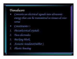
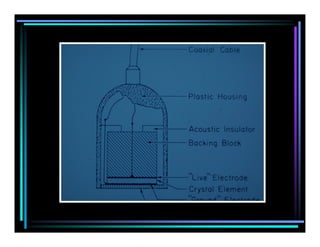
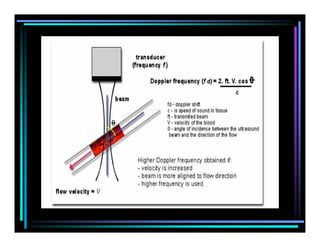
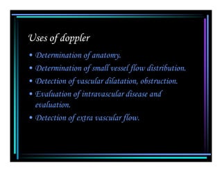
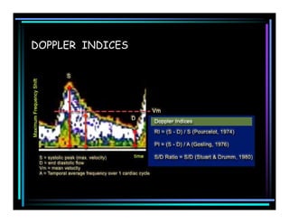
Ultrasound
   Physics
Characteristics of Sound
• Requires medium for its transmission.
• Compression and rarefaction.
• Propagation.
Ultrasound waves
•    Range
•    Longitudinal waves
•    Length of wave
•    Wave once generated continue in original
    direction until it is either reflected, refracted or
    absorbed
Velocity of Sound
• Independent of frequency
• medium
How velocity is related to:
• Compressibility
• Density
• Intensity
Transducers
• Converts an electrical signals into ultrasonic
   energy that can be transmitted to tissues & vice
   versa
• Constituents :-
1. Piezoelectrical crystals
2. Two electrodes
3. Backing block
4. Acoustic insulator(rubber )
5. Plastic housing
• Two basic modes of transducers

1. Continuous mode – doppler studies
2. Pulse mode
Curie temperature
Resonant frequency
Transducer Q factor
•Two characteristics-
       purity of sound & length of sound
•High Q transducer
•Low Q transducer
Advantage of High frequency over low frequency

Depth resolution is superior & near zone is long

Dis-advantage
Tissue absorption increases with increased
  frequency
Interaction between ultrasound & matter
1. Reflection

2. Refraction

3. Absorption
Reflection
• Reflected wave sound produces image

• It depends on

1. Acoustic impedance

2   Angle of incidence
Acoustic impedance
• Impedance is product of tissue density with
  velocity of sound in the material.
 Angle of incidence
  Higher the amount of angle less is the amount
  of sound reflected
  Specular interface Examples
    diaphragm,walls of the vessels
    boundaries of many organs
Refraction
• Bending of waves as they pass from one medium
  to another is called refraction
• Interfaces that are either smaller than the
  wavelength or not smooth are nonspecular
  interface
• Example
  Rbc,liver parenchyma
Absorption
• It means conversion of ultrasonic to thermal
   energy
• It depend on
1. Frequency of sound
2. Viscosity of medium
3. Relaxation time
Ultrasonic display

• A Mode– displayed as spikes projecting from baseline. Spike
  height is proportional to echo intensity. It is static one-
  dimensional mode.
• TM Mode– echoes produced by moving structures as dots.

• B Mode– produces a picture of a slice of tissue. Echoes
  displayed as dots.
• Gray scale Imaging– It’s a B mode type displaying large
  amplitude of echoes arising from tissues as varying shades of
  gray
• Real time imaging–
   produces multiple images in a very short period
• Two types
1. Mechanical– Oscillating & Rotating wheel
2. Electronic array– Linear array & phased or
   steered array
PRINCIPLES
OF
DOPPLER
ULTRASOUND
BASIC PRINCIPLE
• Doppler ultrasound is a technique for making
  non-invasive velocity measurements of blood
  flow.

• Christian Doppler was the first to describe the
  frequency shift that occurs when sound or
  light is emitted from a moving source.

• Ultrasound is transmitted into a vessel and
  the sound that is reflected from the blood is
  detected. Because the blood is moving, the
  sound undergoes a frequency (Doppler) shift.
Uses of doppler
• Determination of anatomy.
• Determination of small vessel flow distribution.
• Detection of vascular dilatation, obstruction.
• Evaluation of intravascular disease and
  evaluation.
• Detection of extra vascular flow.
1) Continuous wave doppler
• Without B MODE imaging
2) Pulsed wave doppler
• Pulsed Doppler ultrasound is a technique for measuring
  the velocity of blood in a small sample volume
• Used in general & obstetric ultrasound & also used to
  provide data for doppler sonograms & colour flow images.
Disadvantage     – “Aliasing”


3) Color Doppler
DOPPLER INDICES

Ultrasound Physics

  • 1.
    Ultrasound Physics
  • 2.
    Characteristics of Sound •Requires medium for its transmission. • Compression and rarefaction. • Propagation.
  • 3.
    Ultrasound waves • Range • Longitudinal waves • Length of wave • Wave once generated continue in original direction until it is either reflected, refracted or absorbed
  • 4.
    Velocity of Sound •Independent of frequency • medium How velocity is related to: • Compressibility • Density • Intensity
  • 6.
    Transducers • Converts anelectrical signals into ultrasonic energy that can be transmitted to tissues & vice versa • Constituents :- 1. Piezoelectrical crystals 2. Two electrodes 3. Backing block 4. Acoustic insulator(rubber ) 5. Plastic housing
  • 8.
    • Two basicmodes of transducers 1. Continuous mode – doppler studies 2. Pulse mode
  • 9.
    Curie temperature Resonant frequency TransducerQ factor •Two characteristics- purity of sound & length of sound •High Q transducer •Low Q transducer
  • 10.
    Advantage of Highfrequency over low frequency Depth resolution is superior & near zone is long Dis-advantage Tissue absorption increases with increased frequency
  • 11.
    Interaction between ultrasound& matter 1. Reflection 2. Refraction 3. Absorption
  • 12.
    Reflection • Reflected wavesound produces image • It depends on 1. Acoustic impedance 2 Angle of incidence
  • 13.
    Acoustic impedance • Impedanceis product of tissue density with velocity of sound in the material. Angle of incidence Higher the amount of angle less is the amount of sound reflected Specular interface Examples diaphragm,walls of the vessels boundaries of many organs
  • 14.
    Refraction • Bending ofwaves as they pass from one medium to another is called refraction • Interfaces that are either smaller than the wavelength or not smooth are nonspecular interface • Example Rbc,liver parenchyma
  • 16.
    Absorption • It meansconversion of ultrasonic to thermal energy • It depend on 1. Frequency of sound 2. Viscosity of medium 3. Relaxation time
  • 17.
    Ultrasonic display • AMode– displayed as spikes projecting from baseline. Spike height is proportional to echo intensity. It is static one- dimensional mode. • TM Mode– echoes produced by moving structures as dots. • B Mode– produces a picture of a slice of tissue. Echoes displayed as dots. • Gray scale Imaging– It’s a B mode type displaying large amplitude of echoes arising from tissues as varying shades of gray
  • 18.
    • Real timeimaging– produces multiple images in a very short period • Two types 1. Mechanical– Oscillating & Rotating wheel 2. Electronic array– Linear array & phased or steered array
  • 19.
  • 20.
    BASIC PRINCIPLE • Dopplerultrasound is a technique for making non-invasive velocity measurements of blood flow. • Christian Doppler was the first to describe the frequency shift that occurs when sound or light is emitted from a moving source. • Ultrasound is transmitted into a vessel and the sound that is reflected from the blood is detected. Because the blood is moving, the sound undergoes a frequency (Doppler) shift.
  • 23.
    Uses of doppler •Determination of anatomy. • Determination of small vessel flow distribution. • Detection of vascular dilatation, obstruction. • Evaluation of intravascular disease and evaluation. • Detection of extra vascular flow.
  • 24.
    1) Continuous wavedoppler • Without B MODE imaging 2) Pulsed wave doppler • Pulsed Doppler ultrasound is a technique for measuring the velocity of blood in a small sample volume • Used in general & obstetric ultrasound & also used to provide data for doppler sonograms & colour flow images. Disadvantage – “Aliasing” 3) Color Doppler
  • 26.