Downloaded 1,080 times


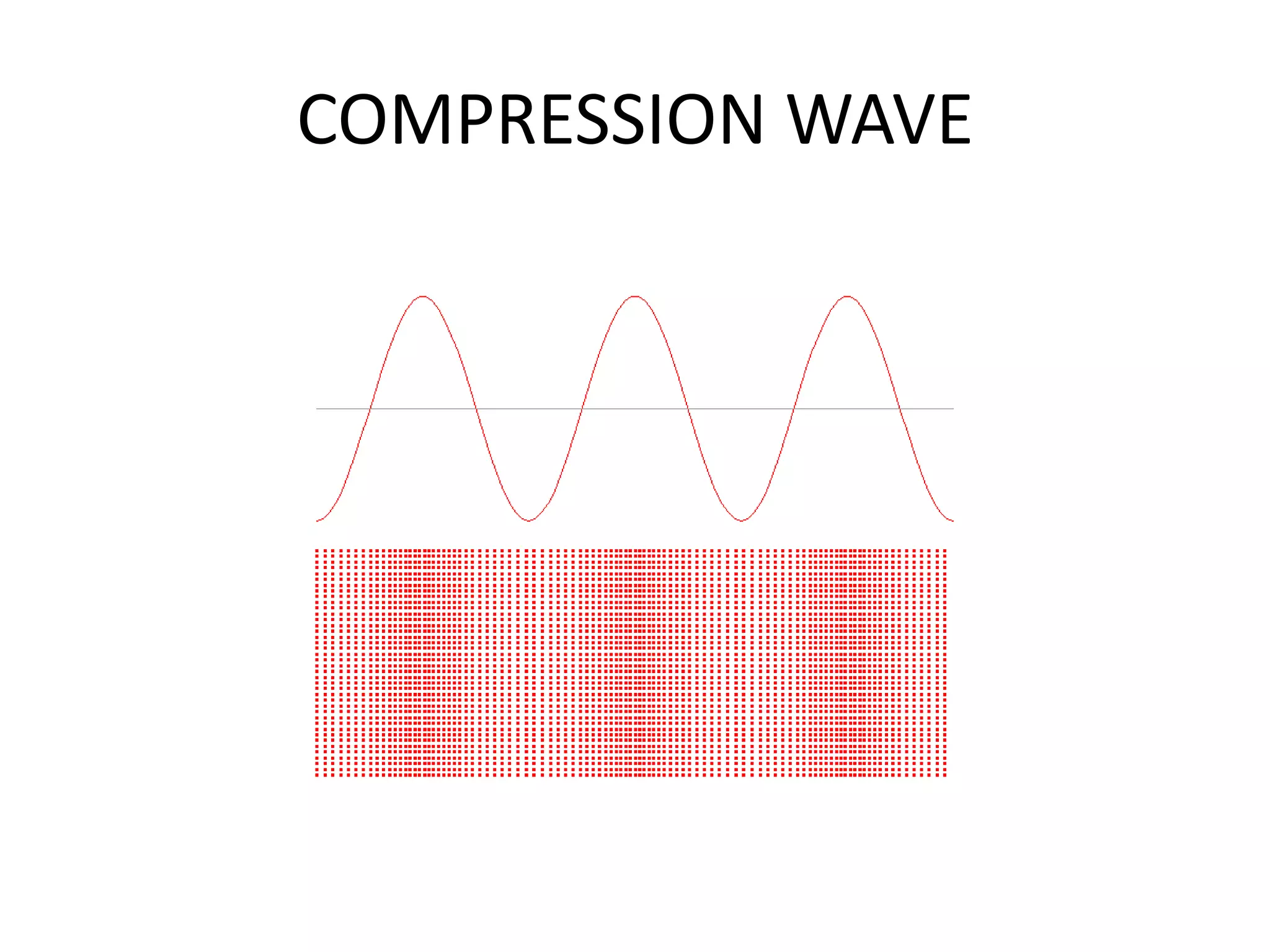
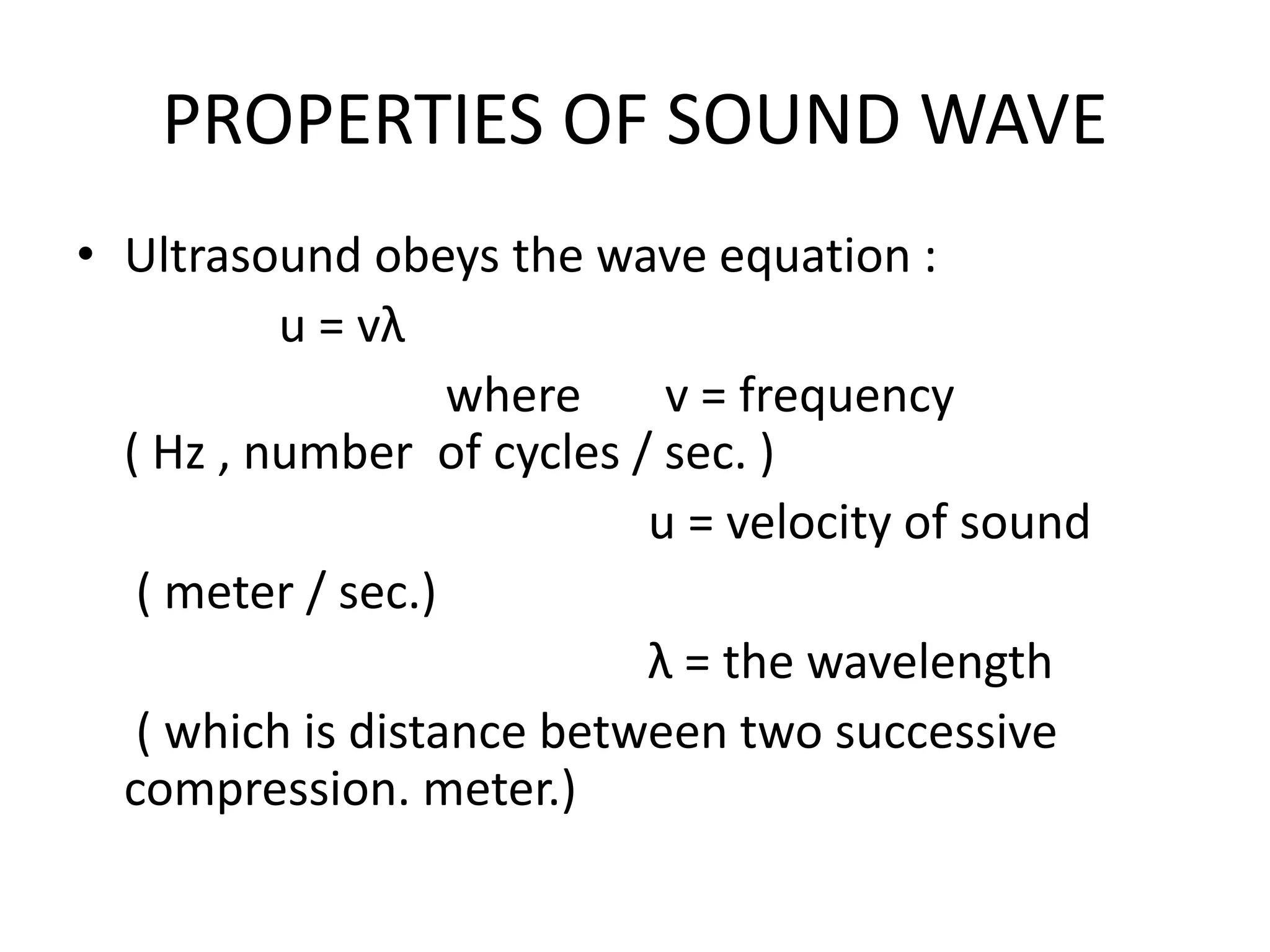


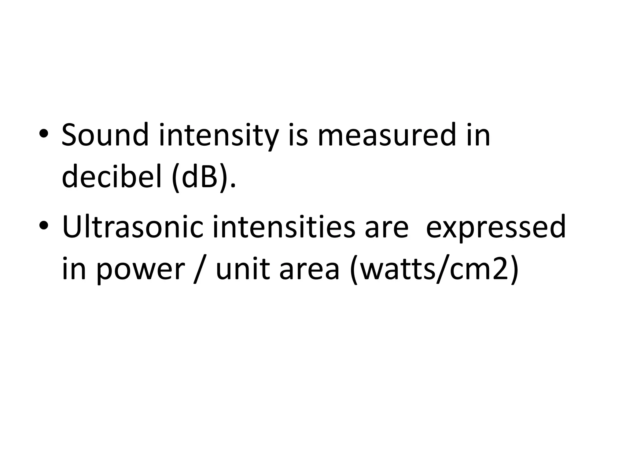


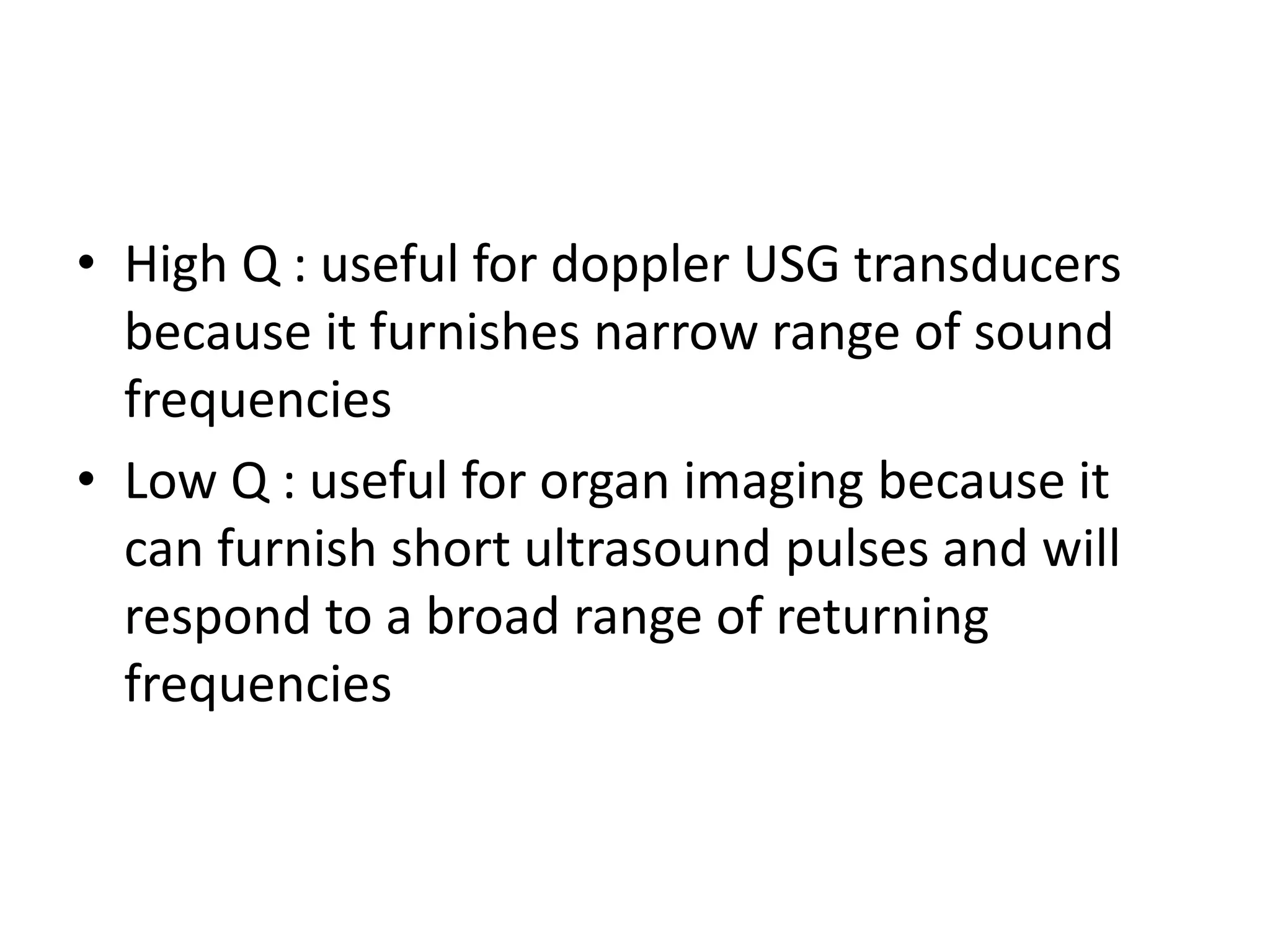


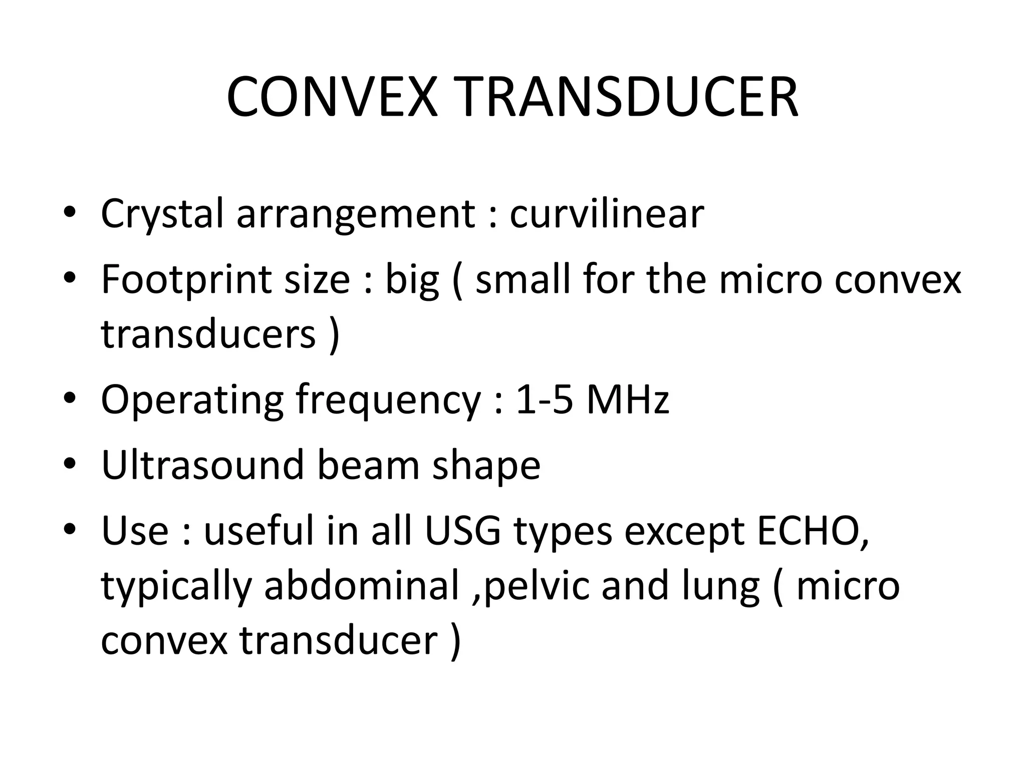


This document provides an overview of ultrasound physics, transducers, and transducer jelly. It discusses the characteristics of sound waves including their generation through mechanical vibration and their transmission through solids, liquids, and gases. The history of ultrasound and piezoelectricity is summarized. Key ultrasound concepts like wavelength, frequency, propagation velocity, amplitude, and absorption are defined. The components and function of ultrasound transducers including the piezoelectric crystal and backing block are described. Finally, the properties and ingredients of transducer jelly used to couple the transducer to the skin are outlined.


