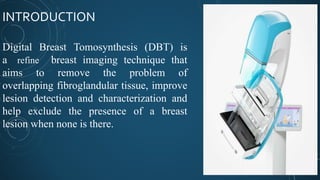
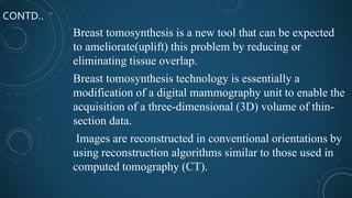
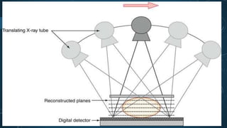
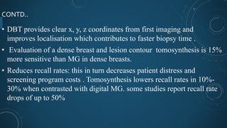
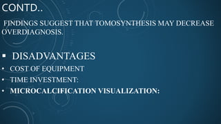
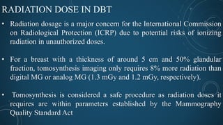
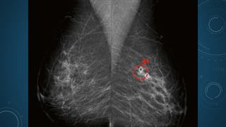
DIGITAL BREASTTOMOSYNTHESIS
Presenter :-Yashawant ku.Yadav
NAMS (Bir Hospital)
Kathmandu , Nepal
OUTLINES
• INTRODUCTION TO DBT
• PRINCIPLE OF DBT
• EQUIPMENT ADVANCEMENT
• WHY DBT OVER FFDM/ DIGITAL MAMMO
• DOSE COMPARISONS AMONG MAMMOGRAPHY MODALITIES
• ADVANCEMENT WITH DBT IN MAMMO SCREENING
• SUMMARY
• REFERENCES
INTRODUCTION
Digital Breast Tomosynthesis (DBT) is
a refine breast imaging technique that
aims to remove the problem of
overlapping fibroglandular tissue, improve
lesion detection and characterization and
help exclude the presence of a breast
lesion when none is there.
CONTD..
• When screening mammograms demonstrate a
questionable finding, the results of follow-up diagnostic
mammography and ultrasonography (US), magnetic
resonance (MR) imaging, or biopsy ultimately determine
whether the finding is significant.
• The process causes anxiety for patients and incurs
additional healthcare costs for findings that frequently are
proved benign.
CONTD..
Breast tomosynthesis is a new tool that can be expected
to ameliorate(uplift) this problem by reducing or
eliminating tissue overlap.
Breast tomosynthesis technology is essentially a
modification of a digital mammography unit to enable the
acquisition of a three-dimensional (3D) volume of thin-
section data.
Images are reconstructed in conventional orientations by
using reconstruction algorithms similar to those used in
computed tomography (CT).
PRINCIPLE
DBT is based on decades old mathematical theory and combines FFDM,
tomographic principles and reconstruction algorithms to produce
multiple thin section images through the breast.
This novel technique may reduce the number of women recalled and
have improved sensitivity compared with FFDM
HOW TO ACHIEVE = ADVANCEMENT
Since the advent of DBT, multiple systems have been designed that vary
in parameters such as geometry, gantry motion, scan angle, angular
sampling and number of projections .
1. The geometry of DBT systems is related to the path and relationship
of the x-ray tube to the detector
a. full isometric geometry
b. Partial isometric geometry
GANATRY MOTION
• The x-ray tube can move in several ways, including in an arc, circular
or linear sweep direction.
• Most DBT systems utilize an arc motion for the x-ray tube.
Images may be acquired by different methods.
• These techniques include a step and shoot or continuous motion
acquisition
CONTD..
• As a result, the overall radiation dose is maintained at an acceptable
level similar to FFDM and well below the Mammography Quality and
Standards Act
 The scan angle
• It is defined as the angular range between the first and last exposure
that is acquired.
• It is typically from 15 to 50 degrees depending on the DBT device and
manufacturer
CONTD..
Furthermore,
1. the scan angle
2. angular sampling
3. number of projections; i.e. number of
projections=(scan angle/angular sampling)+1.
WHY DBT OVER FFDM/ DIGITAL MAMMO
1.Increased sensitivity ? with DBT (compared to DM) indicates that
DBT correctly identifies more breast cancers compared to other
imaging modalities. ( 90%)
2.Increased specificity? would indicate that DBT results in the
correct dismissal of more women with normal or benign findings
without the need for further work-up (i.e., a reduction in false positive
recalls for further assessment). 79%
CONTD..
• DBT provides clear x, y, z coordinates from first imaging and
improves localisation which contributes to faster biopsy time .
• Evaluation of a dense breast and lesion contour tomosynthesis is 15%
more sensitive than MG in dense breasts.
• Reduces recall rates: this in turn decreases patient distress and
screening program costs . Tomosynthesis lowers recall rates in 10%-
30% when contrasted with digital MG. some studies report recall rate
drops of up to 50%
CONTD..
FINDINGS SUGGEST THAT TOMOSYNTHESIS MAY DECREASE
OVERDIAGNOSIS.
 DISADVANTAGES
• COST OF EQUIPMENT
• TIME INVESTMENT:
• MICROCALCIFICATION VISUALIZATION:
RADIATION DOSE IN DBT
• Radiation dosage is a major concern for the International Commission
on Radiological Protection (ICRP) due to potential risks of ionizing
radiation in unauthorized doses.
• For a breast with a thickness of around 5 cm and 50% glandular
fraction, tomosynthesis imaging only requires 8% more radiation than
digital MG or analog MG (1.3 mGy and 1.2 mGy, respectively).
• Tomosynthesis is considered a safe procedure as radiation doses it
requires are within parameters established by the Mammography
Quality Standard Act
AIWITH DBT
• Artificial intelligent can be possible with the digital breast tomosynthesis.
• AI and deep learning are now being used to speed DBT interpretation time
and improve reading workflow.
• These solutions become smarter and more adept at identifying cancer as
they are trained on breast imaging data, automatically highlighting
suspicious areas and enabling radiologists to focus on areas they should
review more closely.
REFERENCES
• https://www.itnonline.com/content/icad-receives-fda-clearance-
powerlook-density-assessment-digital-breast-tomosynthesis
• https://www.icadmed.com/home.html
THANKYOU
Digital breast tomosynthesis

Digital breast tomosynthesis

  • 1.
    DIGITAL BREASTTOMOSYNTHESIS Presenter :-Yashawantku.Yadav NAMS (Bir Hospital) Kathmandu , Nepal
  • 2.
    OUTLINES • INTRODUCTION TODBT • PRINCIPLE OF DBT • EQUIPMENT ADVANCEMENT • WHY DBT OVER FFDM/ DIGITAL MAMMO • DOSE COMPARISONS AMONG MAMMOGRAPHY MODALITIES • ADVANCEMENT WITH DBT IN MAMMO SCREENING • SUMMARY • REFERENCES
  • 3.
    INTRODUCTION Digital Breast Tomosynthesis(DBT) is a refine breast imaging technique that aims to remove the problem of overlapping fibroglandular tissue, improve lesion detection and characterization and help exclude the presence of a breast lesion when none is there.
  • 4.
    CONTD.. • When screeningmammograms demonstrate a questionable finding, the results of follow-up diagnostic mammography and ultrasonography (US), magnetic resonance (MR) imaging, or biopsy ultimately determine whether the finding is significant. • The process causes anxiety for patients and incurs additional healthcare costs for findings that frequently are proved benign.
  • 5.
    CONTD.. Breast tomosynthesis isa new tool that can be expected to ameliorate(uplift) this problem by reducing or eliminating tissue overlap. Breast tomosynthesis technology is essentially a modification of a digital mammography unit to enable the acquisition of a three-dimensional (3D) volume of thin- section data. Images are reconstructed in conventional orientations by using reconstruction algorithms similar to those used in computed tomography (CT).
  • 6.
    PRINCIPLE DBT is basedon decades old mathematical theory and combines FFDM, tomographic principles and reconstruction algorithms to produce multiple thin section images through the breast. This novel technique may reduce the number of women recalled and have improved sensitivity compared with FFDM
  • 7.
    HOW TO ACHIEVE= ADVANCEMENT Since the advent of DBT, multiple systems have been designed that vary in parameters such as geometry, gantry motion, scan angle, angular sampling and number of projections . 1. The geometry of DBT systems is related to the path and relationship of the x-ray tube to the detector a. full isometric geometry b. Partial isometric geometry
  • 9.
    GANATRY MOTION • Thex-ray tube can move in several ways, including in an arc, circular or linear sweep direction. • Most DBT systems utilize an arc motion for the x-ray tube. Images may be acquired by different methods. • These techniques include a step and shoot or continuous motion acquisition
  • 11.
    CONTD.. • As aresult, the overall radiation dose is maintained at an acceptable level similar to FFDM and well below the Mammography Quality and Standards Act  The scan angle • It is defined as the angular range between the first and last exposure that is acquired. • It is typically from 15 to 50 degrees depending on the DBT device and manufacturer
  • 12.
    CONTD.. Furthermore, 1. the scanangle 2. angular sampling 3. number of projections; i.e. number of projections=(scan angle/angular sampling)+1.
  • 13.
    WHY DBT OVERFFDM/ DIGITAL MAMMO 1.Increased sensitivity ? with DBT (compared to DM) indicates that DBT correctly identifies more breast cancers compared to other imaging modalities. ( 90%) 2.Increased specificity? would indicate that DBT results in the correct dismissal of more women with normal or benign findings without the need for further work-up (i.e., a reduction in false positive recalls for further assessment). 79%
  • 15.
    CONTD.. • DBT providesclear x, y, z coordinates from first imaging and improves localisation which contributes to faster biopsy time . • Evaluation of a dense breast and lesion contour tomosynthesis is 15% more sensitive than MG in dense breasts. • Reduces recall rates: this in turn decreases patient distress and screening program costs . Tomosynthesis lowers recall rates in 10%- 30% when contrasted with digital MG. some studies report recall rate drops of up to 50%
  • 16.
    CONTD.. FINDINGS SUGGEST THATTOMOSYNTHESIS MAY DECREASE OVERDIAGNOSIS.  DISADVANTAGES • COST OF EQUIPMENT • TIME INVESTMENT: • MICROCALCIFICATION VISUALIZATION:
  • 17.
    RADIATION DOSE INDBT • Radiation dosage is a major concern for the International Commission on Radiological Protection (ICRP) due to potential risks of ionizing radiation in unauthorized doses. • For a breast with a thickness of around 5 cm and 50% glandular fraction, tomosynthesis imaging only requires 8% more radiation than digital MG or analog MG (1.3 mGy and 1.2 mGy, respectively). • Tomosynthesis is considered a safe procedure as radiation doses it requires are within parameters established by the Mammography Quality Standard Act
  • 18.
    AIWITH DBT • Artificialintelligent can be possible with the digital breast tomosynthesis. • AI and deep learning are now being used to speed DBT interpretation time and improve reading workflow. • These solutions become smarter and more adept at identifying cancer as they are trained on breast imaging data, automatically highlighting suspicious areas and enabling radiologists to focus on areas they should review more closely.
  • 20.
  • 21.

Editor's Notes

  • #4 Mammography is an effective imaging tool for the detection of early-stage breast cancer, and it is the only screening modality proved to reduce mortality from breast cancer. Mammography is not a perfect investigation however: sensitivities are around 70%, lower in women with dense breasts; and false positive recall rates, leading to unnecessary additional imaging and emotional distress, are significant . Up to 10% of women screened will be unnecessarily recalled. Overlapping fibro glandular breast tissue hampering interpretation is the most common reason both for breast cancers to be missed and also for the unnecessary recall of women. Breast cancer detection from screening mammography has been shown to reduce mortality from breast cancer by 30 % among women aged 40–74
  • #5 Overlapping fibro glandular breast tissue hampering interpretation is the most common reason both for breast cancers to be missed and also for the unnecessary recall of women. Mammography is not a perfect investigation however: sensitivities are around 70%, lower in women with dense breasts; and false positive recall rates, leading to unnecessary additional imaging and emotional distress, are significant . Up to 10% of women screened will be unnecessarily recalled.
  • #6 due to the two-dimensional nature of imaging a three-dimensional object such as the breast. Lesion masking by superimposition of breast tissue that lies below or above an abnormality can result in false-negative exam interpretation. The formation of pseudo-lesions due to superimposition of breast tissue resulting in an abnormal appearance of tissue can lead to false-positive interpretations, as well as increase recall rates.
  • #8  The limitations of the technology, such as the visualization and characterization of calcifications, will be discussed, x-ray tube and the detector both move around a pivot axis Hologic models have a detector angular range of±2.1°
  • #10 the x-ray device takes an exposure and then moves to a new position, comes to a halt, obtains another exposure, and then moves to a new position and so on. This process is repeated until the full angular range is completed. In the continuous image acquisition technique, the tube moves constantly at a steady set rate over the prescribed range of motion during the x-ray exposure
  • #14 Sensitivity: the proportion of breast cancers correctly identified by a diagnostic test or the true positive rate. Specificity: the proportion of women correctly identified as not having breast cancer or the true negative rate.
  • #17 breast radiologists went from analyzing four conventional MG projections to hundreds of images using MG with tomosynthesis. A conventional MG usually comprises two images (CC and MLO) on each breast and occasionally up to two additional ones (Eklund or focal compressions); . MG with tomosynthesis significantly increases image analysis time: 35%-65% longer than conventional MG, a time directly correlated with the radiologist’s experience . An average, 5 cm breast requires approximately 50 images per projection. Up until today, microcalcifications are still best viewed using digital MG than tomosynthesis, with latter are reported to have lower sensitivity (72%) for detecting microcalcifications than the former (76%) this may be due to movement artifacts resulting from a relatively longer procedure time in tomosynthesis .