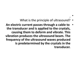
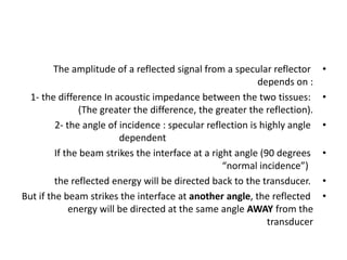
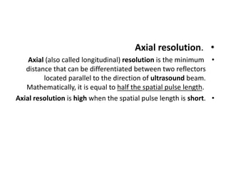
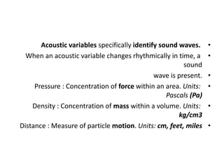
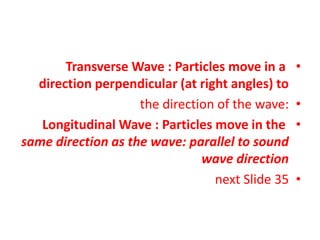
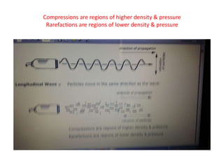
US PHYSICS (4)
Dr. Kamal Sayed MSc US UAA
us principle/reflections/bulk modulus/scientific notations/
OK
.
•
•
What is the principle of ultrasound?
•
An electric current passes through a cable to
the transducer and is applied to the crystals,
causing them to deform and vibrate. This
vibration produces the ultrasound beam. The
frequency of the ultrasound waves produced
is predetermined by the crystals in the
transducer.
Reflections from different frequencies
•
(3 MHz,5 MHz,10 MHz)
•
If transmitted at the same time into same
anatomy
•
would have nearly identical transit times.
•
•
The speed of sound does not vary appreciably with
frequency. Frequency describes the number of cycles
that occur in one second.
•
The speed of sound Is determined by the medium, not
the sound source.
•
Frequency, on the other hand, is determined by the
sound source (the transducer), not by the medium.
•
With specular reflectors, the angle of reflection is
equal to the angle of transmission, so the greatest
reflection will be received back by the transducer
whenever perpendicular Incidence is used.
•
Increasing the amount of received energy (greatest
reflection) enhances the visibility of the reflector
(more bright).
•
1- Reflection and scattering give rise to the
echo signals of organs that are displayed on
the monitor.
•
2- Refletion is sound-tissue interaction which is necessary to
form an ultrasound image
•
The equation for acoustic Impedance is
•
z = pc
•
where Z is the rayl (unit for acoustic impedance),
•
P is density, and C is the speed of sound.
•
Because the speed of sound in tissue is relatively constant
(1540 m/s), the main factor determining acoustic Impedance
Is changes in tissue density.
•
The amplitude of a reflected signal from a specular reflector
depends on :
•
1- the difference In acoustic impedance between the two tissues:
(The greater the difference, the greater the reflection).
•
2- the angle of incidence : specular reflection is highly angle
dependent
•
If the beam strikes the interface at a right angle (90 degrees
“normal incidence”)
•
the reflected energy will be directed back to the transducer.
•
But if the beam strikes the interface at another angle, the reflected
energy will be directed at the same angle AWAY from the
transducer
•
@ 20, 200, and 2000 Hz are in the audible
range (from 20 Hz to 20,000 Hz),
•
@ while 2 Hz is infrasonic.
•
@ Frequencies greater than 20,000 Hz are
ultrasonic.
•
The audible frequency range of sound is from approximately
20 Hz to 20,000 Hz.
•
In physics the term “ultrasound” applies to all acoustic energy
with a frequency above human hearing (20,000 hertz or
20 kilohertz).
•
Typical diagnostic sonographic scanners operate in the
frequency range of 2 to 18 megahertz, hundreds of times
greater than the limit of human hearing
•
Transducers can be operated over a range of
frequencies: the transmitter generally dictates the
actual frequency.
•
The number of electric pulses delivered to the active
element per second is the pulse repetition frequency
(PRF) but does not affect the imaging frequency.
•
The frequency is determined by the propagation speed
and the thickness of the piezoelectric material in the
transducer, and by the center frequency of the drive
signal applied to the transducer.
•
Sound cannot travel through a vacuum.
•
Sound requires a material medium for
propagation.
•
The physical movement of particles within a
medium transports the sound.
•
Since there are no particles to vibrate in a
vacuum, sound cannot propagate
•
Bulk modulus, numerical constant that
describes the elastic properties of a solid or
fluid when it is under pressure on all surfaces.
•
The applied pressure reduces the volume of a
material, which returns to
•
its original volume when the pressure is
removed
•
The applied pressure reduces the volume of a
material, which returns to its original volume
when the pressure is removed.
•
The ratio of the change in pressure to the
fractional volume compression
is called the bulk modulus of the material. ...
The amount of compression of solids and
liquids is seen to be very small.
•
Pulse duration is the time it takes to complete
one pulse.
•
If the number of cycles in the pulse is
increased, it will take more time for one pulse
to occur.
•
Frequency is how many cycles occur in one
second—not how many cycles are contained in
one pulse.
•
Propagation speed (the speed at which sound travels through
a particular medium) is not affected by the number of cycles
in a pulse: it is determined by the medium.
•
Period is the inverse of frequency. It describes the time it
takes for one cycle to occur— not the time it takes for one
pulse to occur.
•
When frequency increases, period decreases, and vice versa.
Bulk modulus is related to media stiffness and helps to
determine propagation speed.
•
AS frequency increases:
•
absorption, scattering, & attenuation all
increase
•
Conventionally expressed numbers & their
equivalents in scientific notation
•
From 1234, move the decimal point to the left until you have a
number between 1 and 10. The number of places you move
the decimal point equals the power of 10 in the scientific
notation representation—in this case 10 .
‫؟‬
•
See next
•
Scientific notation Is a convenient way to write both large and
small numbers while at the same time conveying a sense of
their magnitude.
•
For negative numbers, the decimal point Is moved to the right
until you have a number between 1 and 10.
•
See next
•
Scientific notation Is a convenient way to write
both large and small numbers while at the
same time conveying a sense of their
magnitude. For negative numbers, the decimal
point Is moved to the right until you have a
number between 1 and 10.
•
Terms used to describe the strength of the
sound beam include amplitude & intensity
•
How do you describe an ultrasound?
–
An ultrasound scan is a medical test that uses high-
frequency sound waves to capture live images from the
inside of your body.
–
It is used to help diagnose the causes of pain, swelling
and infection in the body's internal organs and to examine
a baby in pregnant women and the brain and hips in
infants.
–
It's also known as sonography.
–
The technology is similar to that used by sonar and radar,
which help the military detect planes and ships.
•
Intensity in US is the rate at which ultrasound energy is
applied to a specific tissue location within the patient's body.
•
It is the quantity that must be considered with respect to
producing biological effects and safety.
•
Ultrasound intensity is measured in water, at the point of
maximum intensity (spatial peak), averaged over time
(temporal average) and derated by 0.3 dB/MHz/cm
to estimate the 'in‐situ' intensity in tissues.
•
ultrasound beam
•
The area through which the sound energy (emitted from
the ultrasound transducer) travels is known as the ultrasound
beam.
•
The beam is three-dimensional and is symmetrical around its
central axis. ...
•
Most diagnostic applications, however, use pulsed sound,
where the output is a series of short pulses of sound.
•
ADVANTAGES OF US
•
They're generally painless and don't require needles, shots
or cuts (NONINVASIVE).
•
You aren't exposed to ionizing radiation, so the procedure is
safer than X-rays and CT scans. In fact, there are no known
harmful effects when it's used as directed.
•
Ultrasound captures images of soft tissues that don't show
up well on X-rays.
•
Ultrasounds are widely accessible and less expensive than
other methods.
•
•
What are the properties of ultrasound
waves?
•
Ultrasound is sound with a frequency greater than 20,000 Hz.
Humans cannot hear ultrasound but many other animals can,
such as mice, dogs and porpoises.
•
Ultrasound is useful because it has a short wavelength so it
can be focussed into a beam.
•
Ultrasound is defined by the American National
Standards Institute as "sound at frequencies greater than
20 kHz". In air at atmospheric pressure, ultrasonic
waves have wavelengths of 1.9 cm or less.
•
Ultrasound waves have higher frequencies than normal
sound waves, but they also have shorter wavelengths.
•
In other words, the distance between one ultrasound
wave traveling through the air and the one following on
behind it is much shorter than in a normal sound wave.
•
–Ultrasound wavelength decreases with increasing
frequency. –In soft tissue, the ultrasound wavelength is 0.39
mm at 4 MHz and 0.15 mm at 10 MHz. –
•
For sound waves, the relation between velocity (v) measured
in m/s, frequency (f), and wavelength is v = f ×λ (m/s).
•
RESOLUTION
•
Image resolution determines the clarity of the image.
Such spatial resolution is dependent of axial and
lateral resolution.
•
Both of these are dependent on the frequency of
the ultrasound.
•
Axial resolution is the ability to see the two structures
that are side by side as separate and distinct when
parallel to the beam.
•
Axial resolution.
•
Axial (also called longitudinal) resolution is the minimum
distance that can be differentiated between two reflectors
located parallel to the direction of ultrasound beam.
Mathematically, it is equal to half the spatial pulse length.
•
Axial resolution is high when the spatial pulse length is short.
•
Elevational (azimuthal) resolution
•
This represents the extent to which an ultrasound system is
able to resolve objects within an axis perpendicular to the
plane formed by the axial and lateral dimensions.
•
6 Common Types of Ultrasound and How They Are Used
•
Pelvic Ultrasound Imaging
•
Abdominal Ultrasounds
•
Obstetric Ultrasounds
•
Transvaginal Ultrasound
•
Transrectal Ultrasound
•
Carotid and Abdominal Aorta Ultrasound
•
•
The TGC (time gain compensation)
•
is used to amplify echo signals from deeper
structures, which have undergone greater
amounts of attenuation more than echo
signals from shallow structures
•
Acoustic variables specifically identify sound waves.
•
When an acoustic variable changes rhythmically in time, a
sound
•
wave is present.
•
Pressure : Concentration of force within an area. Units:
Pascals (Pa)
•
Density : Concentration of mass within a volume. Units:
kg/cm3
•
Distance : Measure of particle motion. Units: cm, feet, miles
•
Transverse Wave : Particles move in a
direction perpendicular (at right angles) to
•
the direction of the wave:
•
Longitudinal Wave : Particles move in the
same direction as the wave: parallel to sound
wave direction
•
next Slide 35
Compressions are regions of higher density & pressure
Rarefactions are regions of lower density & pressure
Us physics (4)

Us physics (4)

  • 1.
    US PHYSICS (4) Dr.Kamal Sayed MSc US UAA us principle/reflections/bulk modulus/scientific notations/ OK .
  • 2.
    • • What is theprinciple of ultrasound? • An electric current passes through a cable to the transducer and is applied to the crystals, causing them to deform and vibrate. This vibration produces the ultrasound beam. The frequency of the ultrasound waves produced is predetermined by the crystals in the transducer.
  • 3.
    Reflections from differentfrequencies • (3 MHz,5 MHz,10 MHz) • If transmitted at the same time into same anatomy • would have nearly identical transit times.
  • 4.
    • • The speed ofsound does not vary appreciably with frequency. Frequency describes the number of cycles that occur in one second. • The speed of sound Is determined by the medium, not the sound source. • Frequency, on the other hand, is determined by the sound source (the transducer), not by the medium.
  • 5.
    • With specular reflectors,the angle of reflection is equal to the angle of transmission, so the greatest reflection will be received back by the transducer whenever perpendicular Incidence is used. • Increasing the amount of received energy (greatest reflection) enhances the visibility of the reflector (more bright).
  • 6.
    • 1- Reflection andscattering give rise to the echo signals of organs that are displayed on the monitor. • 2- Refletion is sound-tissue interaction which is necessary to form an ultrasound image
  • 7.
    • The equation foracoustic Impedance is • z = pc • where Z is the rayl (unit for acoustic impedance), • P is density, and C is the speed of sound. • Because the speed of sound in tissue is relatively constant (1540 m/s), the main factor determining acoustic Impedance Is changes in tissue density.
  • 8.
    • The amplitude ofa reflected signal from a specular reflector depends on : • 1- the difference In acoustic impedance between the two tissues: (The greater the difference, the greater the reflection). • 2- the angle of incidence : specular reflection is highly angle dependent • If the beam strikes the interface at a right angle (90 degrees “normal incidence”) • the reflected energy will be directed back to the transducer. • But if the beam strikes the interface at another angle, the reflected energy will be directed at the same angle AWAY from the transducer
  • 9.
    • @ 20, 200,and 2000 Hz are in the audible range (from 20 Hz to 20,000 Hz), • @ while 2 Hz is infrasonic. • @ Frequencies greater than 20,000 Hz are ultrasonic.
  • 10.
    • The audible frequencyrange of sound is from approximately 20 Hz to 20,000 Hz. • In physics the term “ultrasound” applies to all acoustic energy with a frequency above human hearing (20,000 hertz or 20 kilohertz). • Typical diagnostic sonographic scanners operate in the frequency range of 2 to 18 megahertz, hundreds of times greater than the limit of human hearing
  • 11.
    • Transducers can beoperated over a range of frequencies: the transmitter generally dictates the actual frequency. • The number of electric pulses delivered to the active element per second is the pulse repetition frequency (PRF) but does not affect the imaging frequency. • The frequency is determined by the propagation speed and the thickness of the piezoelectric material in the transducer, and by the center frequency of the drive signal applied to the transducer.
  • 12.
    • Sound cannot travelthrough a vacuum. • Sound requires a material medium for propagation. • The physical movement of particles within a medium transports the sound. • Since there are no particles to vibrate in a vacuum, sound cannot propagate
  • 13.
    • Bulk modulus, numericalconstant that describes the elastic properties of a solid or fluid when it is under pressure on all surfaces. • The applied pressure reduces the volume of a material, which returns to • its original volume when the pressure is removed
  • 14.
    • The applied pressurereduces the volume of a material, which returns to its original volume when the pressure is removed. • The ratio of the change in pressure to the fractional volume compression is called the bulk modulus of the material. ... The amount of compression of solids and liquids is seen to be very small.
  • 15.
    • Pulse duration isthe time it takes to complete one pulse. • If the number of cycles in the pulse is increased, it will take more time for one pulse to occur. • Frequency is how many cycles occur in one second—not how many cycles are contained in one pulse.
  • 16.
    • Propagation speed (thespeed at which sound travels through a particular medium) is not affected by the number of cycles in a pulse: it is determined by the medium. • Period is the inverse of frequency. It describes the time it takes for one cycle to occur— not the time it takes for one pulse to occur. • When frequency increases, period decreases, and vice versa. Bulk modulus is related to media stiffness and helps to determine propagation speed.
  • 17.
    • AS frequency increases: • absorption,scattering, & attenuation all increase
  • 18.
    • Conventionally expressed numbers& their equivalents in scientific notation • From 1234, move the decimal point to the left until you have a number between 1 and 10. The number of places you move the decimal point equals the power of 10 in the scientific notation representation—in this case 10 . ‫؟‬ • See next
  • 19.
    • Scientific notation Isa convenient way to write both large and small numbers while at the same time conveying a sense of their magnitude. • For negative numbers, the decimal point Is moved to the right until you have a number between 1 and 10. • See next
  • 20.
    • Scientific notation Isa convenient way to write both large and small numbers while at the same time conveying a sense of their magnitude. For negative numbers, the decimal point Is moved to the right until you have a number between 1 and 10.
  • 21.
    • Terms used todescribe the strength of the sound beam include amplitude & intensity
  • 22.
    • How do youdescribe an ultrasound? – An ultrasound scan is a medical test that uses high- frequency sound waves to capture live images from the inside of your body. – It is used to help diagnose the causes of pain, swelling and infection in the body's internal organs and to examine a baby in pregnant women and the brain and hips in infants. – It's also known as sonography. – The technology is similar to that used by sonar and radar, which help the military detect planes and ships.
  • 23.
    • Intensity in USis the rate at which ultrasound energy is applied to a specific tissue location within the patient's body. • It is the quantity that must be considered with respect to producing biological effects and safety. • Ultrasound intensity is measured in water, at the point of maximum intensity (spatial peak), averaged over time (temporal average) and derated by 0.3 dB/MHz/cm to estimate the 'in‐situ' intensity in tissues.
  • 24.
    • ultrasound beam • The areathrough which the sound energy (emitted from the ultrasound transducer) travels is known as the ultrasound beam. • The beam is three-dimensional and is symmetrical around its central axis. ... • Most diagnostic applications, however, use pulsed sound, where the output is a series of short pulses of sound.
  • 25.
    • ADVANTAGES OF US • They'regenerally painless and don't require needles, shots or cuts (NONINVASIVE). • You aren't exposed to ionizing radiation, so the procedure is safer than X-rays and CT scans. In fact, there are no known harmful effects when it's used as directed. • Ultrasound captures images of soft tissues that don't show up well on X-rays. • Ultrasounds are widely accessible and less expensive than other methods. •
  • 26.
    • What are theproperties of ultrasound waves? • Ultrasound is sound with a frequency greater than 20,000 Hz. Humans cannot hear ultrasound but many other animals can, such as mice, dogs and porpoises. • Ultrasound is useful because it has a short wavelength so it can be focussed into a beam. • Ultrasound is defined by the American National Standards Institute as "sound at frequencies greater than 20 kHz". In air at atmospheric pressure, ultrasonic waves have wavelengths of 1.9 cm or less.
  • 27.
    • Ultrasound waves havehigher frequencies than normal sound waves, but they also have shorter wavelengths. • In other words, the distance between one ultrasound wave traveling through the air and the one following on behind it is much shorter than in a normal sound wave. • –Ultrasound wavelength decreases with increasing frequency. –In soft tissue, the ultrasound wavelength is 0.39 mm at 4 MHz and 0.15 mm at 10 MHz. – • For sound waves, the relation between velocity (v) measured in m/s, frequency (f), and wavelength is v = f ×λ (m/s).
  • 28.
    • RESOLUTION • Image resolution determinesthe clarity of the image. Such spatial resolution is dependent of axial and lateral resolution. • Both of these are dependent on the frequency of the ultrasound. • Axial resolution is the ability to see the two structures that are side by side as separate and distinct when parallel to the beam.
  • 29.
    • Axial resolution. • Axial (alsocalled longitudinal) resolution is the minimum distance that can be differentiated between two reflectors located parallel to the direction of ultrasound beam. Mathematically, it is equal to half the spatial pulse length. • Axial resolution is high when the spatial pulse length is short.
  • 30.
    • Elevational (azimuthal) resolution • Thisrepresents the extent to which an ultrasound system is able to resolve objects within an axis perpendicular to the plane formed by the axial and lateral dimensions.
  • 31.
    • 6 Common Typesof Ultrasound and How They Are Used • Pelvic Ultrasound Imaging • Abdominal Ultrasounds • Obstetric Ultrasounds • Transvaginal Ultrasound • Transrectal Ultrasound • Carotid and Abdominal Aorta Ultrasound •
  • 32.
    • The TGC (timegain compensation) • is used to amplify echo signals from deeper structures, which have undergone greater amounts of attenuation more than echo signals from shallow structures
  • 33.
    • Acoustic variables specificallyidentify sound waves. • When an acoustic variable changes rhythmically in time, a sound • wave is present. • Pressure : Concentration of force within an area. Units: Pascals (Pa) • Density : Concentration of mass within a volume. Units: kg/cm3 • Distance : Measure of particle motion. Units: cm, feet, miles
  • 34.
    • Transverse Wave :Particles move in a direction perpendicular (at right angles) to • the direction of the wave: • Longitudinal Wave : Particles move in the same direction as the wave: parallel to sound wave direction • next Slide 35
  • 35.
    Compressions are regionsof higher density & pressure Rarefactions are regions of lower density & pressure