Downloaded 503 times
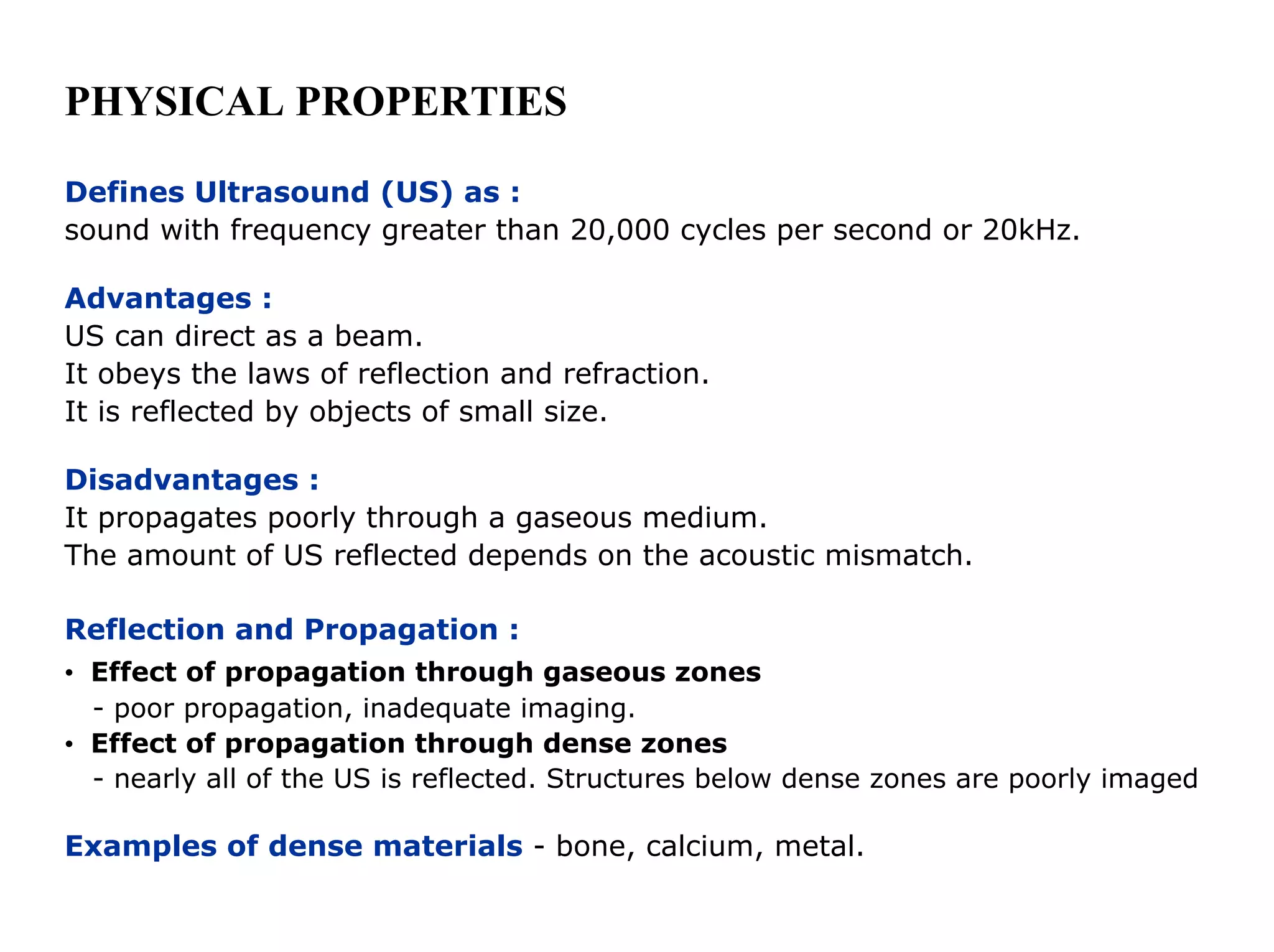
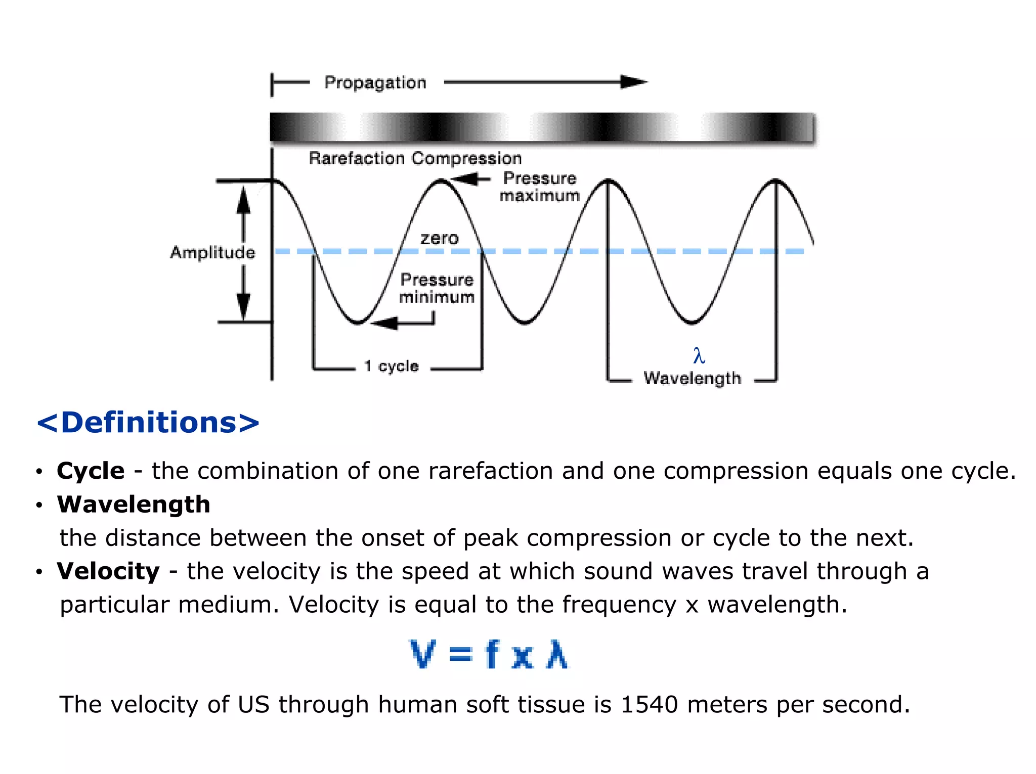
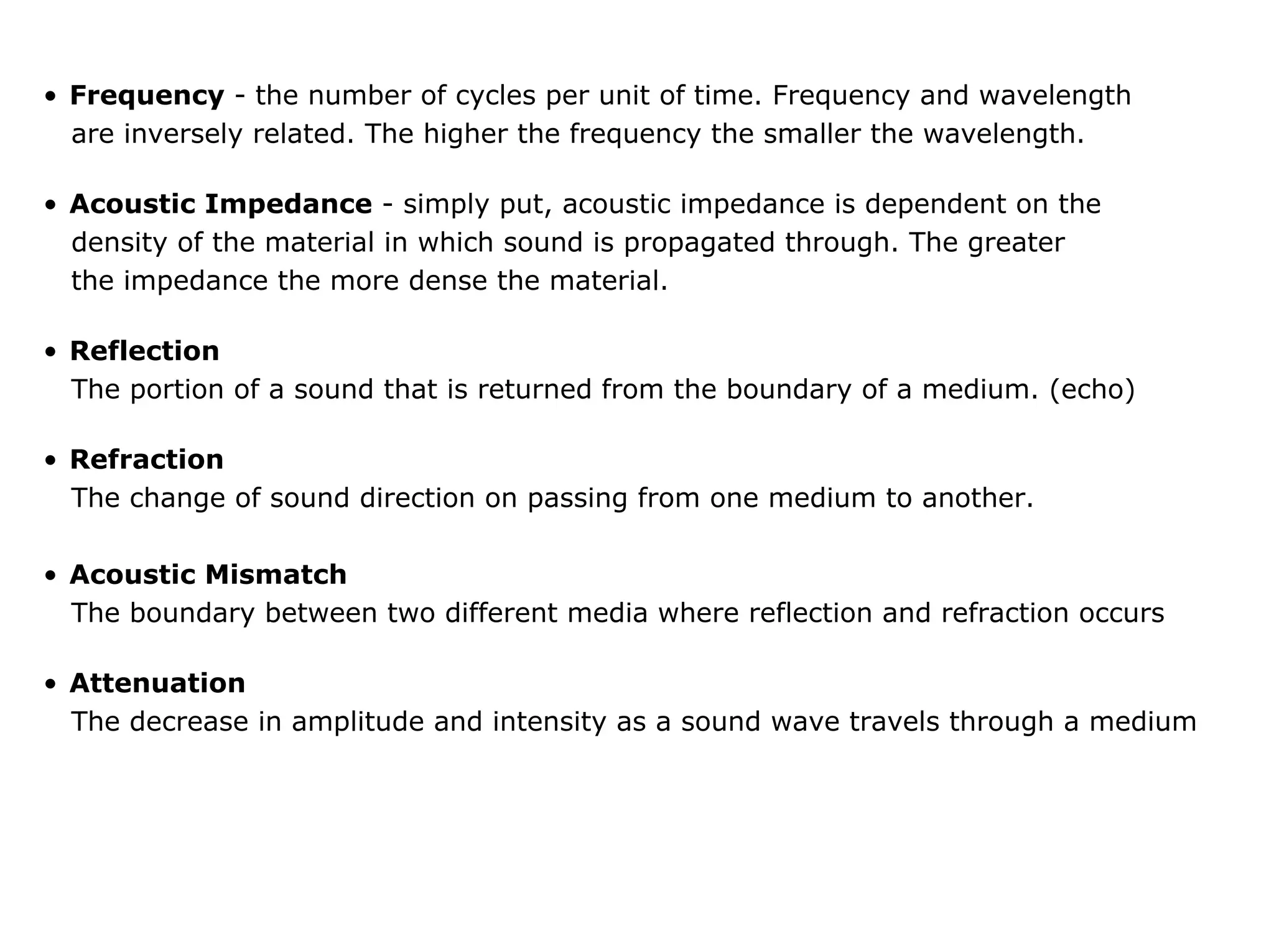
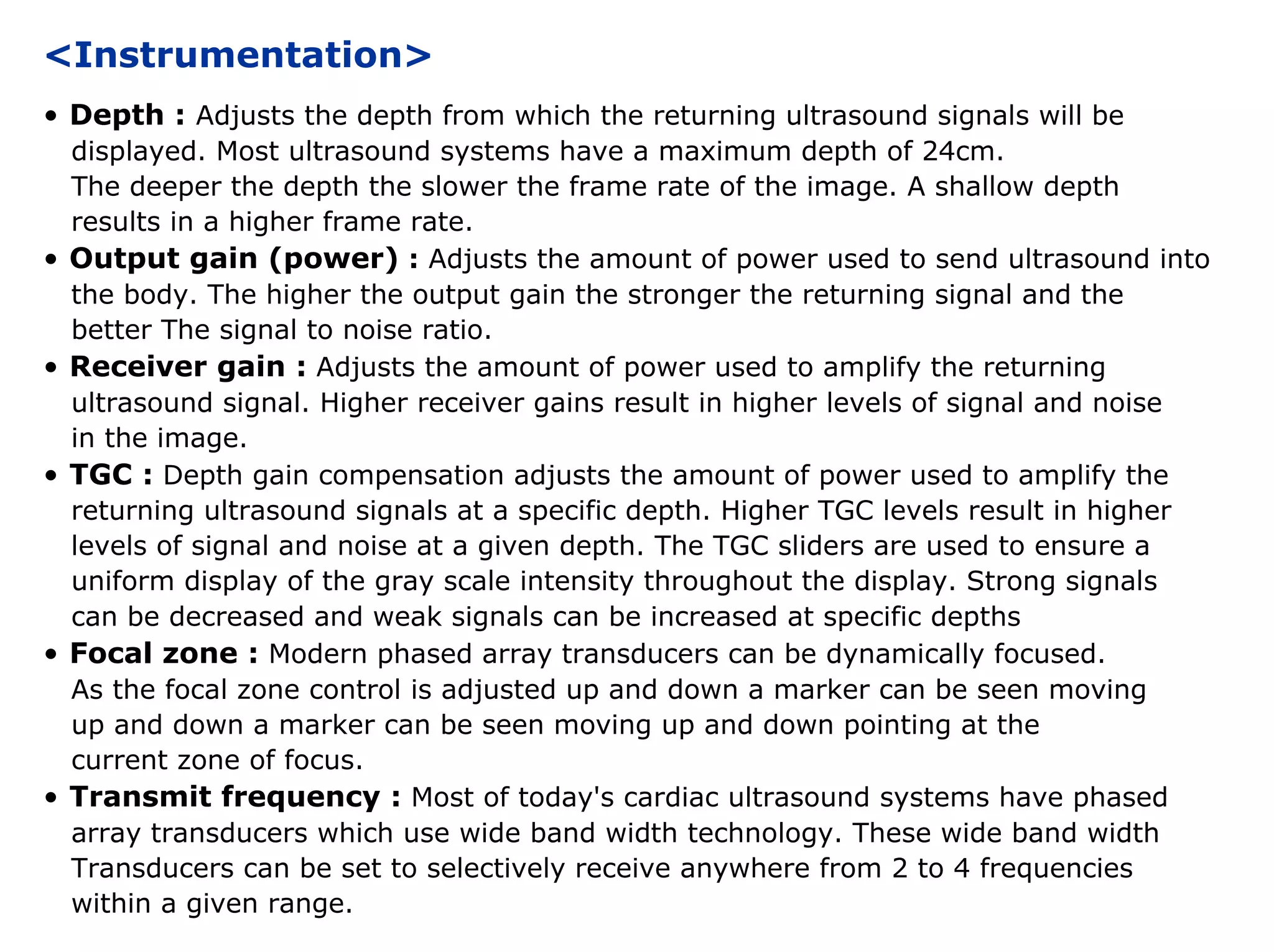
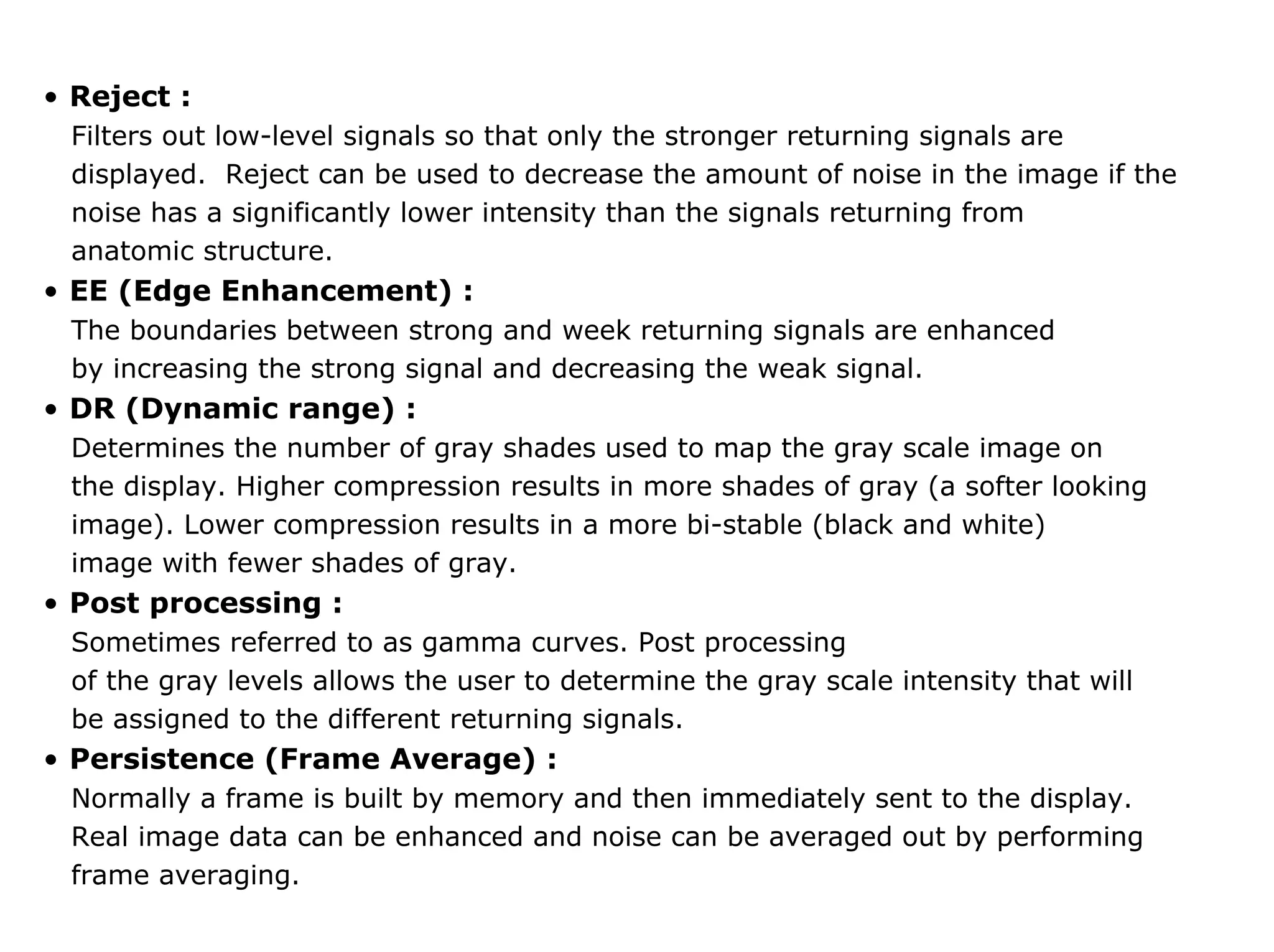
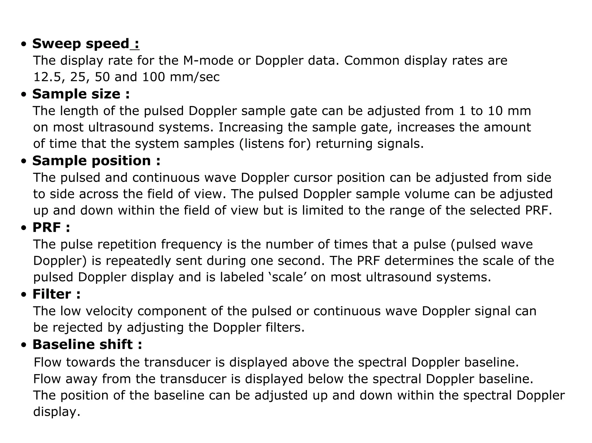
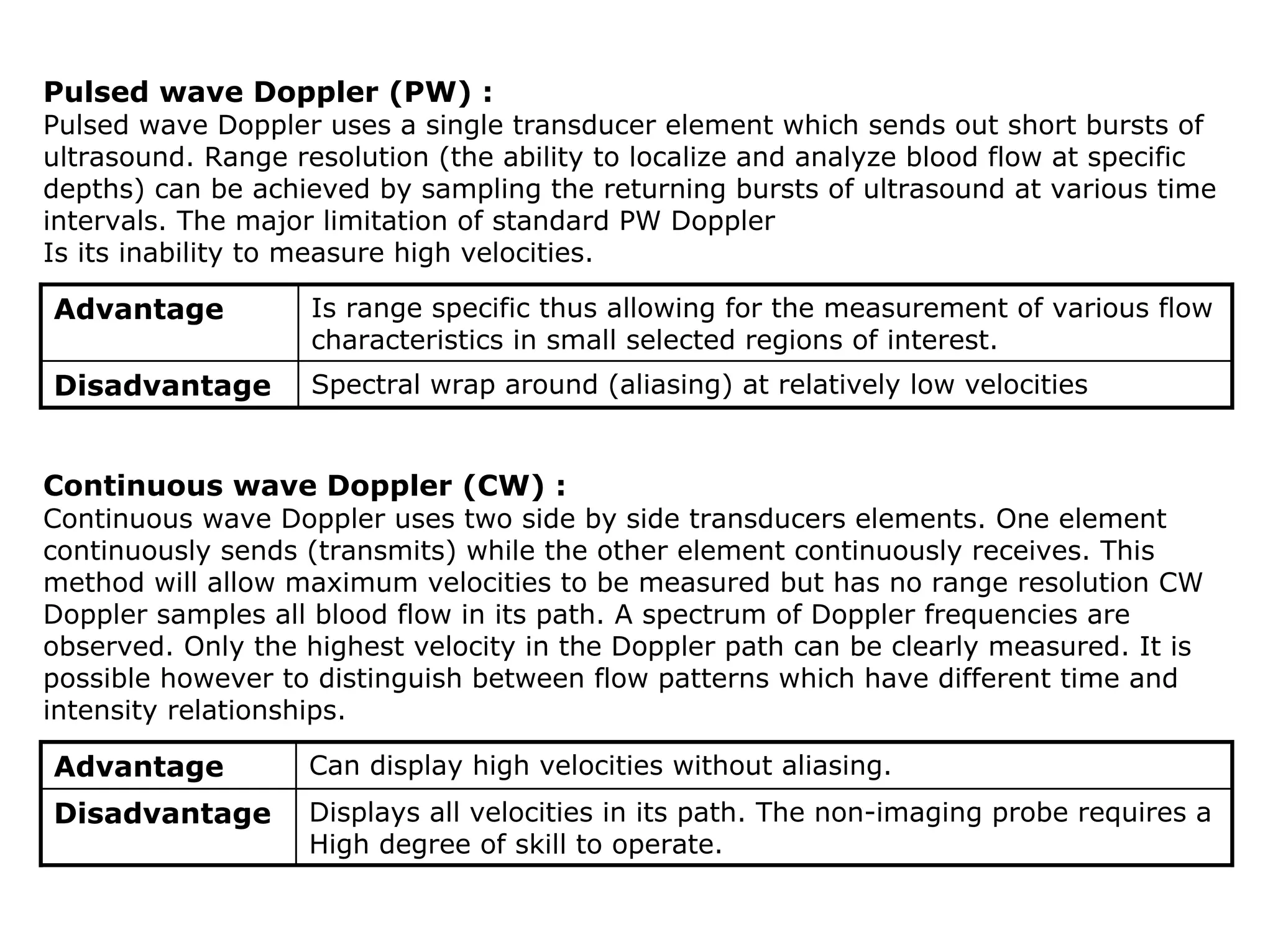

This document provides an overview of basic ultrasound principles and terminology. It defines ultrasound as sound waves with a frequency over 20 kHz. Ultrasound can be directed in a beam and obeys the laws of reflection and refraction. The document describes ultrasound properties such as wavelength, velocity, frequency, and acoustic impedance. It also discusses ultrasound imaging techniques including pulsed wave and continuous wave Doppler, and how settings like depth, focal zone, and filter affect the image.


