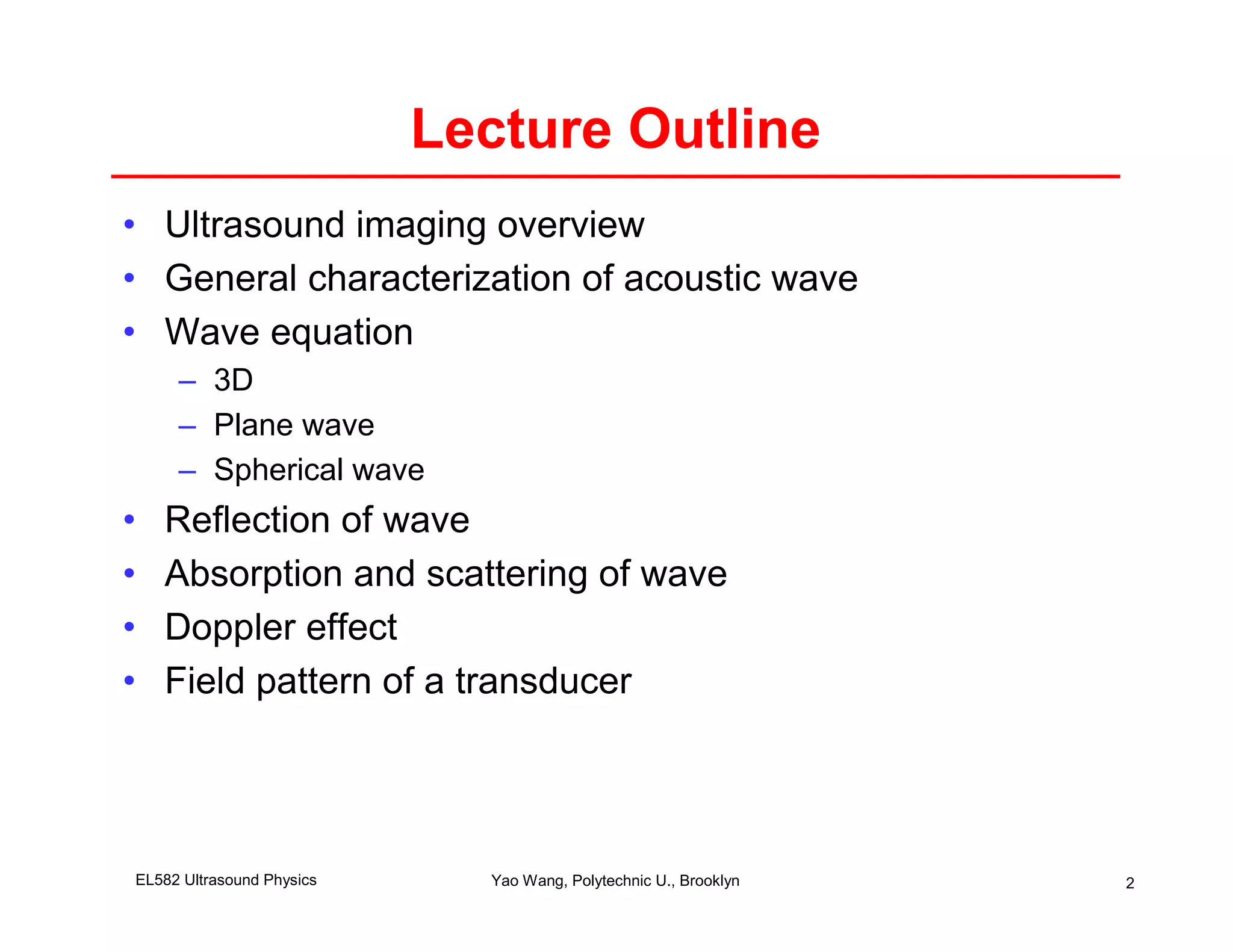
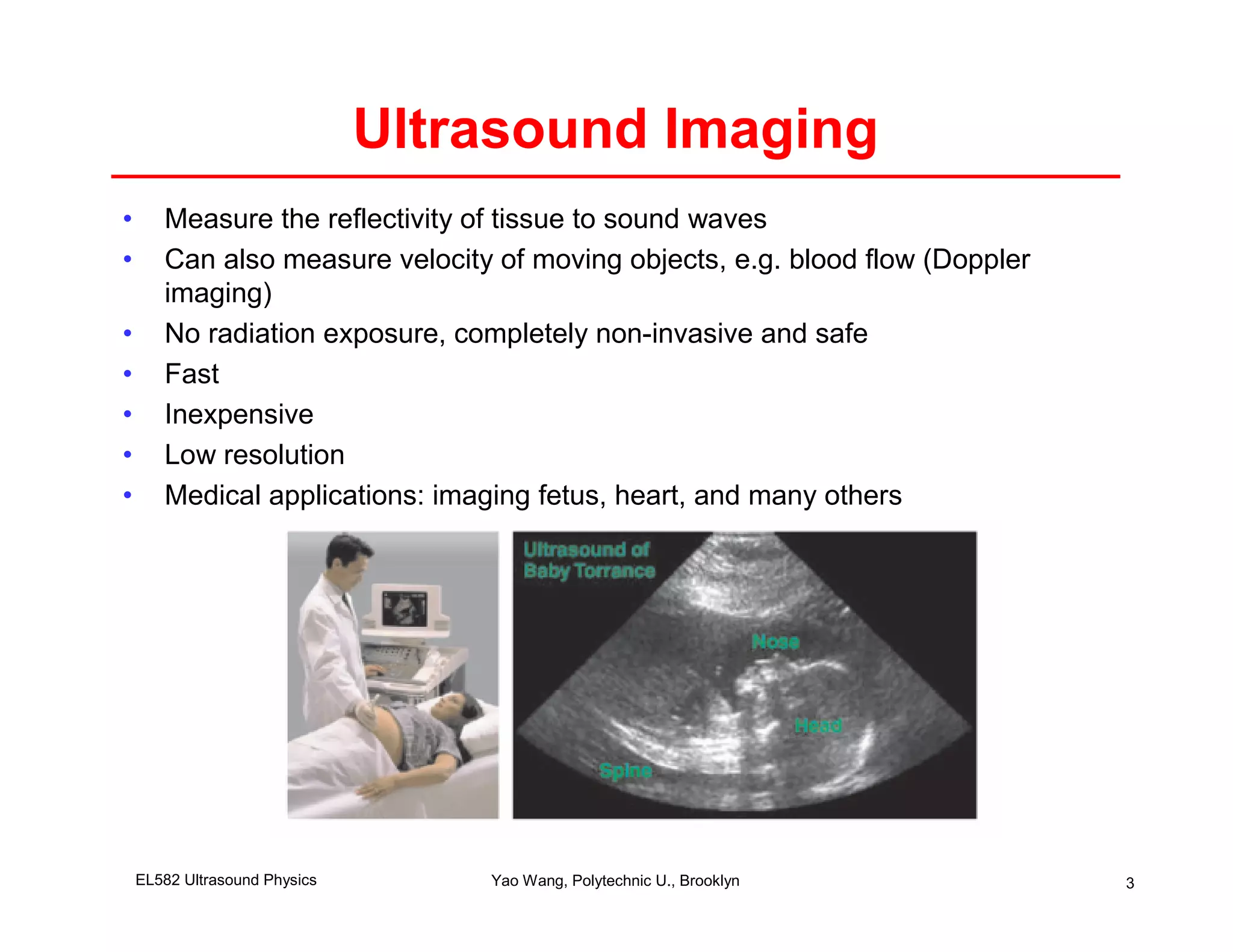
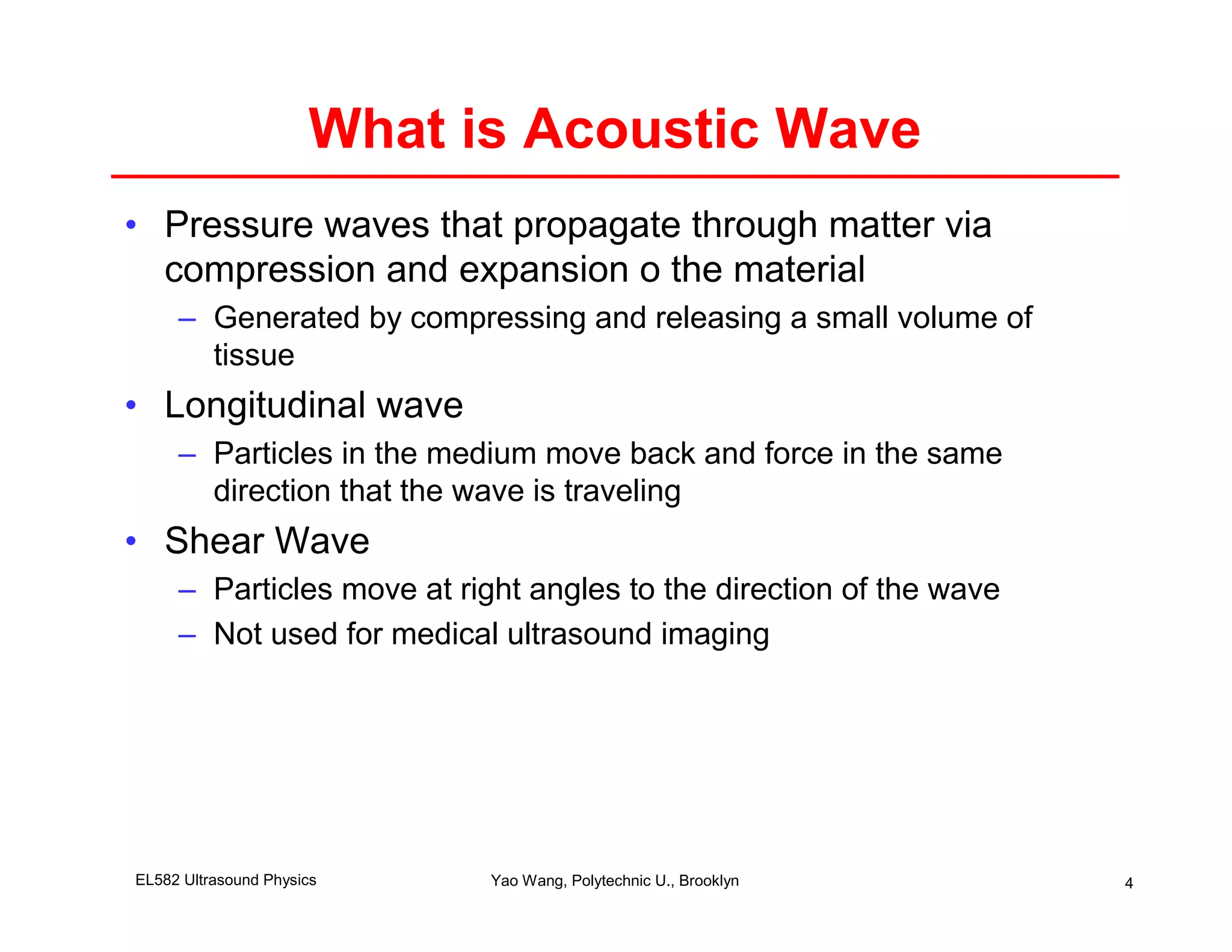
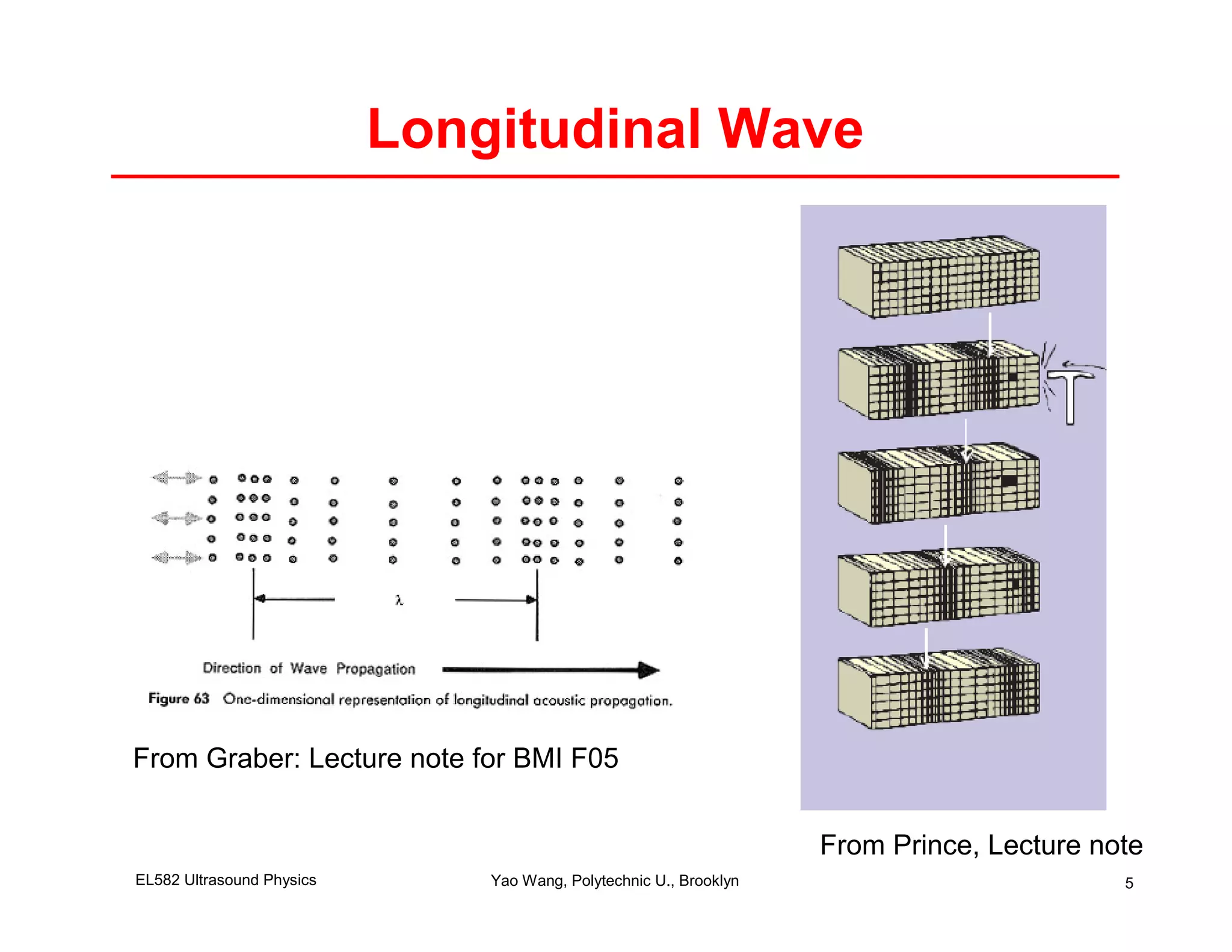
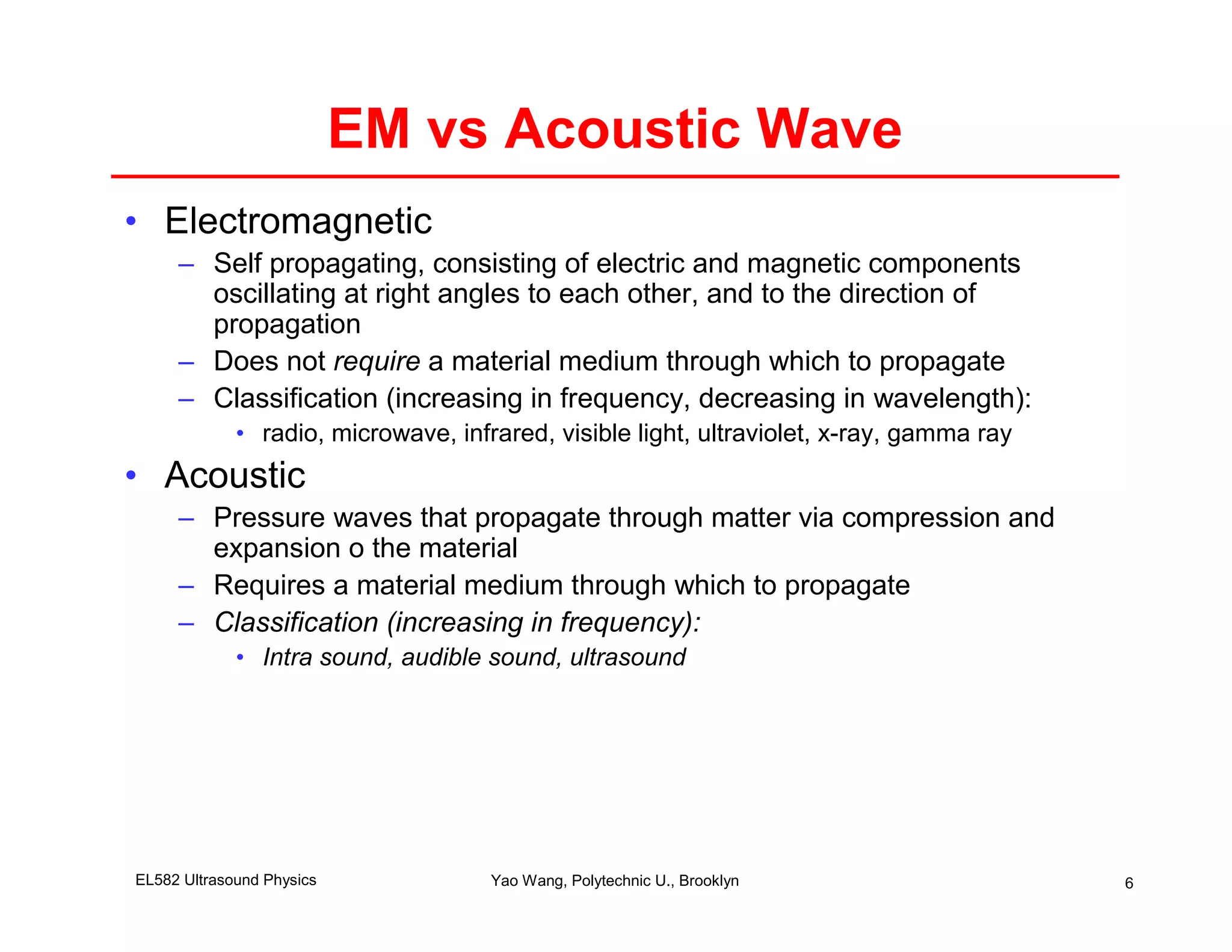
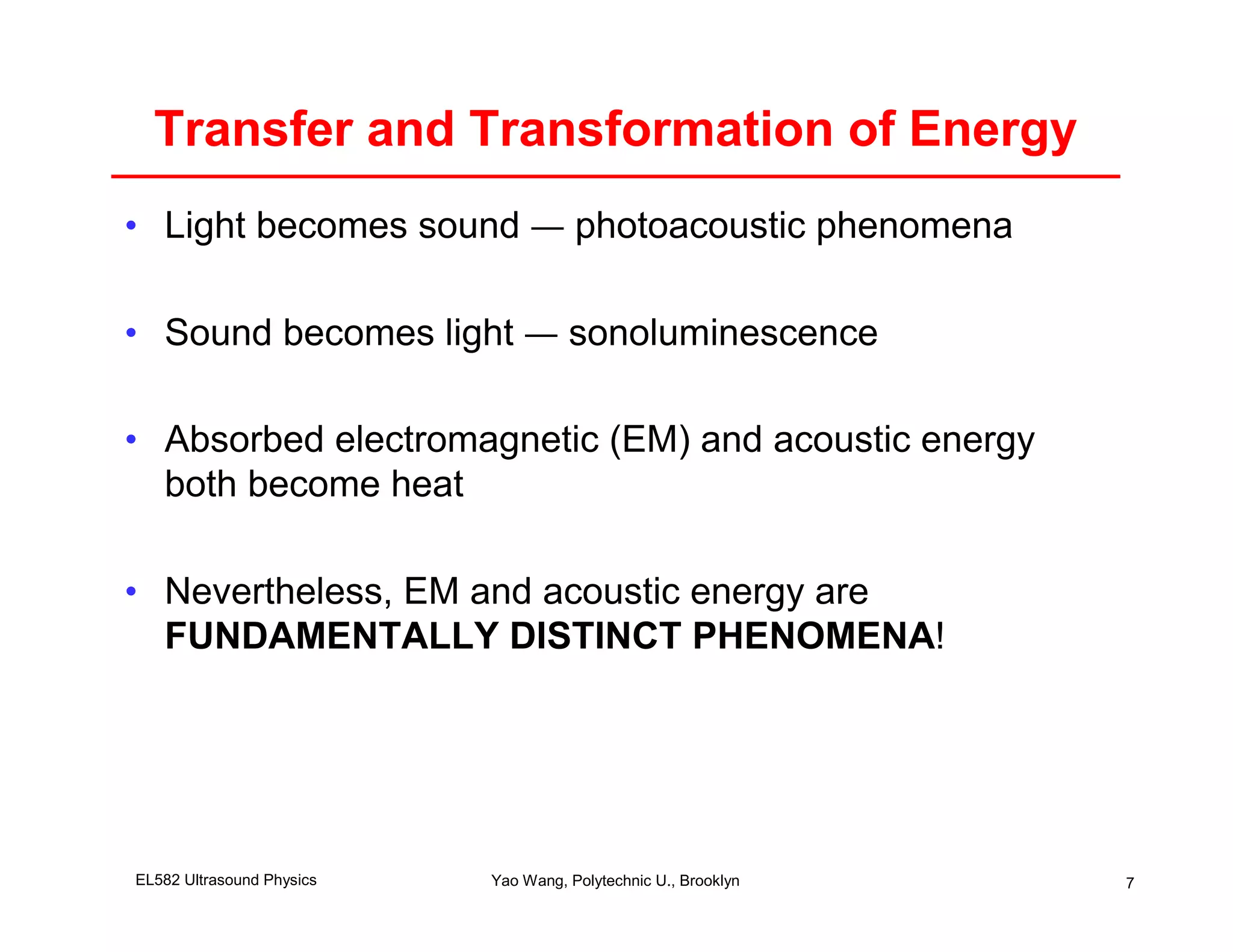
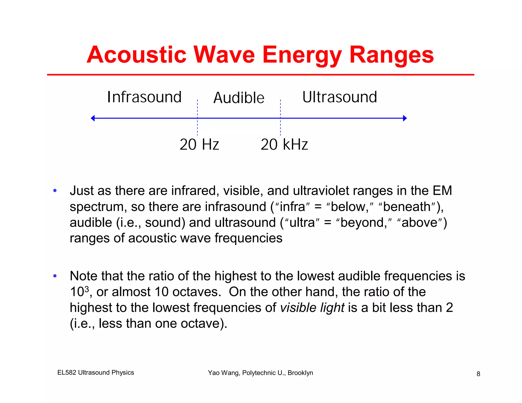
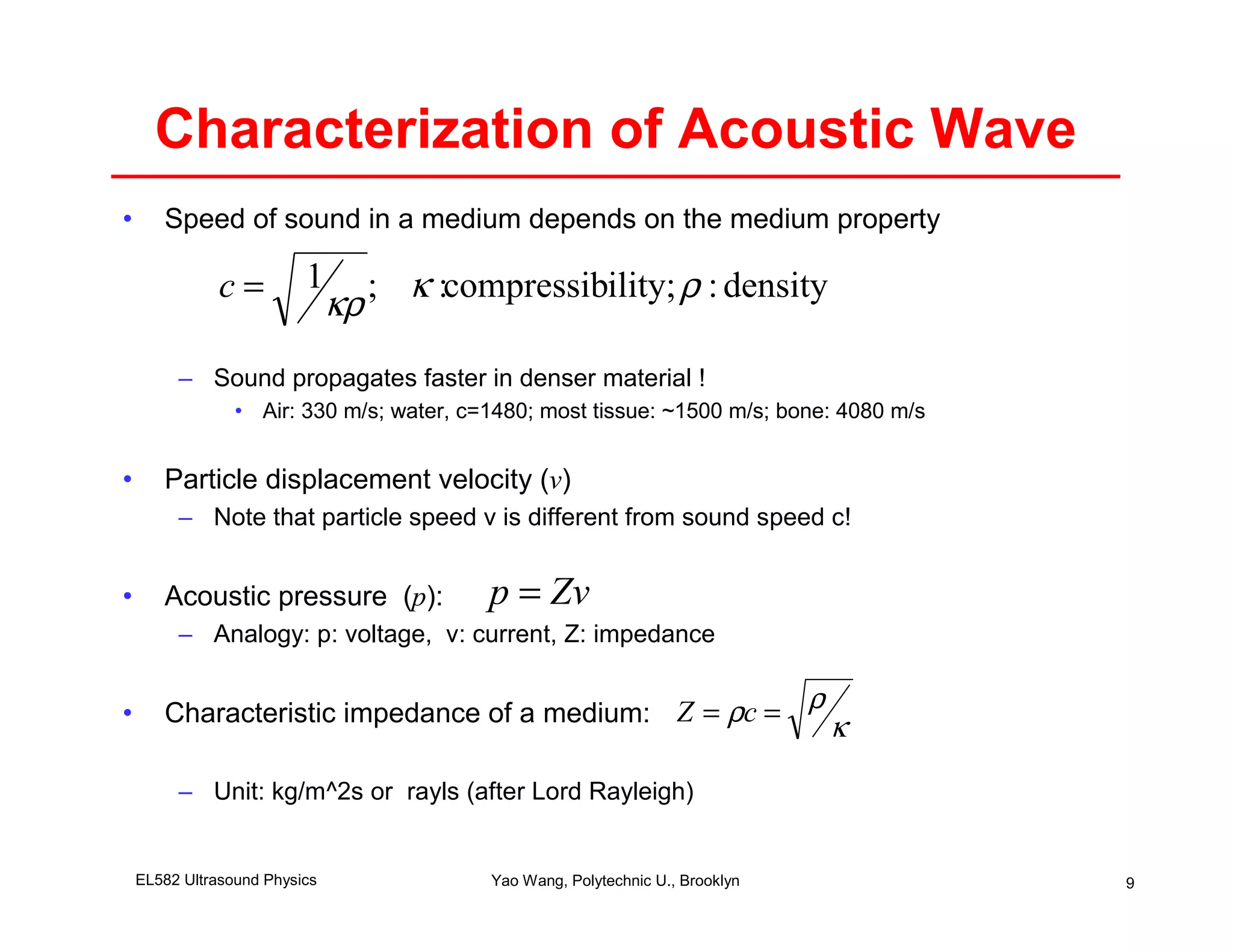
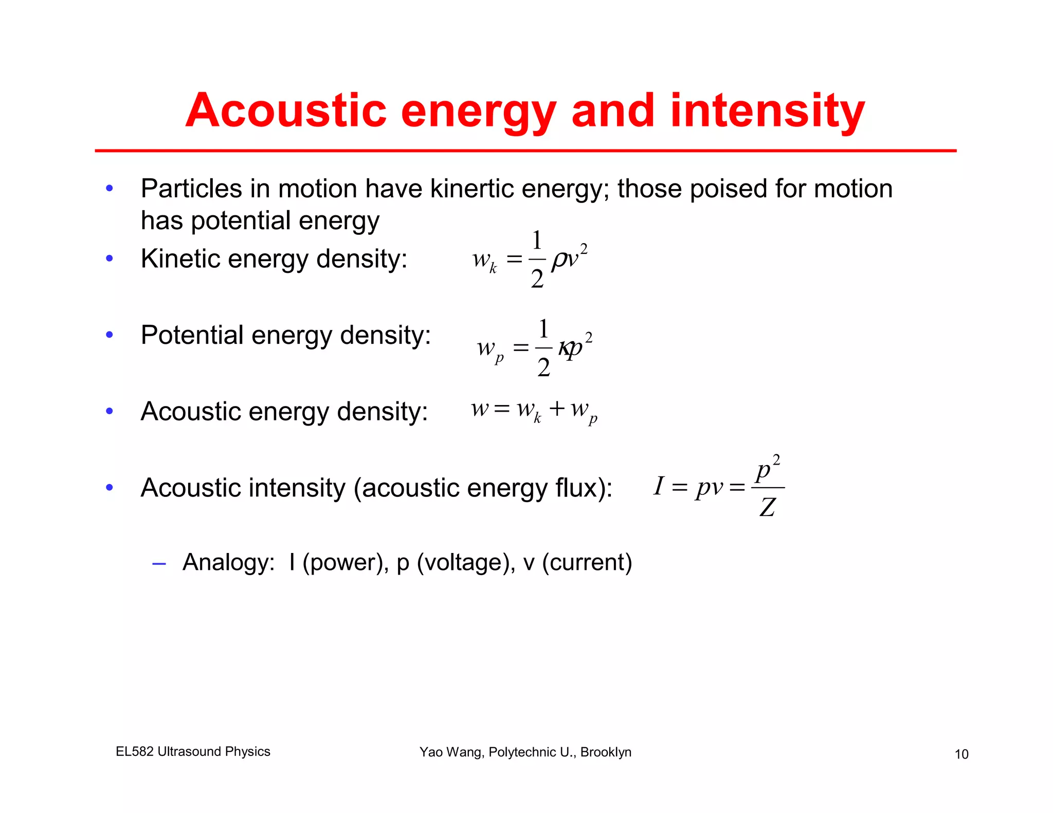
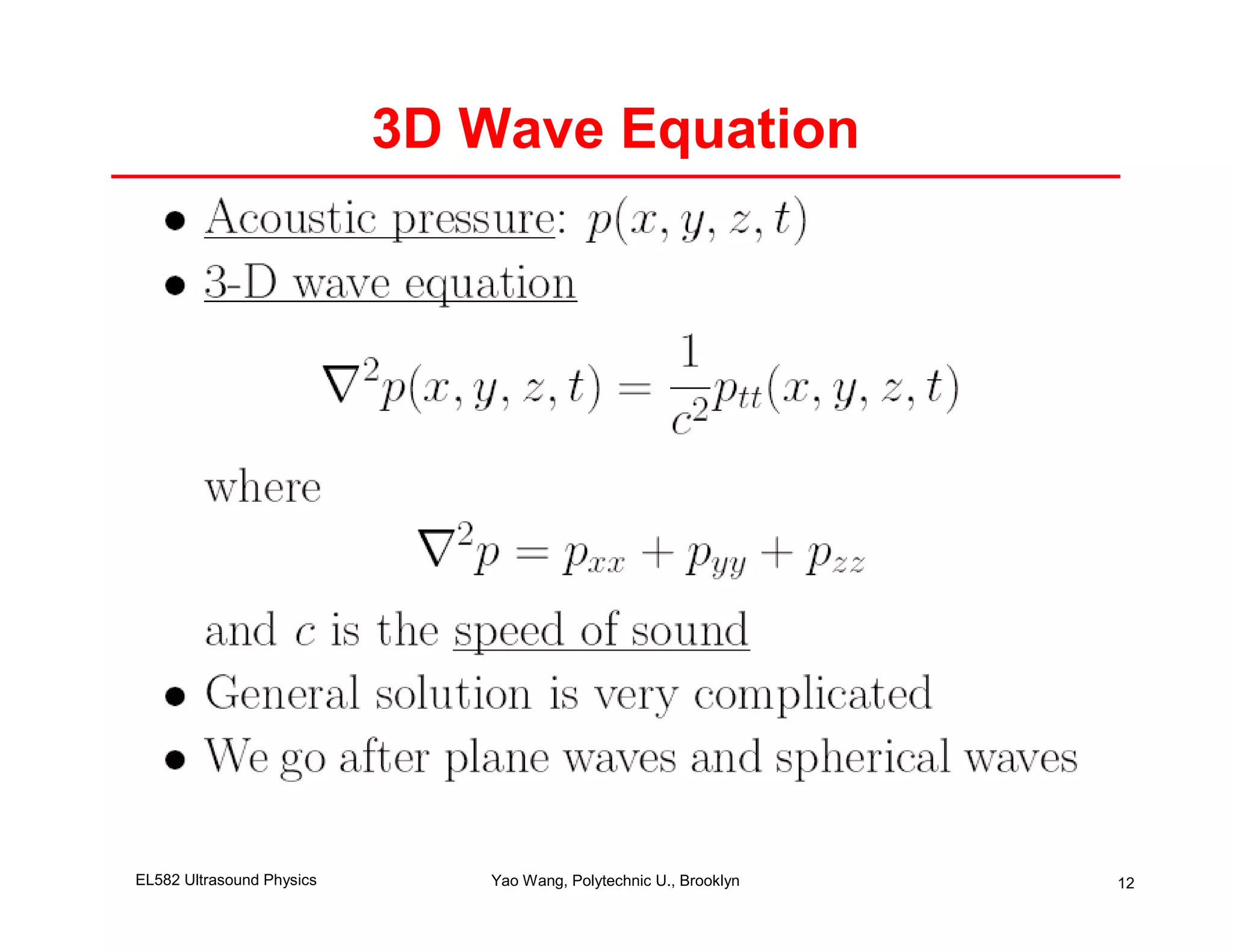
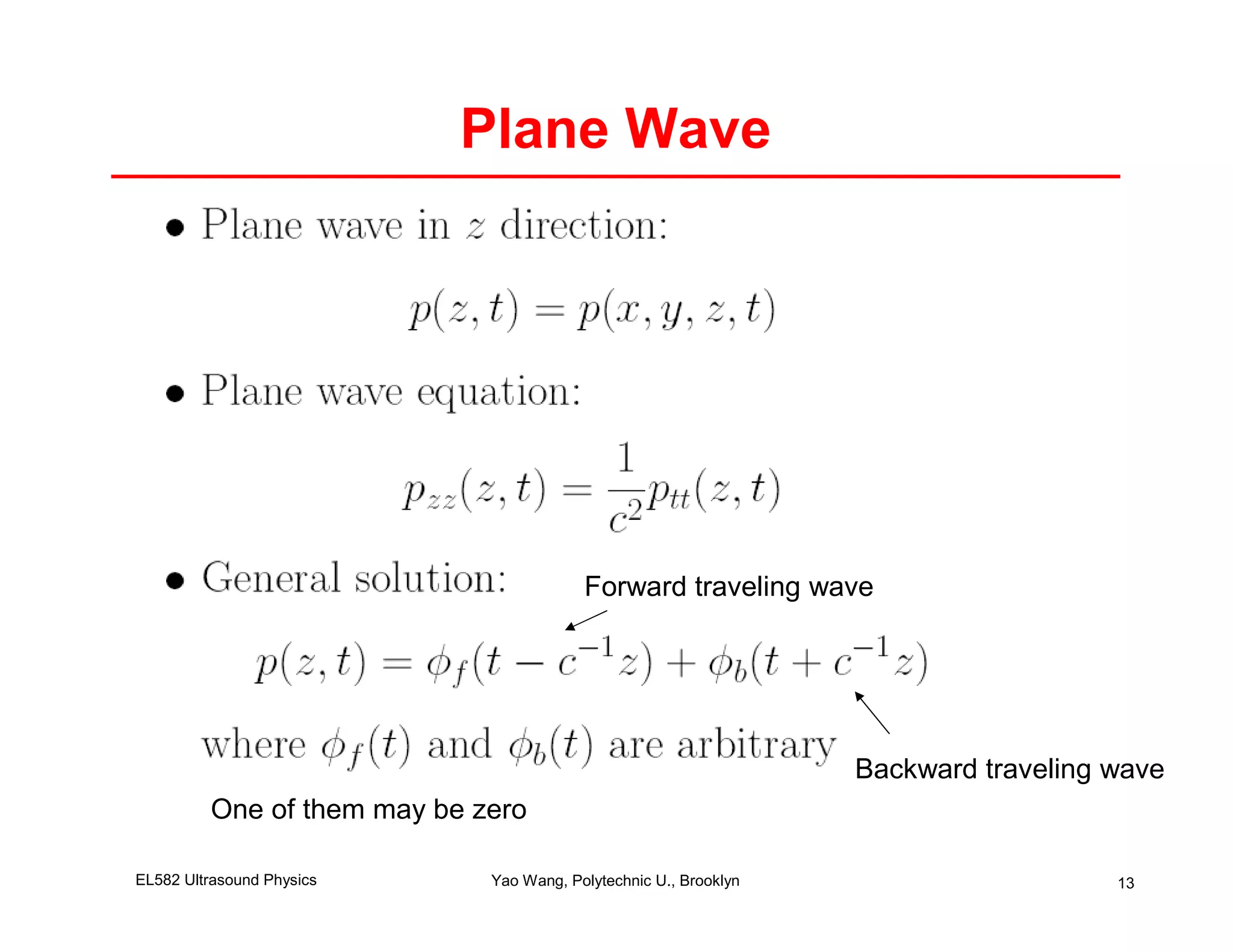
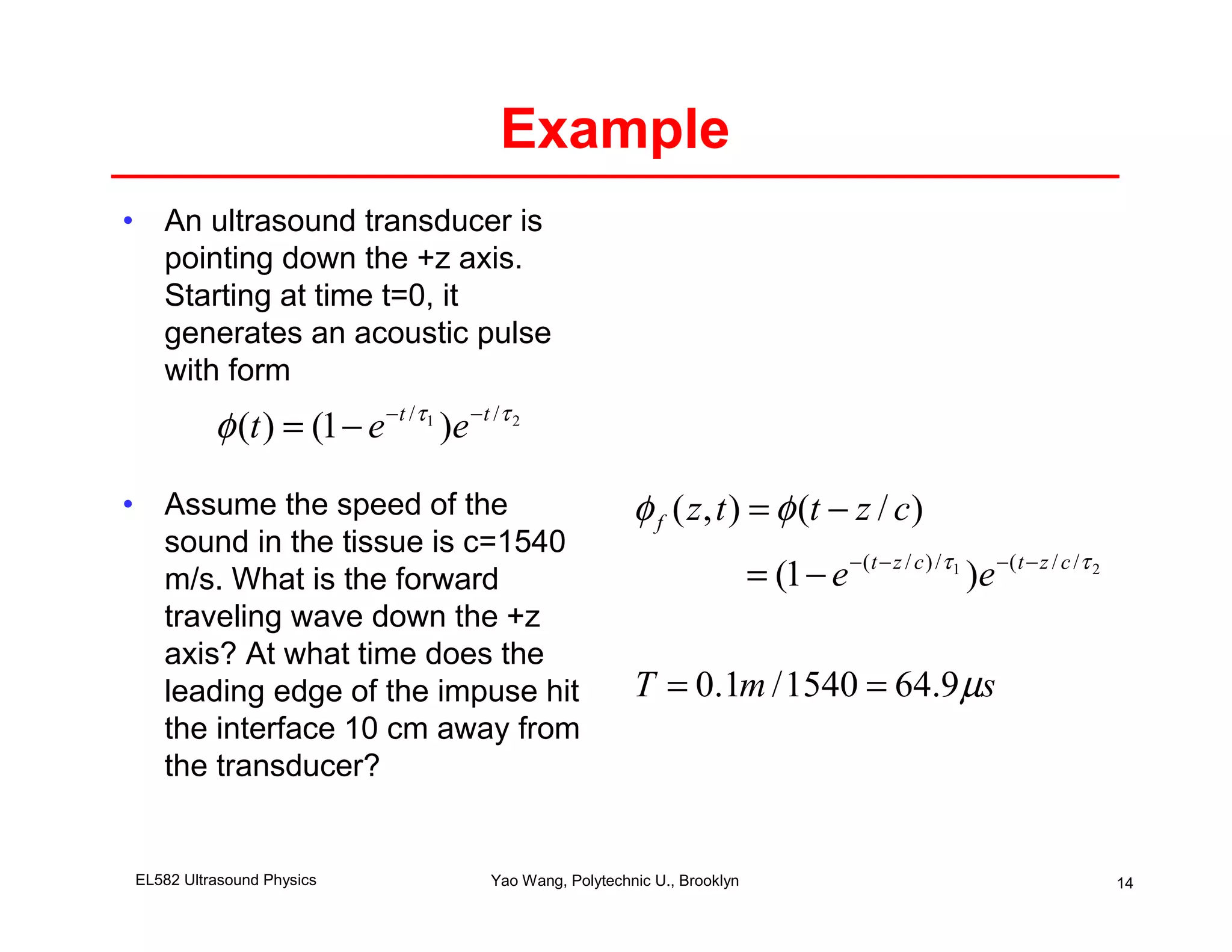
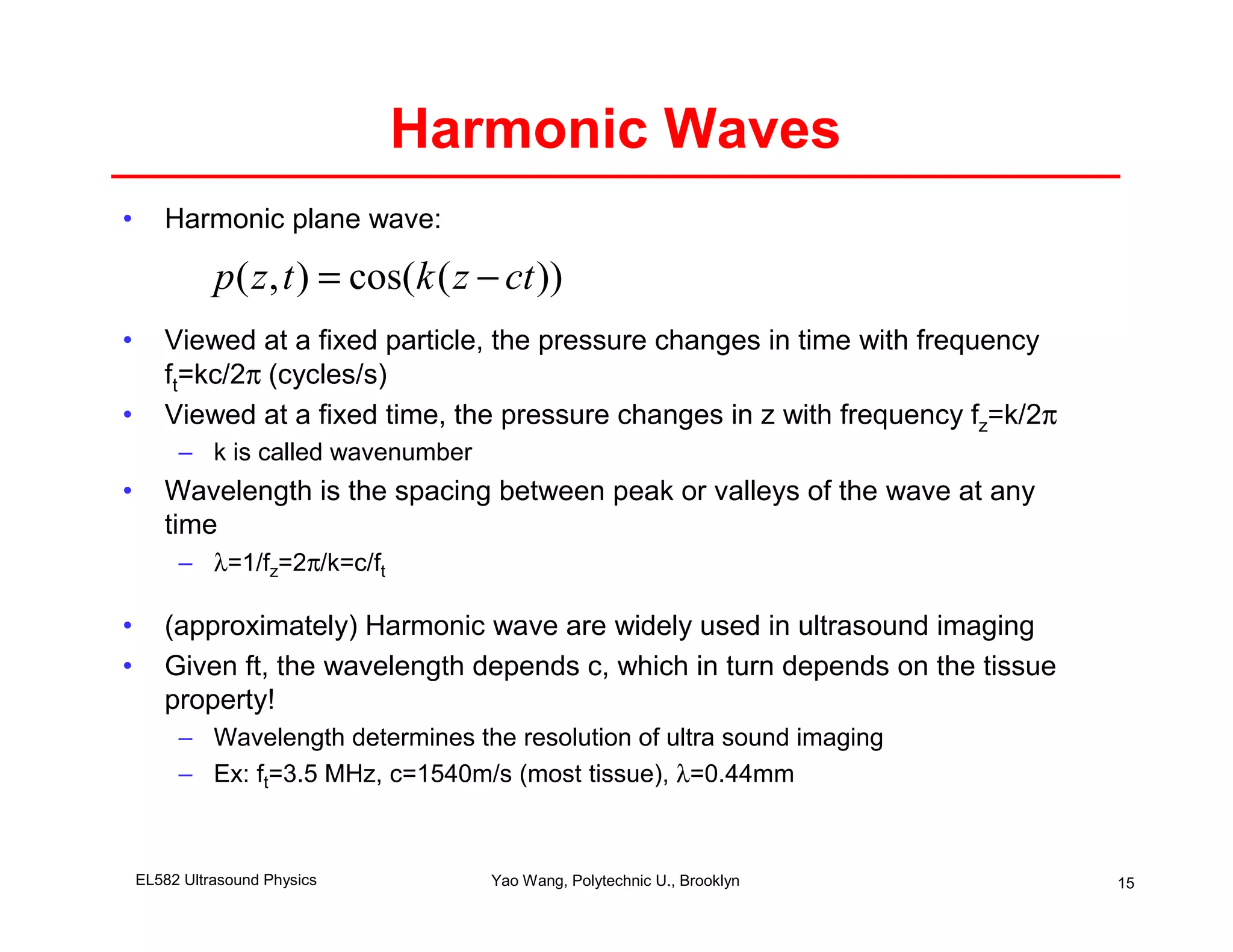
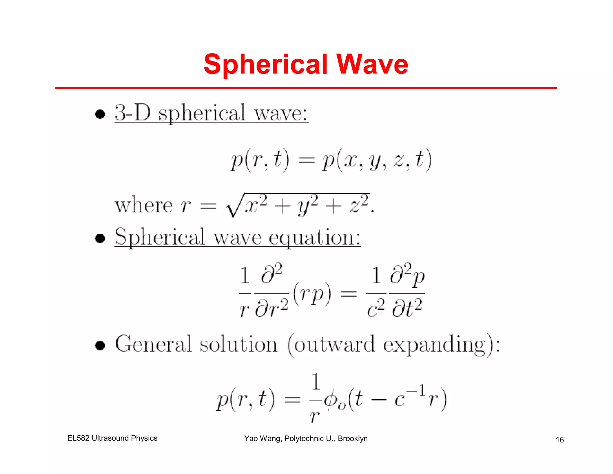
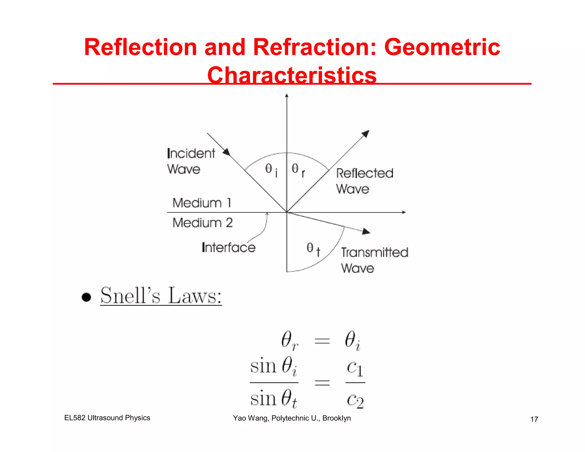
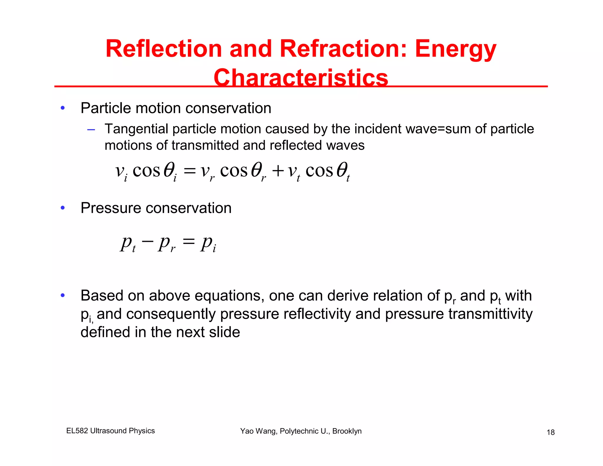
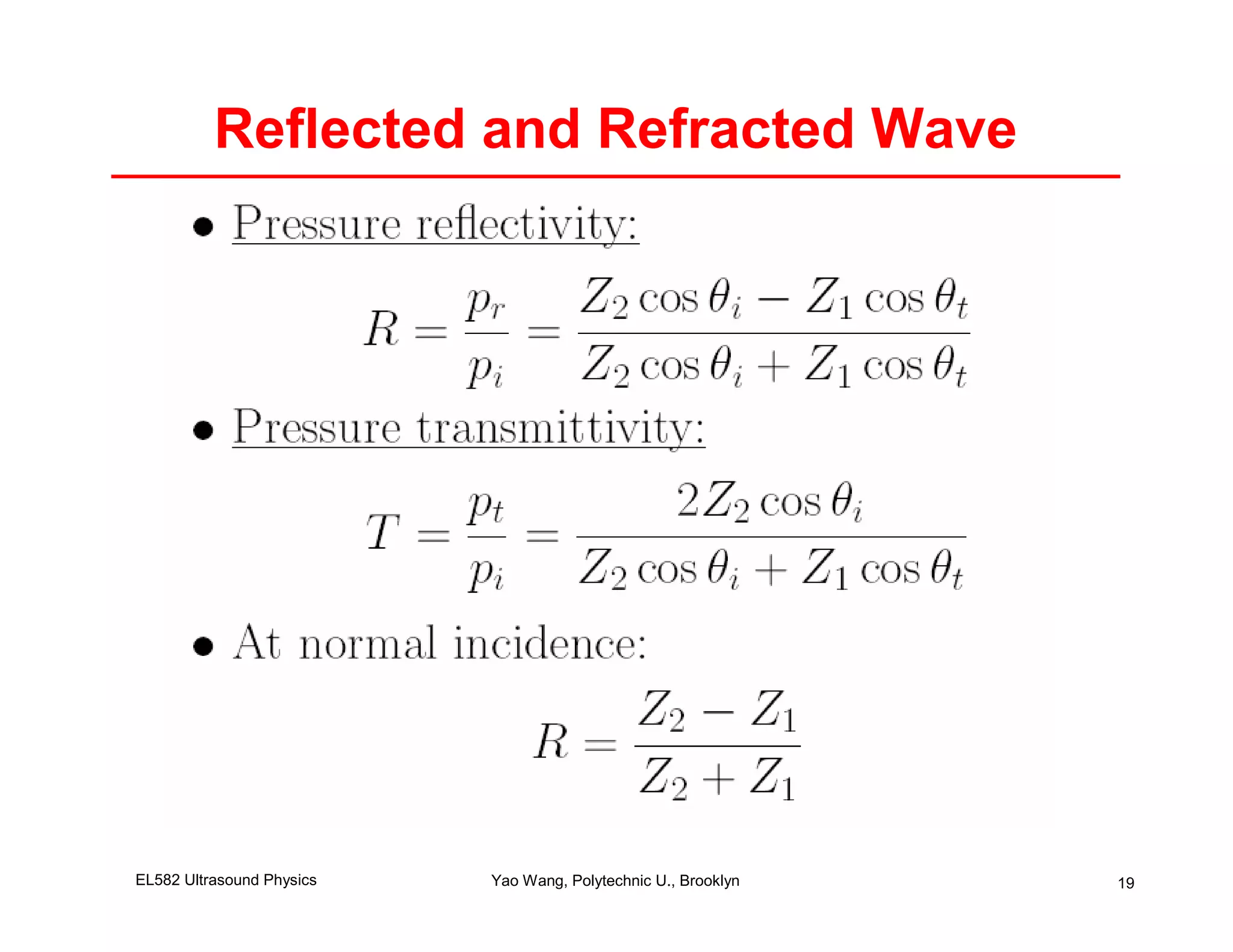
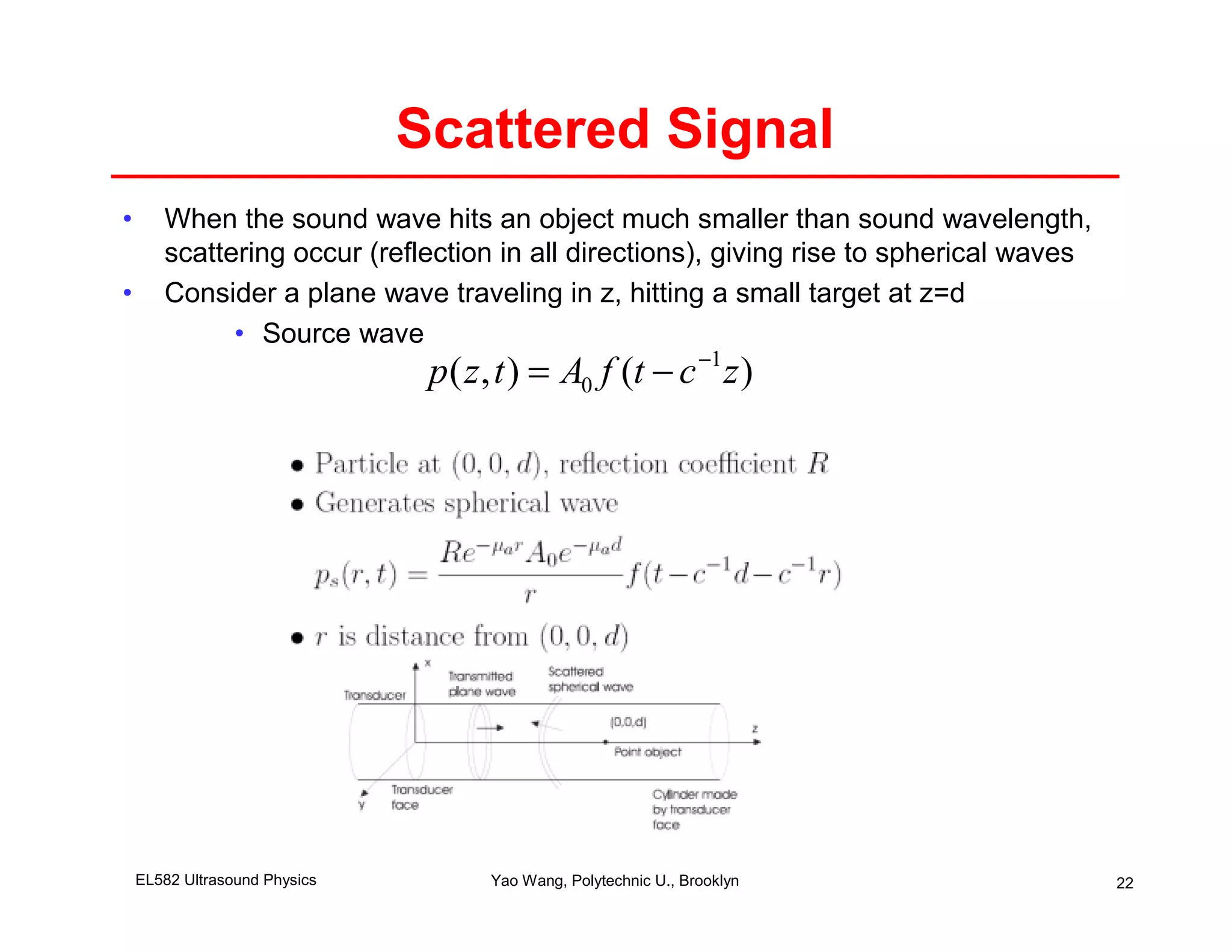
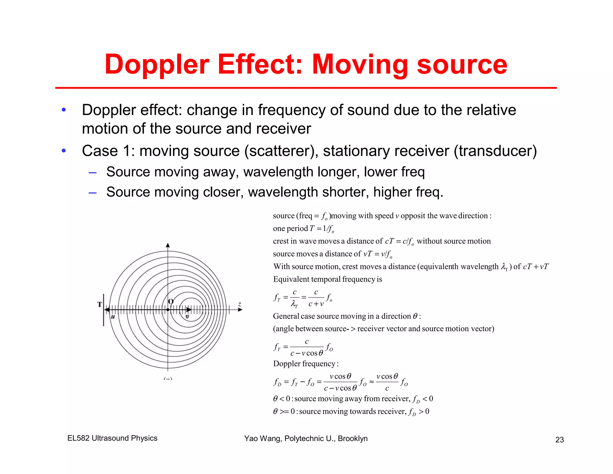
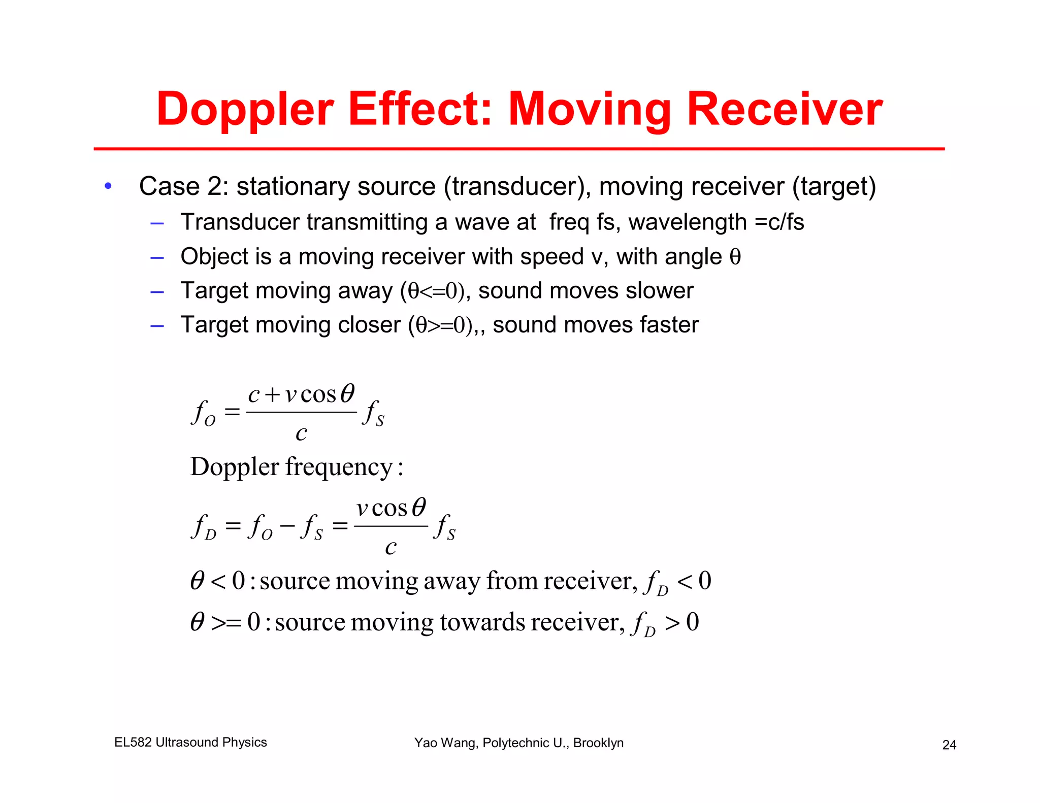
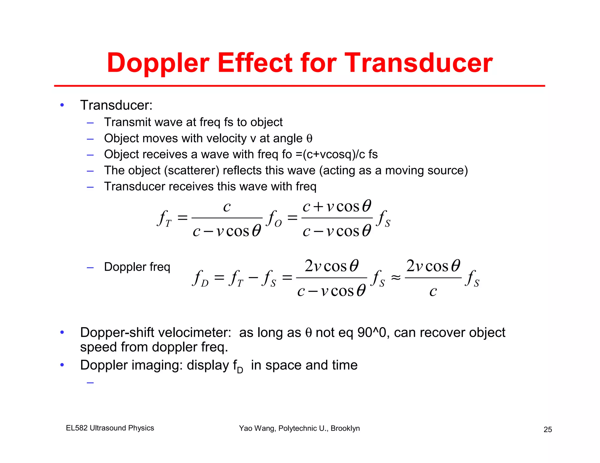
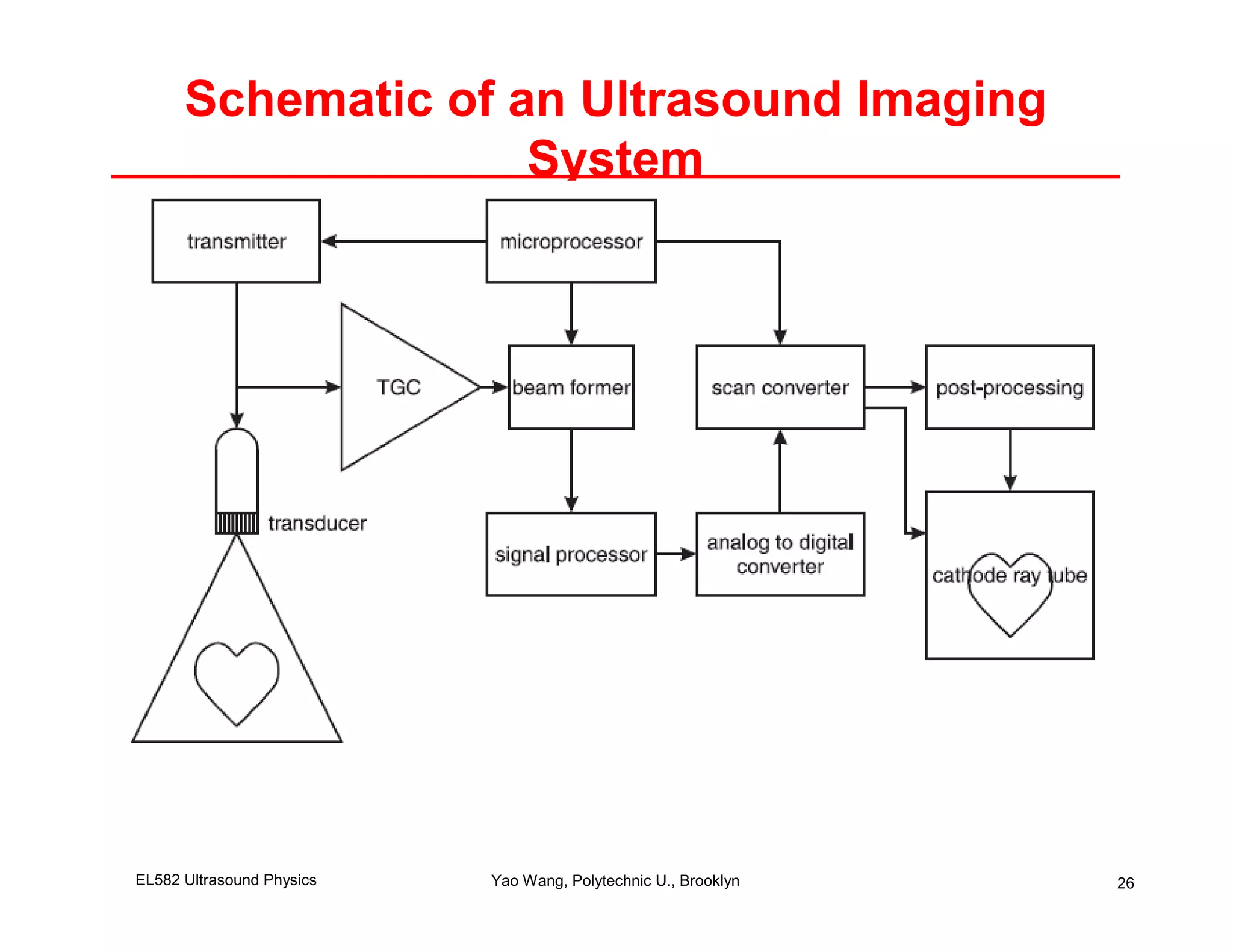
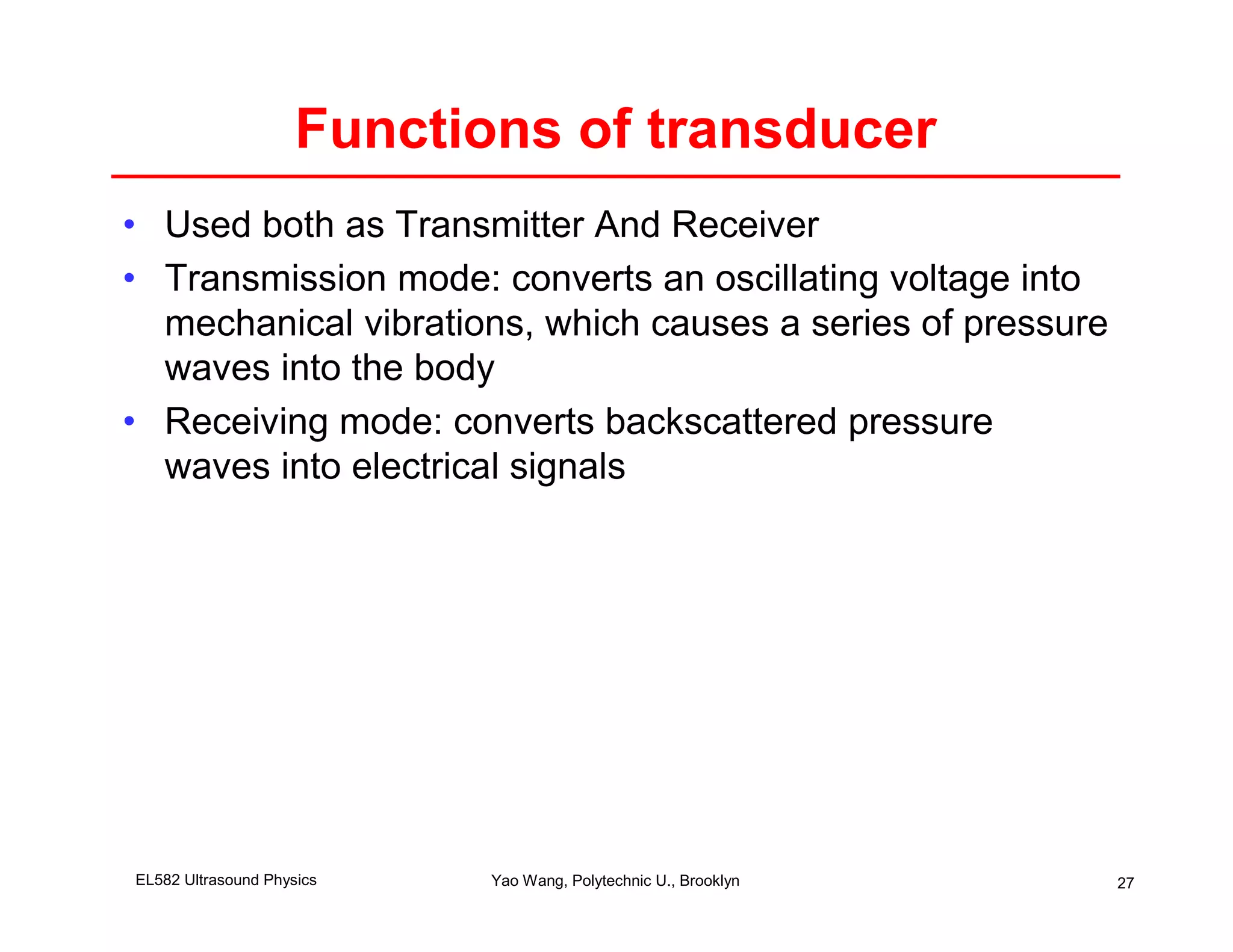
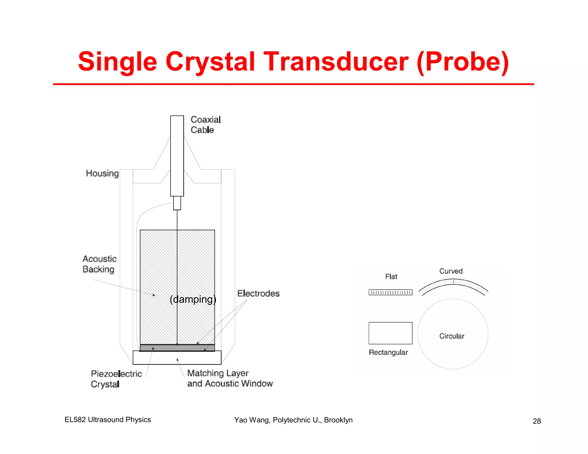
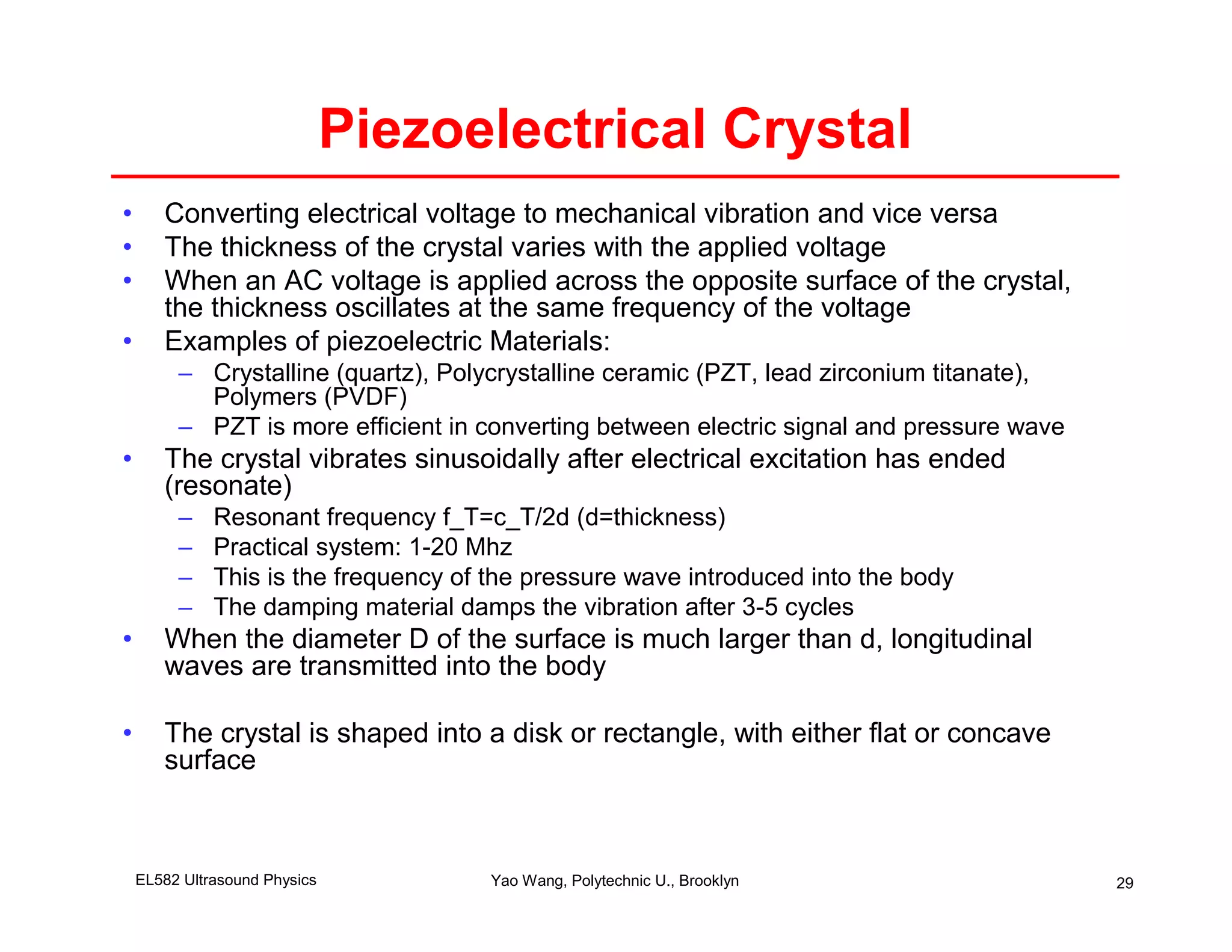
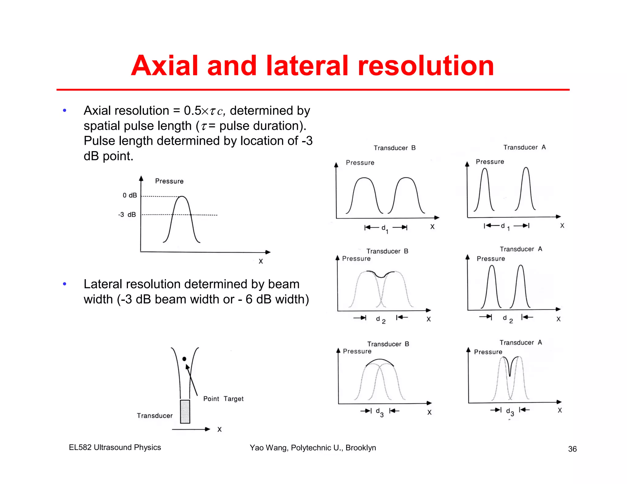
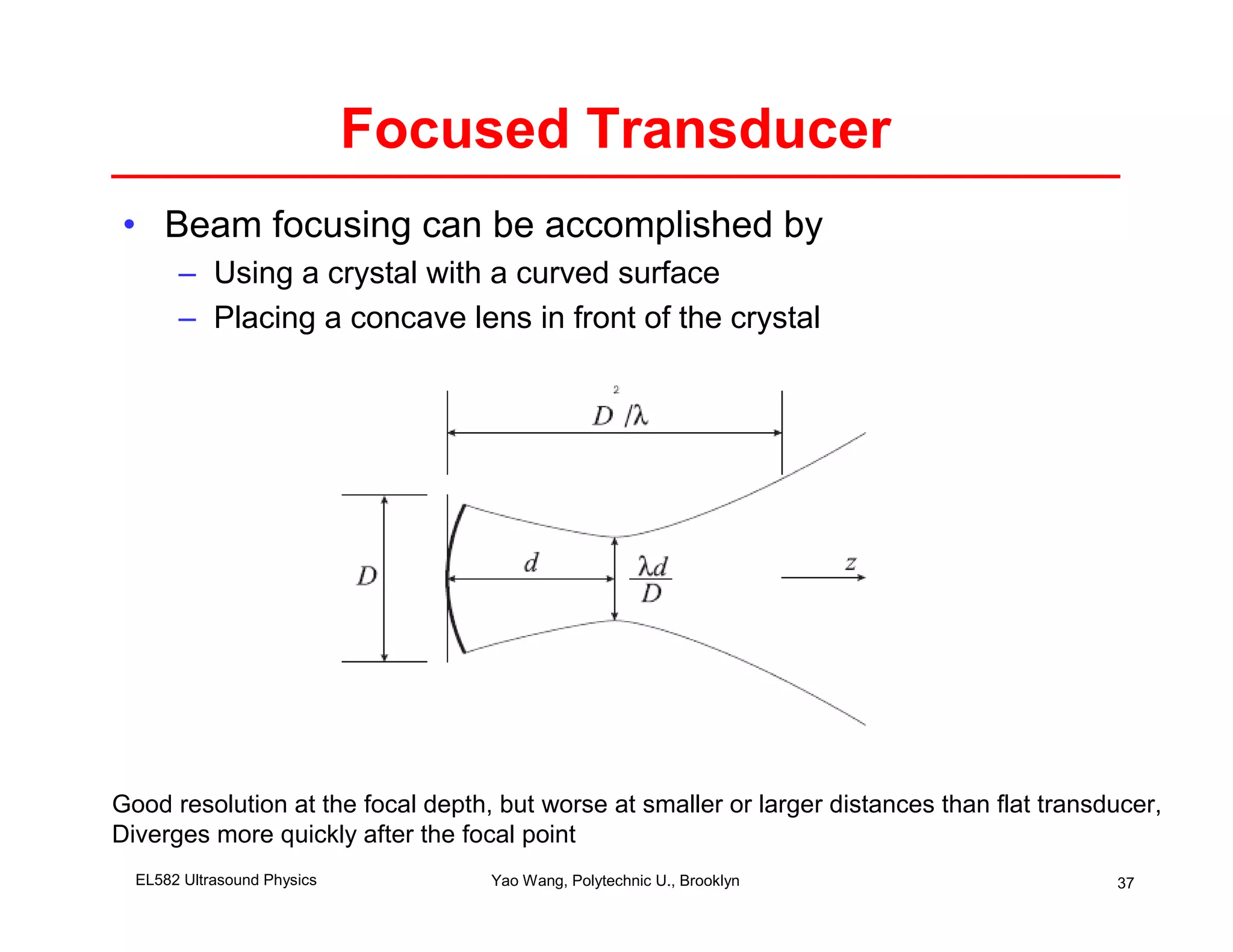
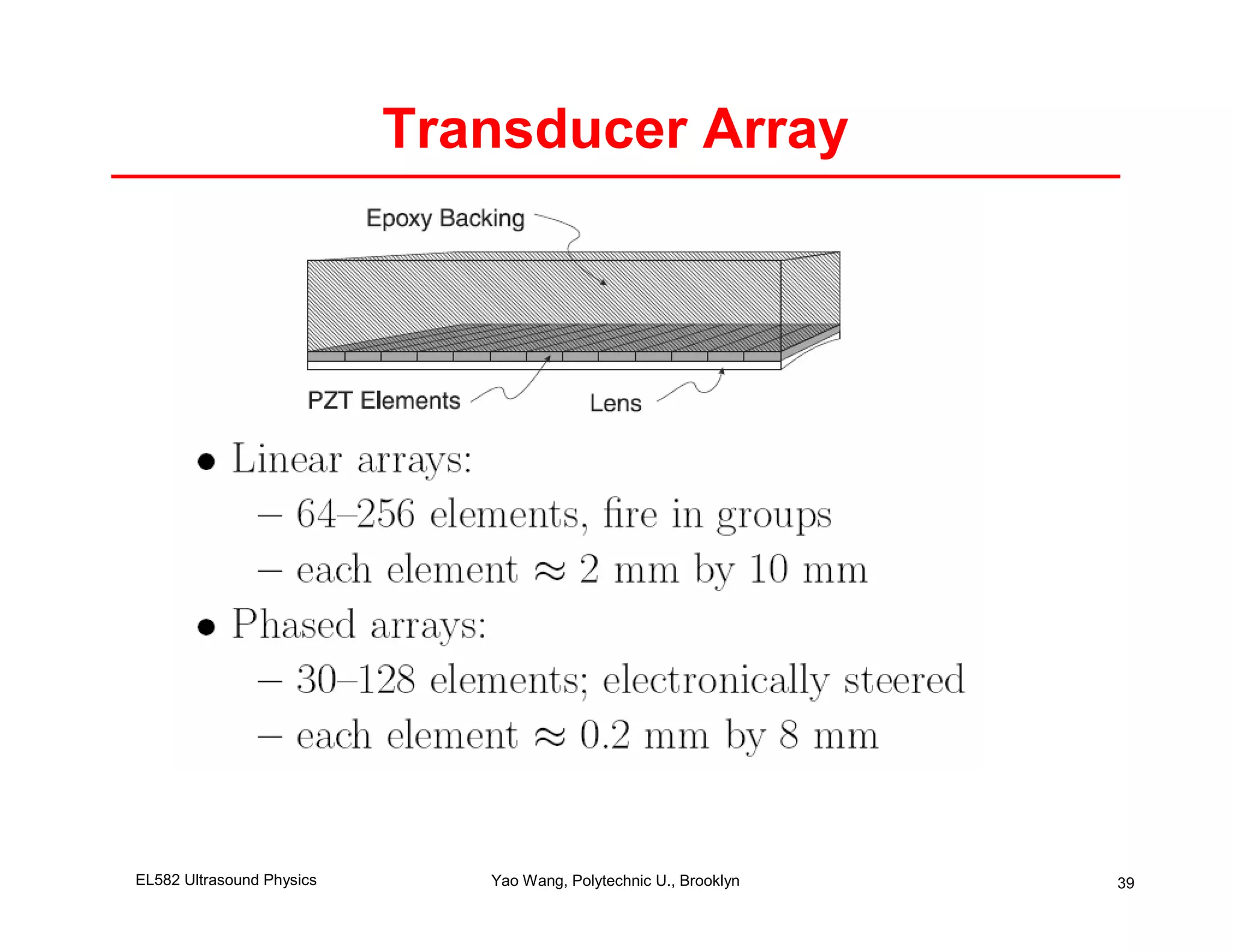
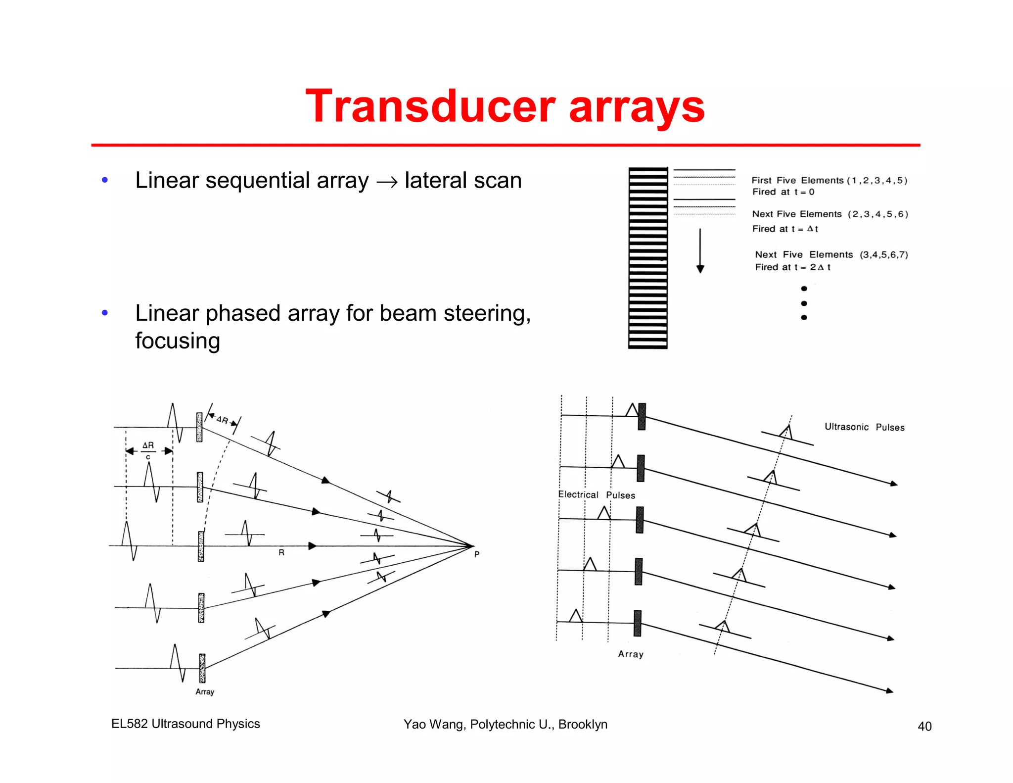
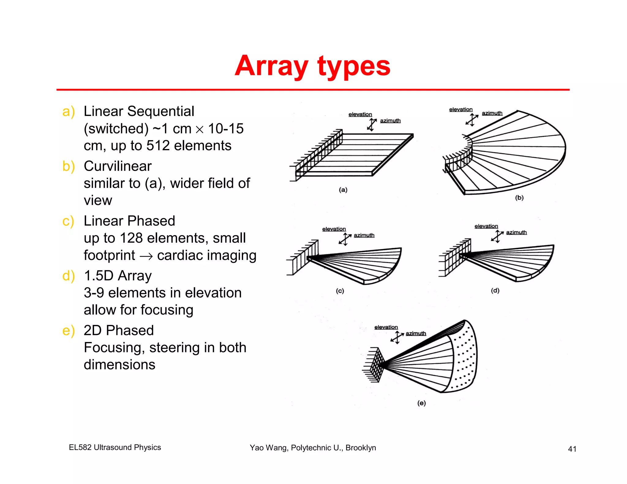
Physics of Ultrasound
        Imaging
                            Yao Wang
            Polytechnic University, Brooklyn, NY 11201

Based on J. L. Prince and J. M. Links, Medical Imaging Signals and
Systems, and lecture notes by Prince. Figures are from the textbook
                      except otherwise noted.
Lecture Outline
• Ultrasound imaging overview
• General characterization of acoustic wave
• Wave equation
     – 3D
     – Plane wave
     – Spherical wave
•   Reflection of wave
•   Absorption and scattering of wave
•   Doppler effect
•   Field pattern of a transducer


EL582 Ultrasound Physics     Yao Wang, Polytechnic U., Brooklyn   2
Ultrasound Imaging
•      Measure the reflectivity of tissue to sound waves
•      Can also measure velocity of moving objects, e.g. blood flow (Doppler
       imaging)
•      No radiation exposure, completely non-invasive and safe
•      Fast
•      Inexpensive
•      Low resolution
•      Medical applications: imaging fetus, heart, and many others


    EL582 Ultrasound Physics       Yao Wang, Polytechnic U., Brooklyn          3
What is Acoustic Wave
• Pressure waves that propagate through matter via
  compression and expansion o the material
     – Generated by compressing and releasing a small volume of
       tissue
• Longitudinal wave
     – Particles in the medium move back and force in the same
       direction that the wave is traveling
• Shear Wave
     – Particles move at right angles to the direction of the wave
     – Not used for medical ultrasound imaging


EL582 Ultrasound Physics    Yao Wang, Polytechnic U., Brooklyn       4
Longitudinal Wave


From Graber: Lecture note for BMI F05


                                                                    From Prince, Lecture note
EL582 Ultrasound Physics       Yao Wang, Polytechnic U., Brooklyn                          5
EM vs Acoustic Wave
• Electromagnetic
     – Self propagating, consisting of electric and magnetic components
       oscillating at right angles to each other, and to the direction of
       propagation
     – Does not require a material medium through which to propagate
     – Classification (increasing in frequency, decreasing in wavelength):
             • radio, microwave, infrared, visible light, ultraviolet, x-ray, gamma ray
• Acoustic
     – Pressure waves that propagate through matter via compression and
       expansion o the material
     – Requires a material medium through which to propagate
     – Classification (increasing in frequency):
             • Intra sound, audible sound, ultrasound


EL582 Ultrasound Physics             Yao Wang, Polytechnic U., Brooklyn                   6
Transfer and Transformation of Energy
• Light becomes sound — photoacoustic phenomena

• Sound becomes light — sonoluminescence

• Absorbed electromagnetic (EM) and acoustic energy
  both become heat

• Nevertheless, EM and acoustic energy are
  FUNDAMENTALLY DISTINCT PHENOMENA!


EL582 Ultrasound Physics   Yao Wang, Polytechnic U., Brooklyn   7
Acoustic Wave Energy Ranges
               Infrasound      Audible                      Ultrasound


                           20 Hz              20 kHz

• Just as there are infrared, visible, and ultraviolet ranges in the EM
  spectrum, so there are infrasound (“infra” = “below,” “beneath”),
  audible (i.e., sound) and ultrasound (“ultra” = “beyond,” “above”)
  ranges of acoustic wave frequencies

• Note that the ratio of the highest to the lowest audible frequencies is
  103, or almost 10 octaves. On the other hand, the ratio of the
  highest to the lowest frequencies of visible light is a bit less than 2
  (i.e., less than one octave).


EL582 Ultrasound Physics      Yao Wang, Polytechnic U., Brooklyn            8
Characterization of Acoustic Wave
•      Speed of sound in a medium depends on the medium property

               c=         1 ; κ :compressibility; ρ : density
                           κρ
         – Sound propagates faster in denser material !
                 • Air: 330 m/s; water, c=1480; most tissue: ~1500 m/s; bone: 4080 m/s


•      Particle displacement velocity (v)
         – Note that particle speed v is different from sound speed c!


•      Acoustic pressure (p):             p = Zv
         – Analogy: p: voltage, v: current, Z: impedance


•      Characteristic impedance of a medium: Z = ρc =
                                                      ρ
                                                                               κ

         – Unit: kg/m^2s or rayls (after Lord Rayleigh)


    EL582 Ultrasound Physics              Yao Wang, Polytechnic U., Brooklyn             9
Acoustic energy and intensity
• Particles in motion have kinertic energy; those poised for motion
  has potential energy
                                     1 2
• Kinetic energy density:      wk = ρv
                                     2

• Potential energy density:                1
                                      w p = κp 2
                                           2
• Acoustic energy density:            w = wk + w p

                                                                            p2
• Acoustic intensity (acoustic energy flux):                       I = pv =
                                                                            Z
     – Analogy: I (power), p (voltage), v (current)


EL582 Ultrasound Physics      Yao Wang, Polytechnic U., Brooklyn                 10
Acoustic Properties of Common Material

                                                                         Notice how similar
                                                                         these values are
       metal                                                             to each other and
         gas                                                             to that for water,
      acrylic
soft tissues                                                             and how different
                                                                         they are from
hard tissue                                                              these.


                       From [Graber: Lecture note for BMI F95]


                 Table 10.1 in [Prince] gives more information, including
                 density and absorption coefficient

EL582 Ultrasound Physics            Yao Wang, Polytechnic U., Brooklyn                        11
3D Wave Equation


EL582 Ultrasound Physics      Yao Wang, Polytechnic U., Brooklyn   12
Plane Wave


                                         Forward traveling wave


                                                                  Backward traveling wave
         One of them may be zero

EL582 Ultrasound Physics     Yao Wang, Polytechnic U., Brooklyn                      13
Example
• An ultrasound transducer is
  pointing down the +z axis.
  Starting at time t=0, it
  generates an acoustic pulse
  with form
          φ (t ) = (1 − e −t /τ )e −t /τ
                               1           2


• Assume the speed of the                                φ f ( z, t ) = φ (t − z / c)
  sound in the tissue is c=1540
  m/s. What is the forward                                                 = (1 − e −(t − z / c ) /τ 1 )e −( t − z / c /τ 2
  traveling wave down the +z
  axis? At what time does the
  leading edge of the impuse hit                         T = 0.1m / 1540 = 64.9 µs
  the interface 10 cm away from
  the transducer?


EL582 Ultrasound Physics              Yao Wang, Polytechnic U., Brooklyn                                                      14
Harmonic Waves
•      Harmonic plane wave:

              p ( z , t ) = cos(k ( z − ct ))
•      Viewed at a fixed particle, the pressure changes in time with frequency
       ft=kc/2π (cycles/s)
•      Viewed at a fixed time, the pressure changes in z with frequency fz=k/2π
         – k is called wavenumber
•      Wavelength is the spacing between peak or valleys of the wave at any
       time
         – λ=1/fz=2π/k=c/ft

•      (approximately) Harmonic wave are widely used in ultrasound imaging
•      Given ft, the wavelength depends c, which in turn depends on the tissue
       property!
         – Wavelength determines the resolution of ultra sound imaging
         – Ex: ft=3.5 MHz, c=1540m/s (most tissue), λ=0.44mm


    EL582 Ultrasound Physics          Yao Wang, Polytechnic U., Brooklyn          15
Spherical Wave


EL582 Ultrasound Physics     Yao Wang, Polytechnic U., Brooklyn   16
Reflection and Refraction: Geometric
                Characteristics


EL582 Ultrasound Physics   Yao Wang, Polytechnic U., Brooklyn   17
Reflection and Refraction: Energy
                   Characteristics
• Particle motion conservation
     – Tangential particle motion caused by the incident wave=sum of particle
       motions of transmitted and reflected waves
             vi cos θ i = vr cos θ r + vt cos θ t
• Pressure conservation

              pt − pr = pi

• Based on above equations, one can derive relation of pr and pt with
  pi, and consequently pressure reflectivity and pressure transmittivity
  defined in the next slide


EL582 Ultrasound Physics        Yao Wang, Polytechnic U., Brooklyn              18
Reflected and Refracted Wave


EL582 Ultrasound Physics   Yao Wang, Polytechnic U., Brooklyn   19
Energy Attenuation due to Absorption
              and Scattering
• The pressure of an acoustic wave decreases as the
  wave propagates due to
     – Absorption
             • The wave energy is converted to thermal energy
             • Two forms
                    – Classical: due to frictions between particles as the wave propagates
                    – Relaxation: due to particle motion to return (relax) to original position
                      after displacement by the wave pressure
     – Scattering
             • When the sound wave hits an object much larger than its
               wavelength, reflection occurs.
             • When the object size <= wavelength, scattering occur (reflection in
               all directions)


EL582 Ultrasound Physics               Yao Wang, Polytechnic U., Brooklyn                         20
Overall Attenuation
•      Absorption and scattering together causes the pressure and intensity of a
       sound wave to decrease exponentially in the propagation distance z
                  Suppose p(0, t ) = A0 f (t )
                  No attenuation : p ( z , t ) = A0 f (t − c −1 z )
                  With attenuation : p( z , t ) = A0 e − µ a z f (t − c −1 z )
                  µ a : Amplitude attenuation factor [cm -1 ]
                  Attenuation coefficient in dB :
                  α = 20(log10 e )µ a ≈ 8.7 µ a [dB/cm]


•      The attenuation coefficient depends on the frequency of the wave,
       generally α = af
                          b

•      Rough approximation (1 MHzα = af
                                     <=f<=10MHz): b=1,
           See Table 10.2 in textbook for “a” value for biological tissues
                   (much larger for bone and lung)

    EL582 Ultrasound Physics                 Yao Wang, Polytechnic U., Brooklyn    21
Scattered Signal
•      When the sound wave hits an object much smaller than sound wavelength,
       scattering occur (reflection in all directions), giving rise to spherical waves
•      Consider a plane wave traveling in z, hitting a small target at z=d
             • Source wave
                                p ( z , t ) = A0 f (t − c −1 z )


    EL582 Ultrasound Physics          Yao Wang, Polytechnic U., Brooklyn                 22
Doppler Effect: Moving source
• Doppler effect: change in frequency of sound due to the relative
  motion of the source and receiver
• Case 1: moving source (scatterer), stationary receiver (transducer)
     – Source moving away, wavelength longer, lower freq
     – Source moving closer, wavelength shorter, higher freq.
                                    source (freq = f o )moving with speed v opposit the wave direction :
                                    one period T = 1/f o
                                    crest in wave moves a distance of cT = c/f o without source motion
                                    source moves a distance of vT = v/f o
                                     With source motion, crest moves a distance (equivalenth wavelength λT ) of cT + vT
                                    Equivalent temporal frequency is
                                             c   c
                                     fT =      =     fo
                                            λT c + v
                                    General case :source moving in a direction θ :
                                    (angle between source- > receiver vector and source motion vector)
                                             c
                                     fT =           fO
                                        c − v cos θ
                                    Doppler frequency :
                                                      v cos θ        v cos θ
                                     f D = fT − f O =           fO ≈         fO
                                                    c − v cos θ         c
                                    θ < 0 : source moving away from receiver, f D < 0
                                    θ >= 0 : source moving towards receiver, f D > 0


EL582 Ultrasound Physics     Yao Wang, Polytechnic U., Brooklyn                                                           23
Doppler Effect: Moving Receiver
• Case 2: stationary source (transducer), moving receiver (target)
     –    Transducer transmitting a wave at freq fs, wavelength =c/fs
     –    Object is a moving receiver with speed v, with angle θ
     –    Target moving away (θ<=0), sound moves slower
     –    Target moving closer (θ>=0),, sound moves faster

                  c + v cos θ
            fO =               fS
                       c
           Doppler frequency :
                              v cos θ
            f D = fO − f S =          fS
                                  c
           θ < 0 : source moving away from receiver, f D < 0
           θ >= 0 : source moving towards receiver, f D > 0


EL582 Ultrasound Physics        Yao Wang, Polytechnic U., Brooklyn      24
Doppler Effect for Transducer
•      Transducer:
         –    Transmit wave at freq fs to object
         –    Object moves with velocity v at angle θ
         –    Object receives a wave with freq fo =(c+vcosq)/c fs
         –    The object (scatterer) reflects this wave (acting as a moving source)
         –    Transducer receives this wave with freq
                                         c           c + v cos θ
                               fT =             fO =             fS
                                    c − v cos θ      c − v cos θ
         – Doppler freq                               2v cos θ        2v cos θ
                                    f D = fT − f S =             fS ≈          fS
                                                     c − v cos θ          c

•      Dopper-shift velocimeter: as long as θ not eq 90^0, can recover object
       speed from doppler freq.
•      Doppler imaging: display fD in space and time
         –


    EL582 Ultrasound Physics               Yao Wang, Polytechnic U., Brooklyn         25
Schematic of an Ultrasound Imaging
                    System


EL582 Ultrasound Physics   Yao Wang, Polytechnic U., Brooklyn   26
Functions of transducer
• Used both as Transmitter And Receiver
• Transmission mode: converts an oscillating voltage into
  mechanical vibrations, which causes a series of pressure
  waves into the body
• Receiving mode: converts backscattered pressure
  waves into electrical signals


EL582 Ultrasound Physics    Yao Wang, Polytechnic U., Brooklyn   27
Single Crystal Transducer (Probe)


                           (damping)


EL582 Ultrasound Physics               Yao Wang, Polytechnic U., Brooklyn   28
Piezoelectrical Crystal
•      Converting electrical voltage to mechanical vibration and vice versa
•      The thickness of the crystal varies with the applied voltage
•      When an AC voltage is applied across the opposite surface of the crystal,
       the thickness oscillates at the same frequency of the voltage
•      Examples of piezoelectric Materials:
         – Crystalline (quartz), Polycrystalline ceramic (PZT, lead zirconium titanate),
           Polymers (PVDF)
         – PZT is more efficient in converting between electric signal and pressure wave
•      The crystal vibrates sinusoidally after electrical excitation has ended
       (resonate)
         –    Resonant frequency f_T=c_T/2d (d=thickness)
         –    Practical system: 1-20 Mhz
         –    This is the frequency of the pressure wave introduced into the body
         –    The damping material damps the vibration after 3-5 cycles
•      When the diameter D of the surface is much larger than d, longitudinal
       waves are transmitted into the body

•      The crystal is shaped into a disk or rectangle, with either flat or concave
       surface


    EL582 Ultrasound Physics           Yao Wang, Polytechnic U., Brooklyn                  29
Piezoelectrical Effect


EL582 Ultrasound Physics         Yao Wang, Polytechnic U., Brooklyn   30
Matching Layer
•      To provide acoustic coupling
       between the crystal and patient
       skin and to protect surface of the                                                                              ZL
       crystal
•      Z of PZT (ZT) is about 15 times of
       Z of skin (ZL)
         – Placing crystal directly over skin                                                             ZT
           would result a large amount of
           energy be reflected back from the
           boundary                                                                                Transducer           Load (tissue)
                 • R= (ZL-ZT)/(ZL+ZT) ~1
•      Matching layer                                                                                                               ZL
         – layer thickness = λ/4
                         Z l = ZT Z L
                                                                                                                            Zl

         – Maximize energy transfer into the
           body                                                                                                 ZT
         – Show as a homework
•      Problems: Finding material with


                                                                                                                                     Load (Target)
       exact Zl value
                                                                                                          Transducer


                                                                                                                         Matching
         – Dual-layer:


                                                                                                                          Layer
                  Z l ,1 = Z T3/ 4 Z 1/ 4 ;
                                     L
                                              Z l ,2 = Z T 4 Z L 4
                                                         1/    3/


    EL582 Ultrasound Physics                                         Yao Wang, Polytechnic U., Brooklyn                                              31
Continuous Wave vs Pulsed
• Continuous wave (CW) Ultrasound
     – The voltage signal applied to the transducer is a continuous
       sinusoid signal
• Pulsed Ultrasound
     – A pulsed signal is introduced (more common), allowing to
       separate reflected signal from targets at different distances
       based on time delay


EL582 Ultrasound Physics    Yao Wang, Polytechnic U., Brooklyn         32
Typical Transmit Pulse


EL582 Ultrasound Physics     Yao Wang, Polytechnic U., Brooklyn   33
Flat (Piston) Plate Transducer
                                          Near field boundary (NFB)
                                   Fresnel region Fraunhofer region
Vibrating flat plate
                                                  (far field, beam spreading)


                              D2
                              4λ
 Geometric
 approximation:
 Wave is confined in
 a cylinder


   EL582 Ultrasound Physics        Yao Wang, Polytechnic U., Brooklyn           34
• At border of the beam width, the signal strength drops by
  a factor of 2, compared to the strength on the z-axis
• Beam width determines the imaging resolution (lateral
  resolution).
• Smaller D is good only before far field
• D=1~5 cm in practice, very poor lateral resolution
• Focused plate is used to produce narrow beam


                           At border of the beam width, the signal strength drops by a factor of 2
                           Beam width determines the imaging resolution (lateral resolution).
                           Smaller D is good only before far field
                           D=1~5 cm in practice, very poor lateral resolution
                           Focused plate is used to produce narrow beam


EL582 Ultrasound Physics               Yao Wang, Polytechnic U., Brooklyn                        35
Axial and lateral resolution
•      Axial resolution = 0.5×τ c, determined by
       spatial pulse length (τ = pulse duration).
       Pulse length determined by location of -3
       dB point.


•      Lateral resolution determined by beam
       width (-3 dB beam width or - 6 dB width)


    EL582 Ultrasound Physics         Yao Wang, Polytechnic U., Brooklyn   36
Focused Transducer
• Beam focusing can be accomplished by
       – Using a crystal with a curved surface
       – Placing a concave lens in front of the crystal


Good resolution at the focal depth, but worse at smaller or larger distances than flat transducer,
Diverges more quickly after the focal point
  EL582 Ultrasound Physics         Yao Wang, Polytechnic U., Brooklyn                        37
Transducer Array
• With a single crystal, manual or mechanical steering of
  the beam is needed to produce a two-dimensional image
• Practical systems today use an array of small
  piezoelectric crystals
     – Allow electronic steering of the beam to optimize the lateral
       resolution


EL582 Ultrasound Physics      Yao Wang, Polytechnic U., Brooklyn       38
Transducer Array


EL582 Ultrasound Physics      Yao Wang, Polytechnic U., Brooklyn   39
Transducer arrays
•      Linear sequential array → lateral scan


•      Linear phased array for beam steering,
       focusing


    EL582 Ultrasound Physics       Yao Wang, Polytechnic U., Brooklyn   40
Array types
a) Linear Sequential
   (switched) ~1 cm × 10-15
   cm, up to 512 elements
b) Curvilinear
   similar to (a), wider field of
   view
c) Linear Phased
   up to 128 elements, small
   footprint → cardiac imaging
d) 1.5D Array
   3-9 elements in elevation
   allow for focusing
e) 2D Phased
   Focusing, steering in both
   dimensions


 EL582 Ultrasound Physics      Yao Wang, Polytechnic U., Brooklyn   41
Summary
• What is acoustic wave?
• Characterization of wave:
         – Material properties: k, ρ, c, Z
         – Wave properties: particle velocity v, pressure p, intensity I
•      Plane wave vs. Spherical wave
•      Reflection and refraction of wave at an interface
•      Scattering of wave
•      Transducer for ultrasound imaging
         –    Piezoelectric crystal
         –    Resonance frequency (determined by thickness of crystal)
         –    Flat surface vs. focused
         –    Beam divergence in both types of transducer
         –    Beam width determines lateral resolution
         –    Pulse period determines axial resolution
         –    Array transducer


    EL582 Ultrasound Physics       Yao Wang, Polytechnic U., Brooklyn      42
Reference
• Prince and Links, Medical Imaging Signals and Systems,
  Chap. 10 (Sec. 10.5 not required),11.2,11.3
• A. Webb, Introduction to Biomedical Imaging, Chap. 3


EL582 Ultrasound Physics   Yao Wang, Polytechnic U., Brooklyn   43
Homework
• Reading:
     – Prince and Links, Medical Imaging Signals and Systems, Chap. 10
       (Sec. 10.5 not required),11.2,11.3
• Note down all the corrections for Ch. 10,11 on your copy of the
  textbook based on the provided errata (see Course website or
  book website for update).
• Problems (Due 11/20):
     –    P10.1
     –    P10.3
     –    P10.6
     –    P10.8
     –    P10.10
     –    P10.12
     –    P10.13
     –    Considering the matching layer in a transducer. Show that, when a
          matching layer of depth λ/4 is used, the transmitted energy into the
          tissue is maximized with an impedance of Z = Z Z           l   T   L


EL582 Ultrasound Physics        Yao Wang, Polytechnic U., Brooklyn               44

Physics of Ultrasound Imaging

  • 1.
    Physics of Ultrasound Imaging Yao Wang Polytechnic University, Brooklyn, NY 11201 Based on J. L. Prince and J. M. Links, Medical Imaging Signals and Systems, and lecture notes by Prince. Figures are from the textbook except otherwise noted.
  • 2.
    Lecture Outline • Ultrasoundimaging overview • General characterization of acoustic wave • Wave equation – 3D – Plane wave – Spherical wave • Reflection of wave • Absorption and scattering of wave • Doppler effect • Field pattern of a transducer EL582 Ultrasound Physics Yao Wang, Polytechnic U., Brooklyn 2
  • 3.
    Ultrasound Imaging • Measure the reflectivity of tissue to sound waves • Can also measure velocity of moving objects, e.g. blood flow (Doppler imaging) • No radiation exposure, completely non-invasive and safe • Fast • Inexpensive • Low resolution • Medical applications: imaging fetus, heart, and many others EL582 Ultrasound Physics Yao Wang, Polytechnic U., Brooklyn 3
  • 4.
    What is AcousticWave • Pressure waves that propagate through matter via compression and expansion o the material – Generated by compressing and releasing a small volume of tissue • Longitudinal wave – Particles in the medium move back and force in the same direction that the wave is traveling • Shear Wave – Particles move at right angles to the direction of the wave – Not used for medical ultrasound imaging EL582 Ultrasound Physics Yao Wang, Polytechnic U., Brooklyn 4
  • 5.
    Longitudinal Wave From Graber:Lecture note for BMI F05 From Prince, Lecture note EL582 Ultrasound Physics Yao Wang, Polytechnic U., Brooklyn 5
  • 6.
    EM vs AcousticWave • Electromagnetic – Self propagating, consisting of electric and magnetic components oscillating at right angles to each other, and to the direction of propagation – Does not require a material medium through which to propagate – Classification (increasing in frequency, decreasing in wavelength): • radio, microwave, infrared, visible light, ultraviolet, x-ray, gamma ray • Acoustic – Pressure waves that propagate through matter via compression and expansion o the material – Requires a material medium through which to propagate – Classification (increasing in frequency): • Intra sound, audible sound, ultrasound EL582 Ultrasound Physics Yao Wang, Polytechnic U., Brooklyn 6
  • 7.
    Transfer and Transformationof Energy • Light becomes sound — photoacoustic phenomena • Sound becomes light — sonoluminescence • Absorbed electromagnetic (EM) and acoustic energy both become heat • Nevertheless, EM and acoustic energy are FUNDAMENTALLY DISTINCT PHENOMENA! EL582 Ultrasound Physics Yao Wang, Polytechnic U., Brooklyn 7
  • 8.
    Acoustic Wave EnergyRanges Infrasound Audible Ultrasound 20 Hz 20 kHz • Just as there are infrared, visible, and ultraviolet ranges in the EM spectrum, so there are infrasound (“infra” = “below,” “beneath”), audible (i.e., sound) and ultrasound (“ultra” = “beyond,” “above”) ranges of acoustic wave frequencies • Note that the ratio of the highest to the lowest audible frequencies is 103, or almost 10 octaves. On the other hand, the ratio of the highest to the lowest frequencies of visible light is a bit less than 2 (i.e., less than one octave). EL582 Ultrasound Physics Yao Wang, Polytechnic U., Brooklyn 8
  • 9.
    Characterization of AcousticWave • Speed of sound in a medium depends on the medium property c= 1 ; κ :compressibility; ρ : density κρ – Sound propagates faster in denser material ! • Air: 330 m/s; water, c=1480; most tissue: ~1500 m/s; bone: 4080 m/s • Particle displacement velocity (v) – Note that particle speed v is different from sound speed c! • Acoustic pressure (p): p = Zv – Analogy: p: voltage, v: current, Z: impedance • Characteristic impedance of a medium: Z = ρc = ρ κ – Unit: kg/m^2s or rayls (after Lord Rayleigh) EL582 Ultrasound Physics Yao Wang, Polytechnic U., Brooklyn 9
  • 10.
    Acoustic energy andintensity • Particles in motion have kinertic energy; those poised for motion has potential energy 1 2 • Kinetic energy density: wk = ρv 2 • Potential energy density: 1 w p = κp 2 2 • Acoustic energy density: w = wk + w p p2 • Acoustic intensity (acoustic energy flux): I = pv = Z – Analogy: I (power), p (voltage), v (current) EL582 Ultrasound Physics Yao Wang, Polytechnic U., Brooklyn 10
  • 11.
    Acoustic Properties ofCommon Material Notice how similar these values are metal to each other and gas to that for water, acrylic soft tissues and how different they are from hard tissue these. From [Graber: Lecture note for BMI F95] Table 10.1 in [Prince] gives more information, including density and absorption coefficient EL582 Ultrasound Physics Yao Wang, Polytechnic U., Brooklyn 11
  • 12.
    3D Wave Equation EL582Ultrasound Physics Yao Wang, Polytechnic U., Brooklyn 12
  • 13.
    Plane Wave Forward traveling wave Backward traveling wave One of them may be zero EL582 Ultrasound Physics Yao Wang, Polytechnic U., Brooklyn 13
  • 14.
    Example • An ultrasoundtransducer is pointing down the +z axis. Starting at time t=0, it generates an acoustic pulse with form φ (t ) = (1 − e −t /τ )e −t /τ 1 2 • Assume the speed of the φ f ( z, t ) = φ (t − z / c) sound in the tissue is c=1540 m/s. What is the forward = (1 − e −(t − z / c ) /τ 1 )e −( t − z / c /τ 2 traveling wave down the +z axis? At what time does the leading edge of the impuse hit T = 0.1m / 1540 = 64.9 µs the interface 10 cm away from the transducer? EL582 Ultrasound Physics Yao Wang, Polytechnic U., Brooklyn 14
  • 15.
    Harmonic Waves • Harmonic plane wave: p ( z , t ) = cos(k ( z − ct )) • Viewed at a fixed particle, the pressure changes in time with frequency ft=kc/2π (cycles/s) • Viewed at a fixed time, the pressure changes in z with frequency fz=k/2π – k is called wavenumber • Wavelength is the spacing between peak or valleys of the wave at any time – λ=1/fz=2π/k=c/ft • (approximately) Harmonic wave are widely used in ultrasound imaging • Given ft, the wavelength depends c, which in turn depends on the tissue property! – Wavelength determines the resolution of ultra sound imaging – Ex: ft=3.5 MHz, c=1540m/s (most tissue), λ=0.44mm EL582 Ultrasound Physics Yao Wang, Polytechnic U., Brooklyn 15
  • 16.
    Spherical Wave EL582 UltrasoundPhysics Yao Wang, Polytechnic U., Brooklyn 16
  • 17.
    Reflection and Refraction:Geometric Characteristics EL582 Ultrasound Physics Yao Wang, Polytechnic U., Brooklyn 17
  • 18.
    Reflection and Refraction:Energy Characteristics • Particle motion conservation – Tangential particle motion caused by the incident wave=sum of particle motions of transmitted and reflected waves vi cos θ i = vr cos θ r + vt cos θ t • Pressure conservation pt − pr = pi • Based on above equations, one can derive relation of pr and pt with pi, and consequently pressure reflectivity and pressure transmittivity defined in the next slide EL582 Ultrasound Physics Yao Wang, Polytechnic U., Brooklyn 18
  • 19.
    Reflected and RefractedWave EL582 Ultrasound Physics Yao Wang, Polytechnic U., Brooklyn 19
  • 20.
    Energy Attenuation dueto Absorption and Scattering • The pressure of an acoustic wave decreases as the wave propagates due to – Absorption • The wave energy is converted to thermal energy • Two forms – Classical: due to frictions between particles as the wave propagates – Relaxation: due to particle motion to return (relax) to original position after displacement by the wave pressure – Scattering • When the sound wave hits an object much larger than its wavelength, reflection occurs. • When the object size <= wavelength, scattering occur (reflection in all directions) EL582 Ultrasound Physics Yao Wang, Polytechnic U., Brooklyn 20
  • 21.
    Overall Attenuation • Absorption and scattering together causes the pressure and intensity of a sound wave to decrease exponentially in the propagation distance z Suppose p(0, t ) = A0 f (t ) No attenuation : p ( z , t ) = A0 f (t − c −1 z ) With attenuation : p( z , t ) = A0 e − µ a z f (t − c −1 z ) µ a : Amplitude attenuation factor [cm -1 ] Attenuation coefficient in dB : α = 20(log10 e )µ a ≈ 8.7 µ a [dB/cm] • The attenuation coefficient depends on the frequency of the wave, generally α = af b • Rough approximation (1 MHzα = af <=f<=10MHz): b=1, See Table 10.2 in textbook for “a” value for biological tissues (much larger for bone and lung) EL582 Ultrasound Physics Yao Wang, Polytechnic U., Brooklyn 21
  • 22.
    Scattered Signal • When the sound wave hits an object much smaller than sound wavelength, scattering occur (reflection in all directions), giving rise to spherical waves • Consider a plane wave traveling in z, hitting a small target at z=d • Source wave p ( z , t ) = A0 f (t − c −1 z ) EL582 Ultrasound Physics Yao Wang, Polytechnic U., Brooklyn 22
  • 23.
    Doppler Effect: Movingsource • Doppler effect: change in frequency of sound due to the relative motion of the source and receiver • Case 1: moving source (scatterer), stationary receiver (transducer) – Source moving away, wavelength longer, lower freq – Source moving closer, wavelength shorter, higher freq. source (freq = f o )moving with speed v opposit the wave direction : one period T = 1/f o crest in wave moves a distance of cT = c/f o without source motion source moves a distance of vT = v/f o With source motion, crest moves a distance (equivalenth wavelength λT ) of cT + vT Equivalent temporal frequency is c c fT = = fo λT c + v General case :source moving in a direction θ : (angle between source- > receiver vector and source motion vector) c fT = fO c − v cos θ Doppler frequency : v cos θ v cos θ f D = fT − f O = fO ≈ fO c − v cos θ c θ < 0 : source moving away from receiver, f D < 0 θ >= 0 : source moving towards receiver, f D > 0 EL582 Ultrasound Physics Yao Wang, Polytechnic U., Brooklyn 23
  • 24.
    Doppler Effect: MovingReceiver • Case 2: stationary source (transducer), moving receiver (target) – Transducer transmitting a wave at freq fs, wavelength =c/fs – Object is a moving receiver with speed v, with angle θ – Target moving away (θ<=0), sound moves slower – Target moving closer (θ>=0),, sound moves faster c + v cos θ fO = fS c Doppler frequency : v cos θ f D = fO − f S = fS c θ < 0 : source moving away from receiver, f D < 0 θ >= 0 : source moving towards receiver, f D > 0 EL582 Ultrasound Physics Yao Wang, Polytechnic U., Brooklyn 24
  • 25.
    Doppler Effect forTransducer • Transducer: – Transmit wave at freq fs to object – Object moves with velocity v at angle θ – Object receives a wave with freq fo =(c+vcosq)/c fs – The object (scatterer) reflects this wave (acting as a moving source) – Transducer receives this wave with freq c c + v cos θ fT = fO = fS c − v cos θ c − v cos θ – Doppler freq 2v cos θ 2v cos θ f D = fT − f S = fS ≈ fS c − v cos θ c • Dopper-shift velocimeter: as long as θ not eq 90^0, can recover object speed from doppler freq. • Doppler imaging: display fD in space and time – EL582 Ultrasound Physics Yao Wang, Polytechnic U., Brooklyn 25
  • 26.
    Schematic of anUltrasound Imaging System EL582 Ultrasound Physics Yao Wang, Polytechnic U., Brooklyn 26
  • 27.
    Functions of transducer •Used both as Transmitter And Receiver • Transmission mode: converts an oscillating voltage into mechanical vibrations, which causes a series of pressure waves into the body • Receiving mode: converts backscattered pressure waves into electrical signals EL582 Ultrasound Physics Yao Wang, Polytechnic U., Brooklyn 27
  • 28.
    Single Crystal Transducer(Probe) (damping) EL582 Ultrasound Physics Yao Wang, Polytechnic U., Brooklyn 28
  • 29.
    Piezoelectrical Crystal • Converting electrical voltage to mechanical vibration and vice versa • The thickness of the crystal varies with the applied voltage • When an AC voltage is applied across the opposite surface of the crystal, the thickness oscillates at the same frequency of the voltage • Examples of piezoelectric Materials: – Crystalline (quartz), Polycrystalline ceramic (PZT, lead zirconium titanate), Polymers (PVDF) – PZT is more efficient in converting between electric signal and pressure wave • The crystal vibrates sinusoidally after electrical excitation has ended (resonate) – Resonant frequency f_T=c_T/2d (d=thickness) – Practical system: 1-20 Mhz – This is the frequency of the pressure wave introduced into the body – The damping material damps the vibration after 3-5 cycles • When the diameter D of the surface is much larger than d, longitudinal waves are transmitted into the body • The crystal is shaped into a disk or rectangle, with either flat or concave surface EL582 Ultrasound Physics Yao Wang, Polytechnic U., Brooklyn 29
  • 30.
    Piezoelectrical Effect EL582 UltrasoundPhysics Yao Wang, Polytechnic U., Brooklyn 30
  • 31.
    Matching Layer • To provide acoustic coupling between the crystal and patient skin and to protect surface of the ZL crystal • Z of PZT (ZT) is about 15 times of Z of skin (ZL) – Placing crystal directly over skin ZT would result a large amount of energy be reflected back from the boundary Transducer Load (tissue) • R= (ZL-ZT)/(ZL+ZT) ~1 • Matching layer ZL – layer thickness = λ/4 Z l = ZT Z L Zl – Maximize energy transfer into the body ZT – Show as a homework • Problems: Finding material with Load (Target) exact Zl value Transducer Matching – Dual-layer: Layer Z l ,1 = Z T3/ 4 Z 1/ 4 ; L Z l ,2 = Z T 4 Z L 4 1/ 3/ EL582 Ultrasound Physics Yao Wang, Polytechnic U., Brooklyn 31
  • 32.
    Continuous Wave vsPulsed • Continuous wave (CW) Ultrasound – The voltage signal applied to the transducer is a continuous sinusoid signal • Pulsed Ultrasound – A pulsed signal is introduced (more common), allowing to separate reflected signal from targets at different distances based on time delay EL582 Ultrasound Physics Yao Wang, Polytechnic U., Brooklyn 32
  • 33.
    Typical Transmit Pulse EL582Ultrasound Physics Yao Wang, Polytechnic U., Brooklyn 33
  • 34.
    Flat (Piston) PlateTransducer Near field boundary (NFB) Fresnel region Fraunhofer region Vibrating flat plate (far field, beam spreading) D2 4λ Geometric approximation: Wave is confined in a cylinder EL582 Ultrasound Physics Yao Wang, Polytechnic U., Brooklyn 34
  • 35.
    • At borderof the beam width, the signal strength drops by a factor of 2, compared to the strength on the z-axis • Beam width determines the imaging resolution (lateral resolution). • Smaller D is good only before far field • D=1~5 cm in practice, very poor lateral resolution • Focused plate is used to produce narrow beam At border of the beam width, the signal strength drops by a factor of 2 Beam width determines the imaging resolution (lateral resolution). Smaller D is good only before far field D=1~5 cm in practice, very poor lateral resolution Focused plate is used to produce narrow beam EL582 Ultrasound Physics Yao Wang, Polytechnic U., Brooklyn 35
  • 36.
    Axial and lateralresolution • Axial resolution = 0.5×τ c, determined by spatial pulse length (τ = pulse duration). Pulse length determined by location of -3 dB point. • Lateral resolution determined by beam width (-3 dB beam width or - 6 dB width) EL582 Ultrasound Physics Yao Wang, Polytechnic U., Brooklyn 36
  • 37.
    Focused Transducer • Beamfocusing can be accomplished by – Using a crystal with a curved surface – Placing a concave lens in front of the crystal Good resolution at the focal depth, but worse at smaller or larger distances than flat transducer, Diverges more quickly after the focal point EL582 Ultrasound Physics Yao Wang, Polytechnic U., Brooklyn 37
  • 38.
    Transducer Array • Witha single crystal, manual or mechanical steering of the beam is needed to produce a two-dimensional image • Practical systems today use an array of small piezoelectric crystals – Allow electronic steering of the beam to optimize the lateral resolution EL582 Ultrasound Physics Yao Wang, Polytechnic U., Brooklyn 38
  • 39.
    Transducer Array EL582 UltrasoundPhysics Yao Wang, Polytechnic U., Brooklyn 39
  • 40.
    Transducer arrays • Linear sequential array → lateral scan • Linear phased array for beam steering, focusing EL582 Ultrasound Physics Yao Wang, Polytechnic U., Brooklyn 40
  • 41.
    Array types a) LinearSequential (switched) ~1 cm × 10-15 cm, up to 512 elements b) Curvilinear similar to (a), wider field of view c) Linear Phased up to 128 elements, small footprint → cardiac imaging d) 1.5D Array 3-9 elements in elevation allow for focusing e) 2D Phased Focusing, steering in both dimensions EL582 Ultrasound Physics Yao Wang, Polytechnic U., Brooklyn 41
  • 42.
    Summary • What isacoustic wave? • Characterization of wave: – Material properties: k, ρ, c, Z – Wave properties: particle velocity v, pressure p, intensity I • Plane wave vs. Spherical wave • Reflection and refraction of wave at an interface • Scattering of wave • Transducer for ultrasound imaging – Piezoelectric crystal – Resonance frequency (determined by thickness of crystal) – Flat surface vs. focused – Beam divergence in both types of transducer – Beam width determines lateral resolution – Pulse period determines axial resolution – Array transducer EL582 Ultrasound Physics Yao Wang, Polytechnic U., Brooklyn 42
  • 43.
    Reference • Prince andLinks, Medical Imaging Signals and Systems, Chap. 10 (Sec. 10.5 not required),11.2,11.3 • A. Webb, Introduction to Biomedical Imaging, Chap. 3 EL582 Ultrasound Physics Yao Wang, Polytechnic U., Brooklyn 43
  • 44.
    Homework • Reading: – Prince and Links, Medical Imaging Signals and Systems, Chap. 10 (Sec. 10.5 not required),11.2,11.3 • Note down all the corrections for Ch. 10,11 on your copy of the textbook based on the provided errata (see Course website or book website for update). • Problems (Due 11/20): – P10.1 – P10.3 – P10.6 – P10.8 – P10.10 – P10.12 – P10.13 – Considering the matching layer in a transducer. Show that, when a matching layer of depth λ/4 is used, the transmitted energy into the tissue is maximized with an impedance of Z = Z Z l T L EL582 Ultrasound Physics Yao Wang, Polytechnic U., Brooklyn 44