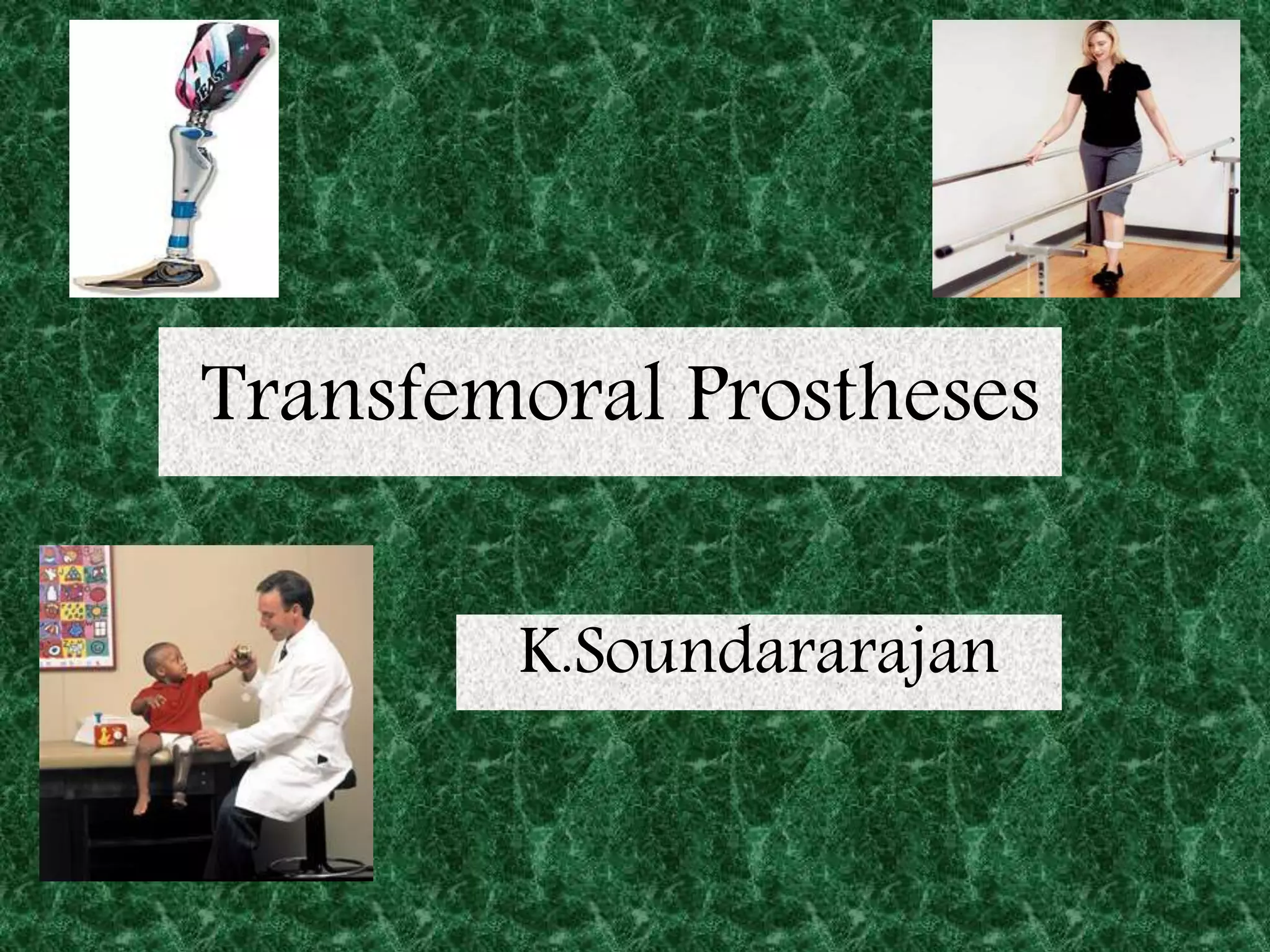
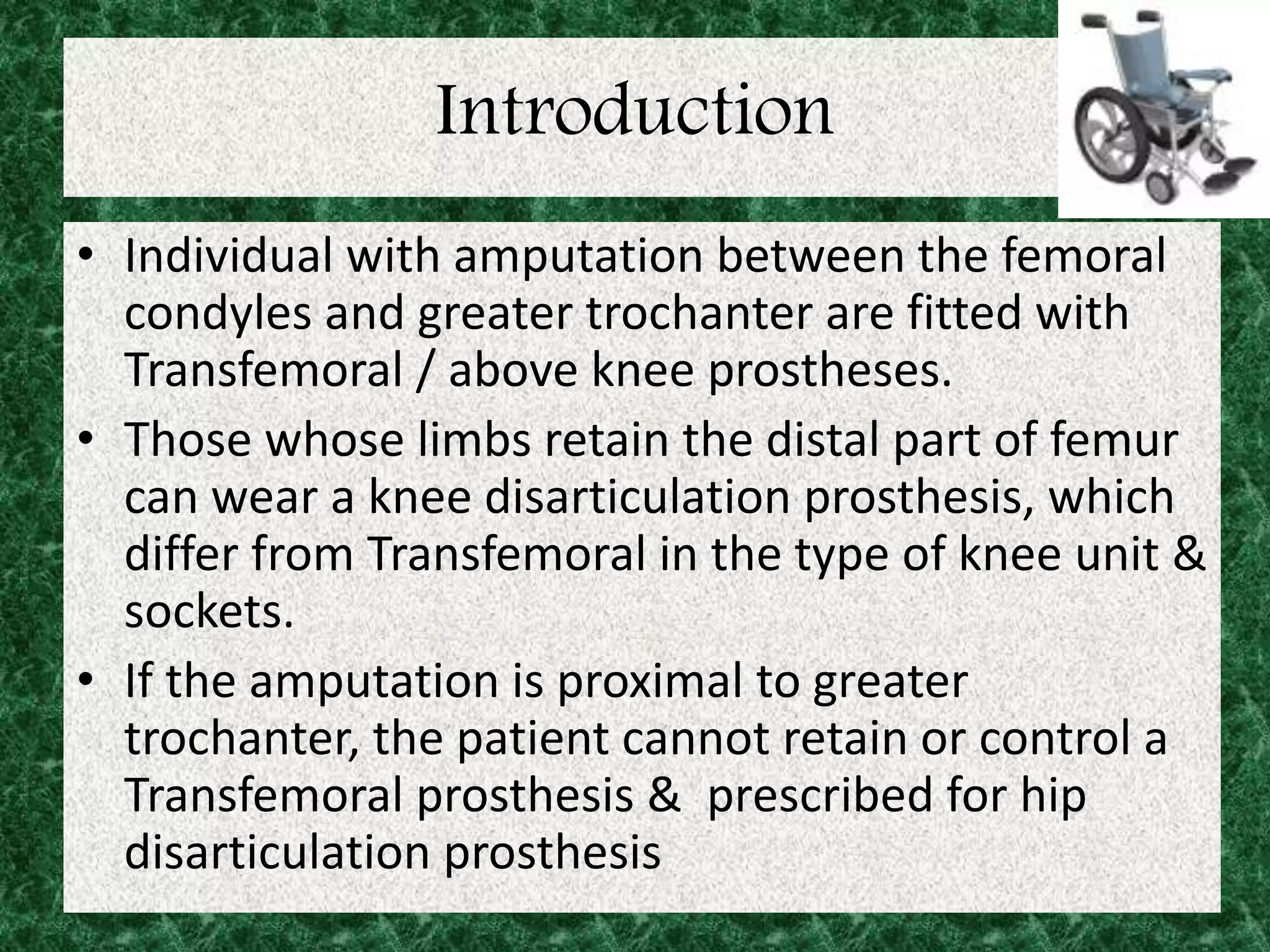
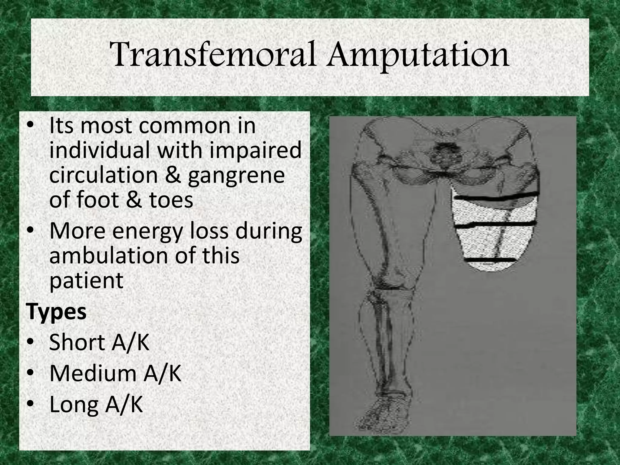
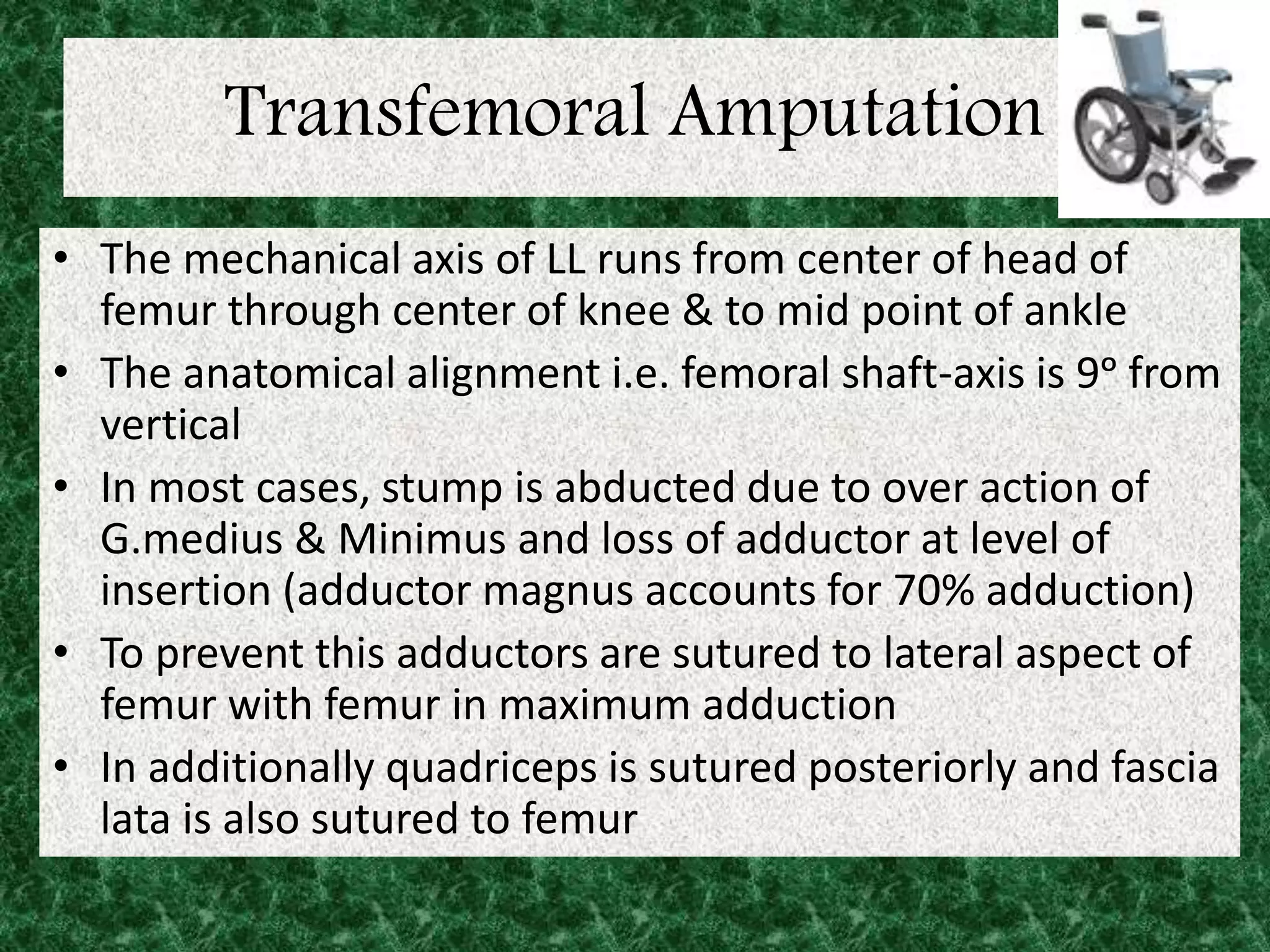
Transfemoral Prostheses
K.Soundararajan
objectives
• Introduction
• Transfemoral amputation
• Rehabilitation of Transfemoral amputation
• Prostheses in Transfemoral amputation
• Conclusion
• Reference
Introduction
• Individual with amputation between the femoral
condyles and greater trochanter are fitted with
Transfemoral / above knee prostheses.
• Those whose limbs retain the distal part of femur
can wear a knee disarticulation prosthesis, which
differ from Transfemoral in the type of knee unit &
sockets.
• If the amputation is proximal to greater
trochanter, the patient cannot retain or control a
Transfemoral prosthesis & prescribed for hip
disarticulation prosthesis
Transfemoral Amputation
• Amputation is defined as surgical removal of
apart or whole of a limb.
• It is considered only if the limb is
1. Dead (gangrenous)
2. Dying (gross ischemic)
3. Dangerous (due to malignancy)
4. Dud (useless e.g. leprosy)
Transfemoral Amputation
• Children – congenital anomalies
• Young – RTA, crush injury
• Elderly – vascular condition (diabetes)
Principle
1. Primary wound healing
2. Preserve as much length as possible
3. Maintain anatomical & mechanical
alignment
Transfemoral Amputation
• Its most common in
individual with impaired
circulation & gangrene
of foot & toes
• More energy loss during
ambulation of this
patient
Types
• Short A/K
• Medium A/K
• Long A/K
Transfemoral Amputation
• The mechanical axis of LL runs from center of head of
femur through center of knee & to mid point of ankle
• The anatomical alignment i.e. femoral shaft-axis is 9ᵒ from
vertical
• In most cases, stump is abducted due to over action of
G.medius & Minimus and loss of adductor at level of
insertion (adductor magnus accounts for 70% adduction)
• To prevent this adductors are sutured to lateral aspect of
femur with femur in maximum adduction
• In additionally quadriceps is sutured posteriorly and fascia
lata is also sutured to femur
Transfemoral Amputation
muscle wasting
• With aim of MRI the amount of atrophy in muscle in
stump after 2 years was assessed & revealed
A. Muscle that are not sectioned like G.medius, minimus,
iliopsoas has 30% atrophy
B. Muscles that lost insertion indirectly like G.maximus &
tensor fascia lata due to non-attachment of fascia lata
showed atrophy 37 to 47%
C. Muscles that are sectioned & attached by myoplasty
or myodesis atrophied 40 to 60%
Rehabilitation of Transfemoral
amputation
Rehabilitation of Transfemoral
amputation
• Rehabilitation can be divided into
• 1. Pre – op period
• 2.Post op period – a) Pre prosthetic stage
b)Prosthetic stage
3.Community &Vocational rehabilitation
4.Life long management &Follow up
Rehabilitation of Transfemoral
amputation
Pre operative period
1.Medical &physical assessment(power of UL, LL,
Trunk, crutch muscles, joint mobility, standing
&sitting balance)
2.Patient education
3. Functional ability
4.Discussion about limb pain, Post-OP Exercise Plan.
5. General conditioning program(cardiopulmonary)
Rehabilitation of Transfemoral
amputation
Exercise management
Breathing exercises – To maintain clear chest
Strengthening exercises – For Shoulder extensors
&adductors. For elbow extensors. Hip extensors ,
flexors , abductors &quadriceps.
Joint mobilization – for hip extension knee flexion
&extension.(mostly expect deformity)
Pelvic bridging , Roll to prone, back to supine
Transfer from bed to wheel chair & for hygienic
activity. (stop, start, turn, control)
 Trunk stability in sitting & standing
Rehabilitation of Transfemoral
amputation
• Post operative period
Pre prosthetic stage:
To prevent
1. Deformity
2. Decubitus contracture
3. Stump edema
4. Phantom limb
Rehabilitation of Transfemoral
amputation
Phantom limb pain
• Phantom limb sensation is the sense that the
amputated limb is still present.
• Phantom limb pain is perception of pain
cortically over an absent body part.
• Major Factor influencing quality of life post
amputation
Rehabilitation of Transfemoral
amputation
Phantom limb treatment
• 1.Heat application
• 2.Biofeedback to reduce muscle tension
• 3.Relaxation techniques
• 4.wound debridement
• 5.physical therapy (TENS)
• 6.neuro stimulation techniques including spinal cord
&brain stimulation
• 7.medications –neuroleptics,anticonvulsants,sodium
channel blockers and NSAIDS.
Rehabilitation of Transfemoral
amputation
Post operative (phase 1 Acute) pre prosthetic
• This phase begins immediately after patient is
discharged from hospital .
• GOALS :1.pain control &promotion of wound
healing
• 2.optimization of ROM, strength whole body,
balance, transfer
• 3.equipment prescription
• 4.General ,Functional mobility
• 5.phantom limb pain education and management
• 6. To re-educate walking
Rehabilitation of Transfemoral
amputation
Prevent post OP complication
1. Breathing EX & Brisk EX for unaffected leg
2. Position in bed: stump parallel to unaffected
limb in neutral position without resting
pillow. Pt lie in prone if possible
3. Encourage to handle stumps as possible , it
help to overcome shock of realization that leg
has actually gone.
Rehabilitation of Transfemoral
amputation
• Isometric exercise
• Control of stump edema
• Functional independence :
pt taught to move up & down bed by pressing
on sole of remaining leg. Encouraged to dress
each day, propel himself to wheel chair ,
axillary crutch.
• Do’s and Don’ts
Transfemoral Amputation PT Management
Treatment Overview
1. Functional Mobility
• Transfers
• Bed Mobility
• Positioning/Stretching
2. Strengthening/Conditioning
• Prepares the patient for prosthetic ambulation
3. Residual Limb Management
• Reduce Limb Swelling using a Shrinker
• Desensitization
• Scar Mobilization
Transfemoral Amputation PT Management
Transfemoral (Above Knee) Exercise Manual
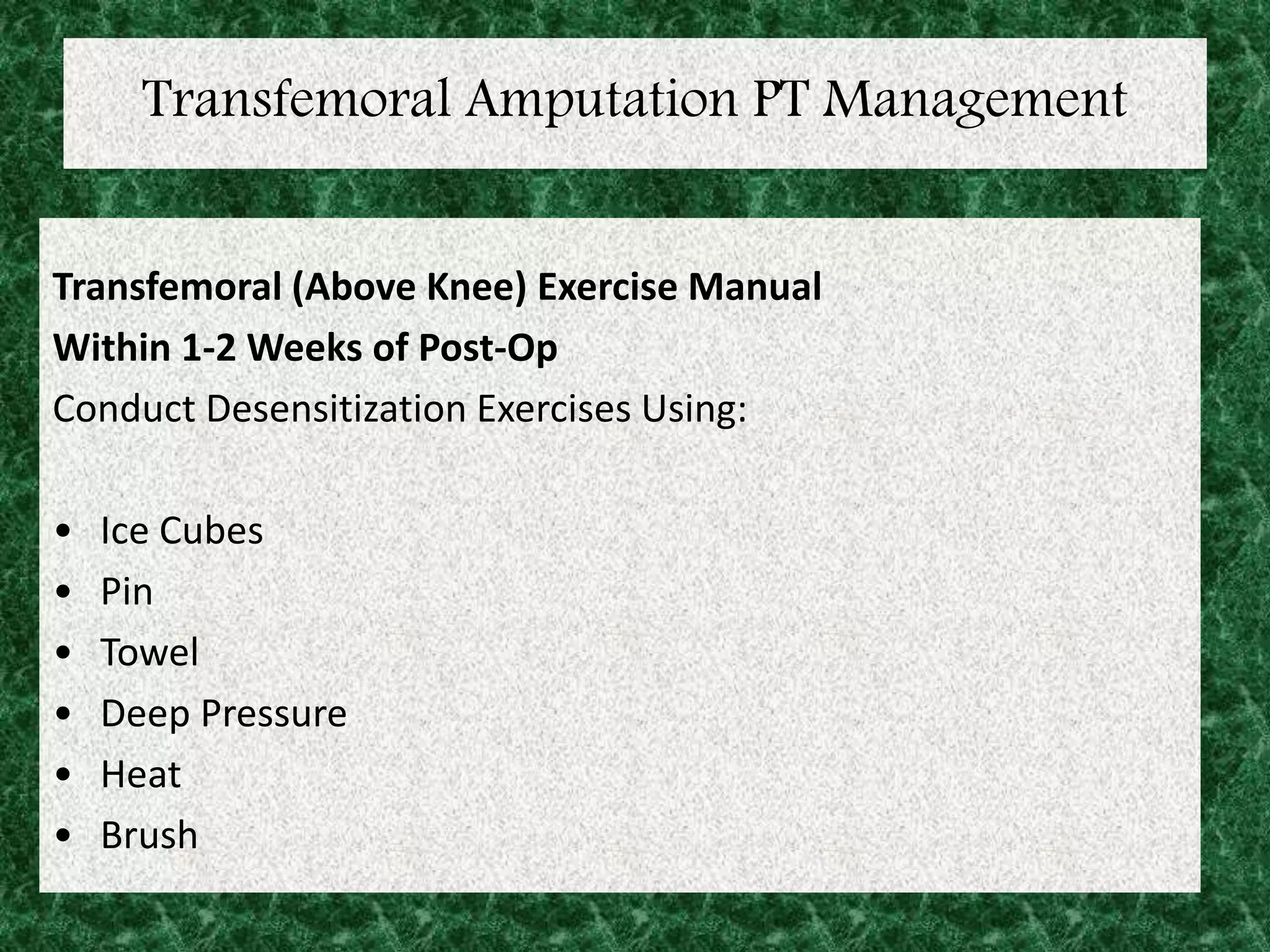
Within 1-2 Weeks of Post-Op
Conduct Desensitization Exercises Using:
• Ice Cubes
• Pin
• Towel
• Deep Pressure
• Heat
• Brush
Transfemoral Amputation PT Management
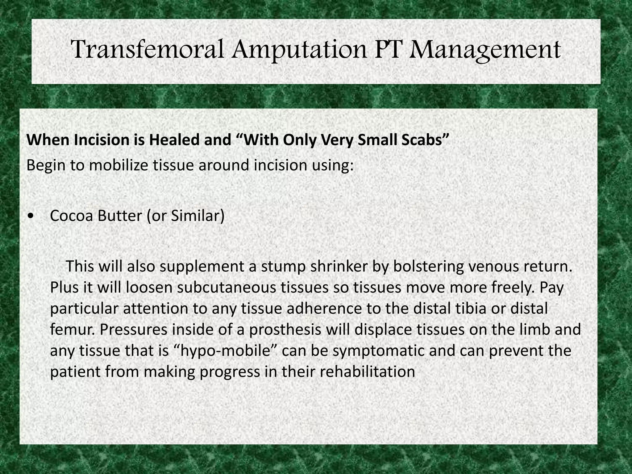
When Incision is Healed and “With Only Very Small Scabs”
Begin to mobilize tissue around incision using:
• Cocoa Butter (or Similar)
This will also supplement a stump shrinker by bolstering venous return.
Plus it will loosen subcutaneous tissues so tissues move more freely. Pay
particular attention to any tissue adherence to the distal tibia or distal
femur. Pressures inside of a prosthesis will displace tissues on the limb and
any tissue that is “hypo-mobile” can be symptomatic and can prevent the
patient from making progress in their rehabilitation
Goals of the Pre-Prosthetic Program
1 Trans-Femoral patients should work towards achieving 0-10 degrees of hip extension.
2 Patient should complete a minimum of 6 weeks of strength/ROM training on the primary
prosthetic gait muscles outlined in this manual.
The patient should be independent with the exercise program and also understand the
importance of being diligent with the program. Educate your patient so that they understand
these exercises will require a lifestyle change.
3 The patient should be able to tolerate a minimum of 30 minutes of physical activity.
4 Following sufficient soft tissue mobilization, the patient should be able to tolerate pressure
and tissue displacement on the distal end and incision without pain.
5 The incision should be fully healed and good soft tissue mobility should be established.
6 It is imperative that patients continue an independent exercise program even after
prosthetic gait training has been initiated. Preferably, the patient will continue their
exercises on their own, outside of therapy, so that therapy appointments can focus on gait
training. This will encourage further muscle development that will supplement any gait
training conducted during regular therapy appointments.
7 They should be independent with transfers and bed mobility; this includes the independent
ability to roll prone.
Amputee Exercises - Bridging Over Bolster
Amputee Exercises
Single Leg Bridging
Amputee Exercises
Short Arc Quadriceps
Amputee Exercises
Straight Leg Raise
Amputee Exercises
Ankle Pumps
Amputee Exercises
Side Lying Hip Abduction - Modified
Amputee Exercises
Side Lying Hip Abduction - Advanced
Amputee Exercises
Prone Hip Extension
Amputee Exercises
Prone Hip Extension (Sound Limb)
Amputee Exercises
Prone Knee Flexion
Amputee Exercises
Prone Abductor Squeeze
Amputee Exercises
Push-ups
Amputee Exercises
Quadruped Leg Lift
Amputee Exercises
Abdominal Curl-up
Amputee Exercises
Bosu Head Twists
Amputee Exercises
Bosu Ball Exercises
Transfemoral Amputation
PT Management
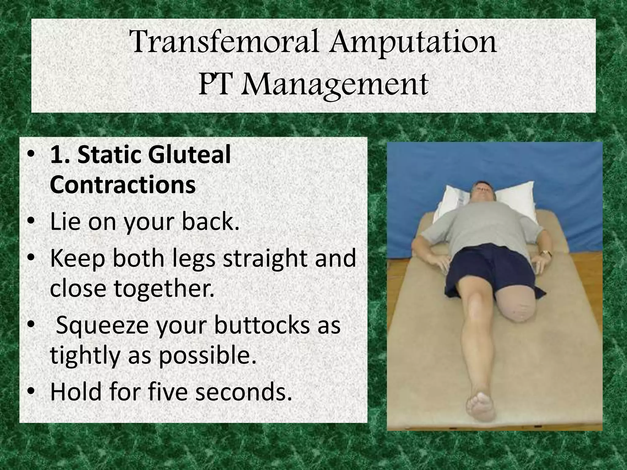
• 1. Static Gluteal
Contractions
• Lie on your back.
• Keep both legs straight and
close together.
• Squeeze your buttocks as
tightly as possible.
• Hold for five seconds.
Transfemoral Amputation
PT Management
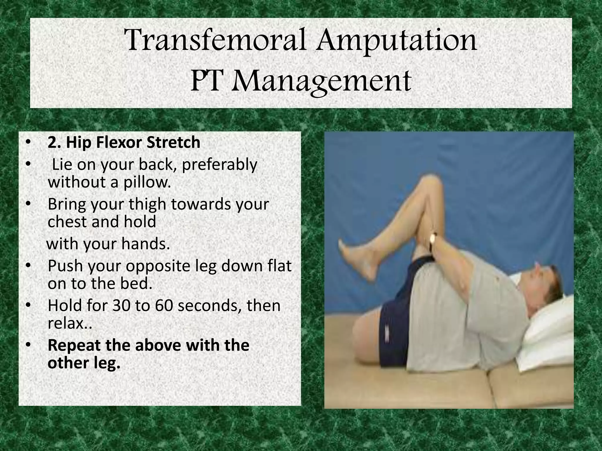
• 2. Hip Flexor Stretch
• Lie on your back, preferably
without a pillow.
• Bring your thigh towards your
chest and hold
with your hands.
• Push your opposite leg down flat
on to the bed.
• Hold for 30 to 60 seconds, then
relax..
• Repeat the above with the
other leg.
Transfemoral Amputation PT Management
3. Hip Hitching
• Lie on your back.
• Keep both legs flat on the bed.
• Hitch one hip up towards you on one side and push away on the other
(shortening one side & stretching the other).
• Hold for three seconds Repeat on the other side.
Transfemoral Amputation PT Management
4. Bridging
• Lie on your back with your arms at the side.
• Place a couple of firm pillows or rolled up blankets under your thighs.
• Pull in your stomach, tighten your buttocks & lift your bottom up off the bed.
• Hold for five seconds.
• To make this exercise more difficult, place your arms across your chest as
shown in the picture.
Transfemoral Amputation PT Management
• 5. Hip Flexion and Extension in Side Lying
• Lie on your side.
• Bend the bottom leg.
• Lift your top leg slightly.
• Bend your thigh fully towards your chest.
• Push your leg backwards as far as you can.
NB: Try not to let your hips roll forwards or backwards
Repeat the above with the other leg.
Transfemoral Amputation PT Management
6. Hip Abduction in Side Lying
• Lie on your side.
• Bend the bottom leg.
• Keep hips and top leg in line with your body.
• Slowly lift your top leg up.
• Slowly lower.
NB: Try not to let your hips roll forwards or backwards.
Repeat the above with the other leg.
Transfemoral Amputation PT Management
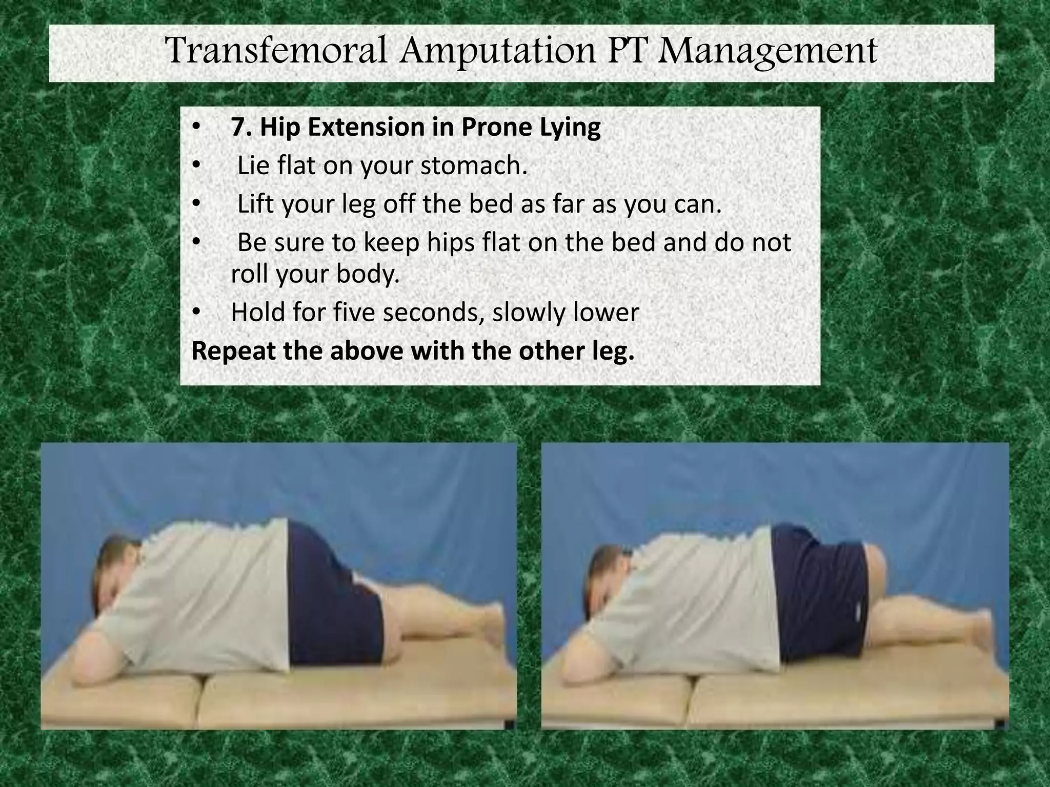
• 7. Hip Extension in Prone Lying
• Lie flat on your stomach.
• Lift your leg off the bed as far as you can.
• Be sure to keep hips flat on the bed and do not
roll your body.
• Hold for five seconds, slowly lower
Repeat the above with the other leg.
Transfemoral Amputation
PT Management
8. Hip Adduction with Resistance
• Sit with both legs out in front of
you.
• Place a pillow or rolled up towel
between your thighs.
• Squeeze your legs together.
• Hold for five seconds.
• Repeat ___ times.
• NB: This exercise can also be
performed when sitting in a
wheelchair or at the edge of a bed.
Prosthesis in Transfemoral Amputation
Prosthesis period
Prosthesis in Transfemoral Amputation
• Prosthetics in Greek means ‘in addition to’.
• Prosthesis is defined as a replacement or
substitution of a missing or a diseased part .
• Prosthetics includes the theory &practice of
the prescription ,fitting ,design, assessment
&production of prosthesis.
Prosthesis in Transfemoral Amputation
• Classification (based on structure)
ENDOPROSTHESIS EXOPROSTHESIS
PROSTHESIS
Prosthesis in Transfemoral Amputation
ENDOPROSTHESIS EXOPROSTHESIS
PROSTHESIS
• Classification(based on function)
• Temporary prosthesis: these are implants used
in orthopedics surgery to replace
joints.eg:Austin moore prosthesis
• Permanent prosthesis : this is fitted after
making a final clinical assessment.
Prosthesis in Transfemoral Amputation
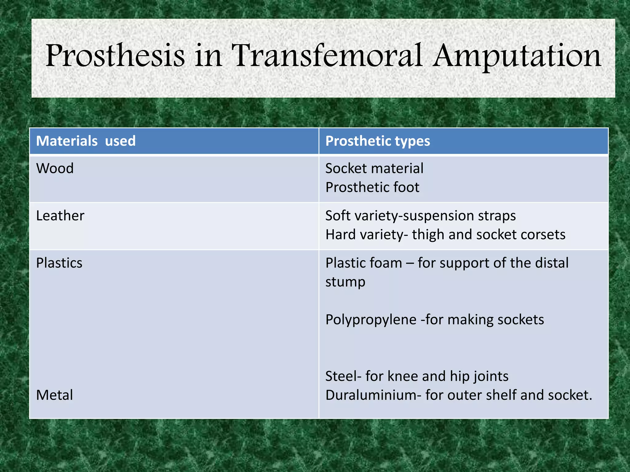
Materials used Prosthetic types
Wood Socket material
Prosthetic foot
Leather Soft variety-suspension straps
Hard variety- thigh and socket corsets
Plastics
Metal
Plastic foam – for support of the distal
stump
Polypropylene -for making sockets
Steel- for knee and hip joints
Duraluminium- for outer shelf and socket.
Prosthesis in Transfemoral Amputation
• Difficulties in making effective
prosthesis is due to
1. Weight bearing area (ischial tuberosity
is proximal to propulsive structure)
(thigh).
2. Passive mechanical replacement of
two major joint (knee, ankle) and foot
3. Limitation in prosthetics technology
Prosthesis in Transfemoral Amputation
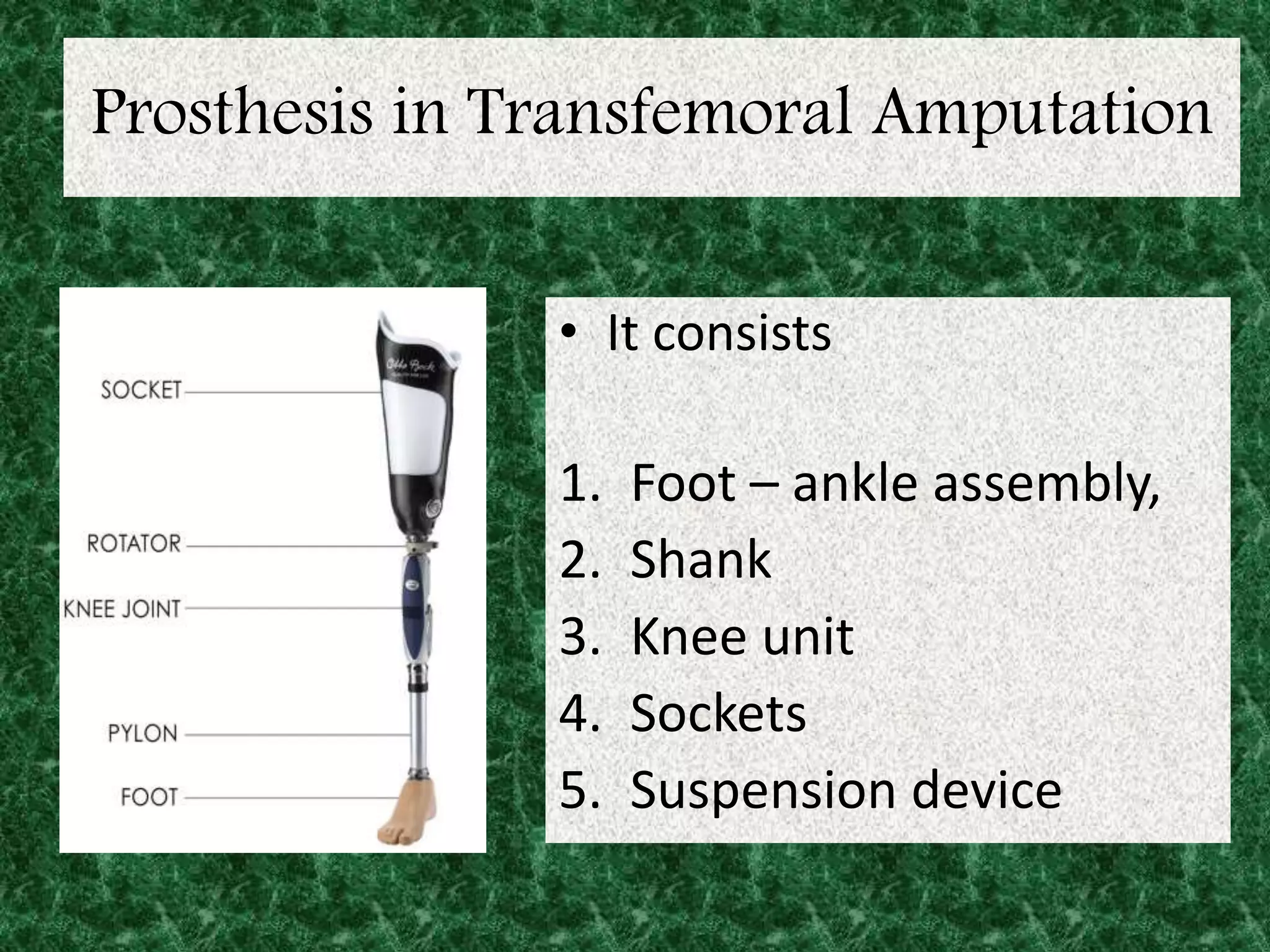
• It consists
1. Foot – ankle assembly,
2. Shank
3. Knee unit
4. Sockets
5. Suspension device
Prosthesis in Transfemoral Amputation
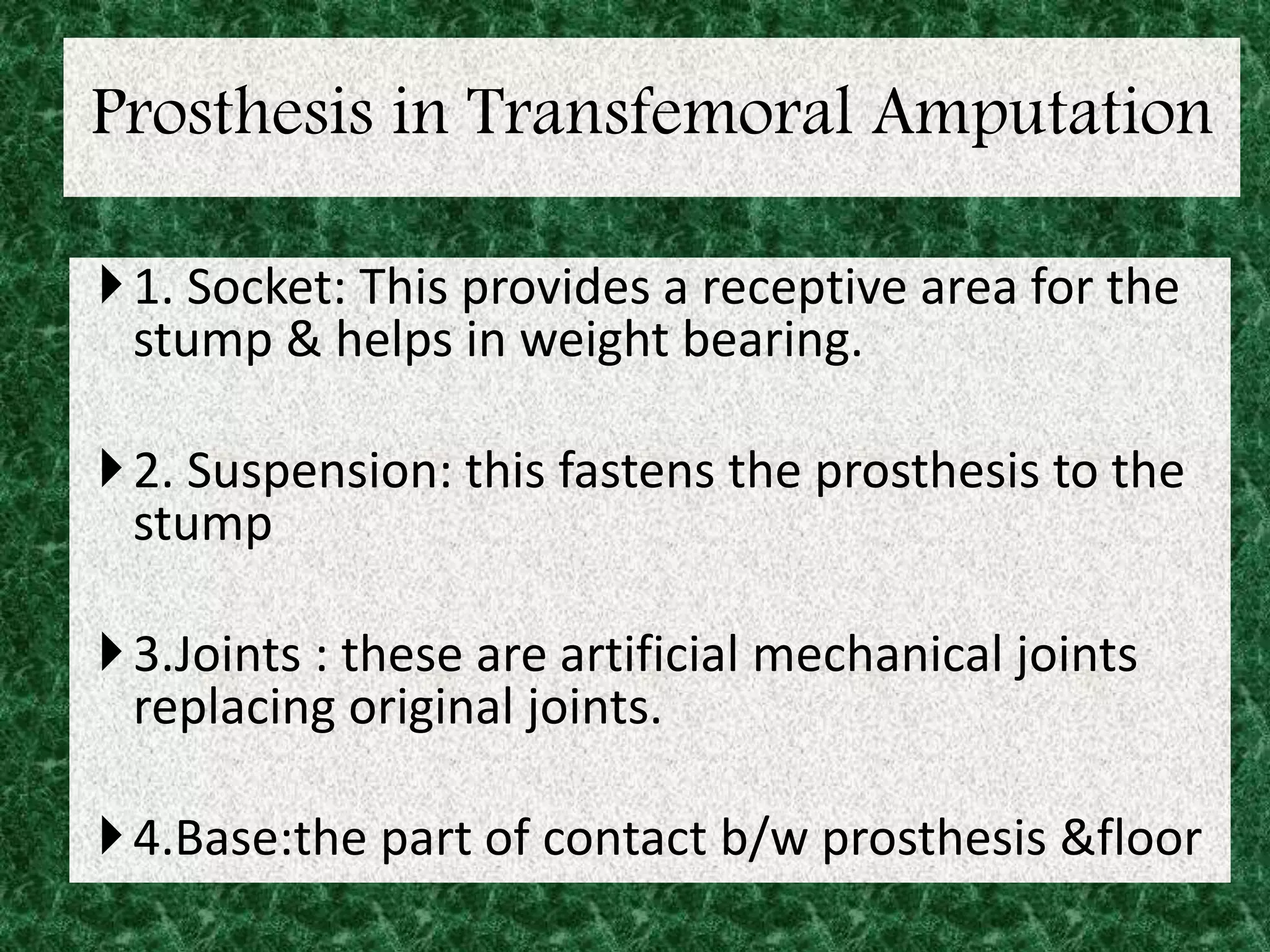
1. Socket: This provides a receptive area for the
stump & helps in weight bearing.
2. Suspension: this fastens the prosthesis to the
stump
3.Joints : these are artificial mechanical joints
replacing original joints.
4.Base:the part of contact b/w prosthesis &floor
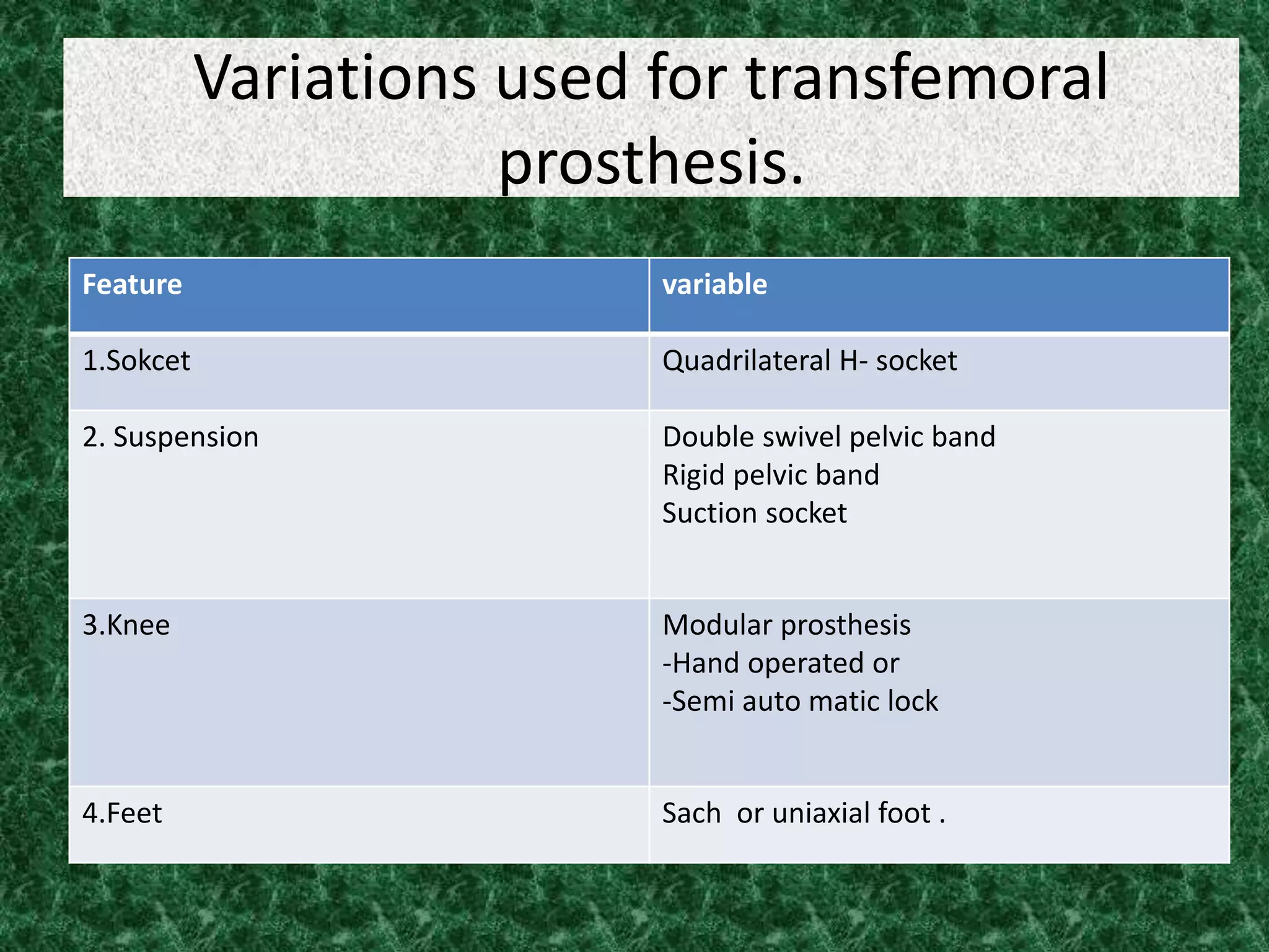
Variations used for transfemoral
prosthesis.
Feature variable
1.Sokcet Quadrilateral H- socket
2. Suspension Double swivel pelvic band
Rigid pelvic band
Suction socket
3.Knee Modular prosthesis
-Hand operated or
-Semi auto matic lock
4.Feet Sach or uniaxial foot .
Prosthesis in Transfemoral Amputation
• Sockets
Two types of sockets commonly used are
1. Quadrilateral Sockets
2. Ischial Containment Sockets
Quadrilateral Sockets
• Developed late in 1950 & is named for its four walls. Distally the
socket is contoured for total contact of residual limb
• Posterior wall : provide major weight bearing area. It has ischial set
for ischial tuberosity & G.muscle which is thicker medially & thin
laterally. Internally provide relief for hamstring muscle. Height is at
level of ischial tuberosity
• Anterior wall: It extends 5cm above height of posterior wall with
anteromedial inward femoral bulge called scrap’s projection. It keep
ischial tuberosity situ anterior wall is convex laterally
• Lateral wall: normally it extend as high as anterior wall. For short
stump it is in trimmed above trochanter to increase stability & control.
The wall inclines medially with 10ᵒ adduction ( normal adduction of
femur)
• Medial wall : it’s vertical & parallel to sagittal plane. Relief is given
internally add. longus anteromedially & hamstrings posteromedially
Ischial Containment Sockets
• Developed in early 1990 & shaped differently from quadrilateral
socket. The ischium and a part of pubic rambus are enclosed in socket.
It contains more area side socket so more area of weight distribution.
Narrow mediolateral dimension helps in keeping up ischial tuberosity
within posteromedial wall of sockets
• The lateral wall cover trochanter to provide more stability. The distal
socket is of total contact socket
• The 2 types of ischial containment sockets are:
1. CATCAM
2. NSANA
Contour Adducted Trochanteric Controlled Alignment Method
Normal Shape Normal Alignment
Comparison between sockets
Quadrilateral
1. With four walls
2. Ischial seat for WB
3. Femoral bulge present
4. Area of WB is less
5. Long lateromedial dimension
6. Narrow anterioposterior
dimension
7. Fair pelvic control
8. Fair rotational stability
9. Less energy efficient
10. Indicated for standard stumps
11. Lateral wall normally extends
below trochanter except in short
stumps
Ischail Containment
1. No walls
2. No Ischial seat
3. Femoral bulge absent
4. Area of WB is more
5. Narrow mediolateral dimension
6. Long anterioposterior
dimension
7. Good pelvic control
8. Good rotational stability
9. More energy efficient
10. Indicated for short stumps & G.
Medius weakness
11. Extends above greater
trochanter
Transfemoral prosthesis.
STATISTICS
• 1,285,000 People Living with Limb Loss in
The U.S.
• 4.9 per 1,000 people
• Of Those 1,285,000 People:
• 36,478 were Transfemoral Amputations (American
Amputee Coalition of America ‘96)
Prosthesis in Transfemoral Amputation
Biomechanical principles
• Can be analyzed in three phases-
ABSORPTION PHASE
ACCELERATION
DECELERATION
Prosthesis in Transfemoral Amputation
Absorption
• Reduction in ground reaction force
• Significant difference in knee angles found at
heel strike.
• Prosthetic absorbs and generates less energy
which results in
– A more passive limb
– Absorption by soft tissue in socket
– Presence of isometric contraction by muscles
– So as foot strikes, a backward force is instantly
created by prosthetic-side hip muscles.
Prosthesis in Transfemoral Amputation
Deceleration
• Hip abductors and adductors and knee
extensors muscles are main source of
absorption. (Sadeghi 01’)
• Fewer gait problems are involved in the swing
phase than with the stance
Prosthesis in Transfemoral Amputation
Acceleration
• Hip extensor effort is main compensation of
propulsion reduction. (Pailler 04’)
• Amputation of ankle reduces the ability of
power to be produced through plantar flexion
Prosthesis in Transfemoral Amputation
Biomechanics Summary
• Longer motions for amputated side
– Step length
– Step time
– Swing time
• Shorter motions for amputated side
– Stance time
– Single support time
Prosthesis in Transfemoral Amputation
ENERGY EXPENDITURE
Amputation
Level
Energy
Above
Baseline, %
Speed, m/min Oxygen Cost,
ml/kg/m
Transfemoral 65 40 .28
Wheelchair 0-8 70 .16
Prosthesis in Transfemoral Amputation
• Energy cost depends on
– Gait speed
– Efficiency
– Not on displacement of center of mass (Detrembleur 05’)
• Energy consumption
– For Transfemoral amputees is more significant than that of
transtibial amputees.
– Is affected by prosthetic alignment
– Is not affected by the use of different prosthetic feet
(Schmalz 02’)
Prosthesis in Transfemoral Amputation
Balance &Stability
• Significant differences found between TTA and controls
during equilibrium and movement studies.
– Transition from bipedal to monopedal
– High failure rate for TTA
– Same difficulty on sound and prosthetic limb (Viton 00’)
• Utilize remaining muscles
• Work on speed of contraction, not maximal force of
contraction (Gailey 03’)
Prosthesis in Transfemoral Amputation
Fitting
• An exact mold of the residual limb does not
make a good socket
• Indent in the region around the patellar tendon
• Many different types of sockets
• Foam of silicone
• Hard
• Soft (Smith, ’03)
Prosthesis in Transfemoral Amputation
Alignment-1
• Alignment is the spatial relationship between the
prosthetic socket and foot.
• Purpose:
– Position the prosthetic socket with respect to the foot so that
adverse patterns of force applied to the residual limb are
avoided
– Produce a normal pattern of gait (Noelle, ’03)
Prosthesis in Transfemoral Amputation
Transtibial vs. Transfemoral
• Transfemoral Amputations:
• Known as above the knee amputations
• Surgeon’s goal is to leave as much residual limb as
possible, preserve the adductor muscles, and the
remaining soft tissue. (Biomed, ’03)
• Transtibial Amputations:
• Known as Below The Knee Amputations
• The Surgeon’s goal is to leave a cylindrical shaped
well-padded residual limb.
• Using the gastrocnemius and soleus muscles to create
a muscular flap.
Prosthesis in Transfemoral Amputation
Prosthetic rehabilitation
• Involves –
• Stump shaping &shrinking
• Desensitization
• ROM & muscle strengthening
• THIS IS DONE (6-8 WEEKS POST OP)WITH SOFT
DRESSINGS OR 3-6 WEEKS POST OP WITH AN
IPOP (IMMEDIATE POST OP PROSTHESIS).
Prosthesis in Transfemoral Amputation
Prosthetic rehabilitation
PROSTHETIC STAGE MANAGEMENT
• DONE AFTER 8 WEEKS
• INCLUDES :GAIT TRAINING- POSTURE, EVEN
WEIGHT BEARING,PROPRIOCEPTION &WEIGHT
SHIFTING, WEIGHT TRANSFER IN STANCE.
Prosthesis in Transfemoral Amputation
Prosthetic rehabilitation
• ADVANCED GAIT TRAINING
• AFTER 10 WKS
• INCLUDES- STAIR CLIMBING,WALKING ON
SLOPE OR UNEVEN FLOOR.
• PROGRESSION- CROWDED AREAS, PUBLIC
TRANSPORT EDUCATION.
COMMUNITY INTEGRATION
• INVOLVES-
• Resumption of familial &community roles
• Addressing emotional needs
• Developing healthy coping strategies
• Resumption of previous recreational activities.
• Vocational rehabilitation by vocational
counsellor .(including previous jobs)
Follow up
• Includes-lifelong prosthetic support, functional
and medical assistance, psychological support.
• Patient should be sent for follow up to the team
members once in 3 months for first 18 months
following which they can go once in 6 months.
• Support groups and Ngo’s
Conclusion
• Responsibility of surgeon doesn't end with
healing of wound but must include restoration
of locomotion by correct limb fitting
(prosthesis) and re-education
• Prosthesis must fit amputee not only
physically but also physiologically.
• It must be integrated emotionally with
patients and be socially acceptable
Reference
• Susan B.O’ Sullivan etal ,Amputation, Physical
Rehabilitation,2014, 1000-1030;
• Susan B.O’ Sullivan etal ,Prosthetics, Physical
Rehabilitation,2014, 1375-1379;
• R Chinnathurai etal, Prosthesis in Transfemoral
Amputation, Short Textbook book of
prosthetics & orthotics 2006,44-47;
• S. Sundar , Amputation & Prosthesis , Text book
of Rehabilitation , 2002, 131-162;
Transfemoral protheses