Orthopaedic operations
• The art and skill of orthopaedic surgery is
directed not simply to reshaping or
constructing a particular arrangement of
parts but to restoring function to the whole.
•(LIFE IS MOVEMENT AND
MOVEMENT IS LIFE)
preoperative preparation
1-General assessment of the patient :
It is important also to evaluate the risk of complications such
as thromboembolism and infection in the particular
individual and, where necessary, to start prophylactic
treatment before the operation LMWH and ABs.
2-Planning the operation:
Operations must be carefully planned in advance,when accurate
measurements can be made and the bones and joints can be
compared for
symmetry with those of the opposite side.
X-rays, magnetic resonance imaging (MRI) and computed
tomography (CT) (if necessary with threedimensional re-
formation) are helpful; templates may be needed to help
select the appropriate shape and size of a prosthetic implants.
3-The operating environment:
Short operating times and limiting the number
of people in the theatre will reduce the likelihood of infection
ultra-clean laminar airfl ow theatres and
prophylactic antibiotics should be administered
before, during and after the operation.
4-Surgical equipment:
The minimum requirements for orthopaedic operations are
drills (for boring holes), osteotomes (for cutting cancellous
bone), saws (for cutting cortical bone), chisels (for shaping
bone), gouges (for removing bone) and plates, screws, wires
and screwdrivers (for fi xing bone). Operations such as joint
replacement, spinal fusion and internal fi xation require
special implants and instruments
• 5-Intraoperative radiography(flouroscopy):
Intraoperative radiography and image intensification
are often helpful and sometimes essential.
Fracture reduction, osteotomy alignments and the
positioning of implants and fixation devices can be
checked during the operation and often again at the end
of the procedure.
6-Magnification(Microscopic ):
Magnification is an integral part of peripheral nerve
and hand surgery like nerve repair or vascular
anastomosis.
6-The ‘bloodless field’
Many operations on limbs can be performed
more rapidly and accurately if bleeding is
prevented by the application of a tourniquet.
Only a pneumatic cuff is suitable. Rubber
bandages are potentially dangerous; the
pressure beneath the bandage cannot be
controlled and there is a risk of damage to the
underlying nerves and muscle.
Principle of Pneumatic Tourniquet
1-Adequate exsanguination of the tissues can usually be
achieved by elevating the limb to 60 degrees above
horizontal for 30 seconds before inflating the
tourniquet cuff.
2-rubber tubular exsanguinator is equally effective.
Tourniquet pressure should not exceed 150 mmHg
above systolic for the lower limb and 100 mmHg above
systolic for the upper limb.
3-Tourniquet time should not exceed 2 hours, Excessive
or prolonged pressure can cause permanent nerve or
muscle damage.
A-OPERATIONS ON BONES
1-OSTEOTOMY:Osteotomy may be used to correct deformity, to change the
shape of the bone, or to relieve pain in arthritis by redirecting the load.
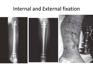
2-BONE FIXATION (internal or external fixation).
3-BONE GRAFT: Bone grafts are both osteoinductive and osteoconductive, i.e.
they are able to stimulateosteogenesis and they also provide linkage
across defects and a scaffold upon which new bone can form.
• 1-Autografts In these, bone is taken from one site to another in the
individual.
• 2-Allografts (homografts) This bone is transferred from an individual
(alive or dead) to another of the same species. The graft must always be
harvested under sterile conditions and the donor must be cleared for
malignancy, venereal disease, hepatitis and human immunodeficiency
virus (HIV).
• 3-Synthetic substitutes Calcium-derived pastes or granules are available
• for use to stimulate healing across defects. These usually consist of
calcium sulphate, calcium phosphate or calcium hydroxyapatite. Bone
morphogenetic protein (BMP) is a synthetic osteoinductive agent.
Internal and External fixation
B-OPERATIONS ON JOINTS
ARTHROTOMY
ARTHRODESIS
ARTHROPLASTY
AMPUTATIONS
• INDICATIONS: -
‘Three Ds’:
(1) Dead.
(2) Dangerous .
(3) Damned nuisance.
• 1-Dead (or dying) Peripheral vascular disease accounts
for almost 90 per cent of all amputations. Other causes
of limb death are severe trauma, burns and frostbite.
• 2-Dangerous ‘Dangerous’ disorders are malignant
tumours, potentially lethal sepsis and crush injury. In
crush injury, releasing the compression may result in
renal failure (the crush syndrome).
• 3-Damned nuisance Retaining the limb may be worse
than having no limb at all. This may be because of: (1)
pain; (2) gross malformation; (3) recurrent sepsis or (4)
severe loss of function. The combination of deformity
and loss of sensation is particularly trying, and in the
lower limb is likely to result in pressure ulceration.
AMPUTATIONS AT SITES OF ELECTION
• Most lower limb amputations are for ischaemic
disease and are performed through the site of
election below the most distal palpable pulse. The
selection of amputation level can be aided by
Doppler US.
• The sites of election are determined also by the
demands of prosthetic design and local function.
Too short a stump may tend to slip out of the
prosthesis.
• Too long a stump may have inadequate circulation
and can become painful, or ulcerate; moreover, it
complicates the incorporation of a joint in the
prosthesis
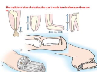
The traditional sites of election;the scar is made terminalbecause these are
not endbearing stumps.
PRINCIPLES OF TECHNIQUE
A tourniquet is used unless there is arteria
insufficiency. Skin flaps are cut so that their
combined length equals 1.5 times the width of the
limb at the site of amputation. As a rule anterior
and posterior flaps of equal length are used for the
upper limb and for transfemoral
(above-knee) amputations; below the knee a
long posterior flap is usual. Muscles are divided
distal to the proposed site of bone section.
It is also helpful to pass the sutures that anchor the
opposing muscle groups through drill-holes in the
bone end, creating an osteomyodesis. Nerves are
divided proximal to the bone cut to ensure a cut
nerve end will not bear weight
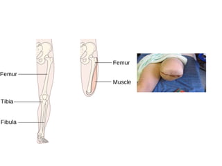
• The bone is sawn across at the proposed level. In
trans-tibial amputations the front of the tibia is
usually bevelled and filed to create a smoothly
rounded contour; the fibula is cut 3 cm shorter.
• The main vessels are tied, the tourniquet is
removed and every bleeding point meticulously
ligated.The skin is sutured carefully without
tension. Suction drainage is advised and the
stump covered without constricting passes of
bandage; figure-of eight passes are better suited
and prevent the creation of a venous tourniquet
proximal to the stump.
AFTERCARE
• If a haematoma forms, it is evacuated as soon
as possible. After satisfactory wound healing,
gradualcompression stump socks are used to
help shrink the stump and produce a conical
limb-end. The muscles must be exercised, the
joints kept mobile and the patient taught to
use his prosthesis.
AMPUTATIONS OTHER THAN AT
SITES OF ELECTION
1-Interscapulo-thoracic (forequarter amputation)
2-Disarticulation at the shoulder
3-Amputation in the forearm
4-Amputations in the hand
5-Hemipelvectomy (hindquarter amputation)
6-Disarticulation through the hip
7-Transfemoral amputations (Above KneeAmputation)
8-Around the knee
9-Transtibial (below-knee) amputations
10-Above the ankle Syme’s amputation
11-Partial foot amputation
COMPLICATIONS OF AMPUTATION
STUMPS
A-EARLY COMPLICATIONS
1-Breakdown of skin flaps This may be due to
ischaemia,suturing under excess tension or (in
below-knee amputations) an unduly long tibia
pressing against the flap.
• 2-Gas gangrene Clostridia and spores from the
perineum may infect a high above-knee
amputation (or reamputation), especially if
performed through ischaemic tissue.
• 3-secondary haemorrhage.
LATE COMPLICATIONS
1-Skin Eczema is common, and tender purulent lumps may
develop in the groin. A rest from the prosthesis is
indicated.
2-Muscle If too much muscle is left at the end of the
stump, the resulting unstable ‘cushion’ induces a
feeling of insecurity that may prevent proper use of a
prosthesis; if so, the excess soft tissue must be excised.
3-Blood supply Poor circulation gives a cold, blue stump
that is liable to ulcerate. This problem chiefly arises
with below-knee amputations and often re-amputation
is necessary.
• 4-Nerve A cut nerve always forms a neuroma and
occasionally this is painful and tender. Excising 3 cm of
the nerve above the neuroma sometimes succeeds.
• Phantom limb’ This term is used to describe the
feeling that the amputated limb is still present.
• 5-Joint The joint above an amputation may be stiff or
deformed. A common deformity is fixed flexion and
fixed abduction at the hip in above-knee stumps
(because the adductors and hamstring muscles have
been divided). It should be prevented by exercises.
Fixed flexion at the knee makes it difficult to walk properly and
should also be prevented.
6-Bone a spur often forms at the end of the bone, but
is usually painless. If there has been infection, however,
the spur may be large and painful and it may be
necessary to excise the end of the bone with the spur.
Orthopedics Operations and Instrument.pptx