AMPUTATIONS OF
LOWER LIMB
DR.DARSHAN N(MS ORTHOPAEDICS)
Introduction
• Latin amputare, "to cut away”
• “Surgical removal of limb or part of the limb through a bone or
multiple bones”
• Should not be viewed as a failure of treatment but as the first step in
Rehabilitation
INDICATIONS
1. Peripheral Vascular Disease
2. Trauma
3. Burns
4. Frostbite
5. Infections
6. Tumours
Peripheral Vascular Disease
• Most common indication for amputation
• Peripheral vascular disease with or without diabetes
• Age-50 to 75years
• Disease processes in the cerebral vasculature, coronary arteries, and
kidneys
• Vascular surgery consultation for improved techniques and
salvageable
• If amputation becomes necessary
• Infection and Nutrition and Immune status should be evaluated.
Trauma
• Leading indication in younger patients
• More common in men
• The only absolute indication for primary amputation is an irreparable
vascular injury
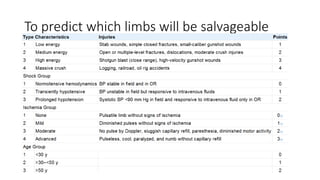
To predict which limbs will be salvageable
• Severely injured limb may lead to metabolic overload and secondary
organ failure
• Multiple injuries and in the elderly
BURNS
• Thermal or electrical injury
• Tissue damage may not be apparent, especially with electrical injury
• Treatment involves early débridement of devitalized tissue and
wound care
• Delayed amputation - increased risk of local infection, systemic
infection ,AKI
Frostbite
• Freezing of tissue in the extremities
• Common problem for high-altitude climbers, skiers, and hunters
• WHY IT HAPPENS
• Mechanisms:
1. Ice crystals in the extracellular fluid
2. Ischemic damage to vascular endothelium, clot formation, and increased
sympathetic tone
• Amputation for frostbite routinely should be delayed 2 to 6 months.
• Clear demarcation of viable tissue may take this long.
• Triple-Phase Technetium Bone Scan
Infection
• Acute or chronic infection that is unresponsive to antibiotics and
surgical débridement.
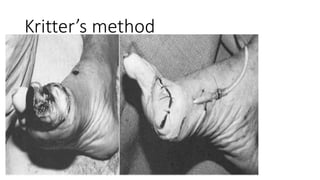
• Open amputation is indicated:
1. A guillotine amputation
2. Definitive level by initially inverting the flaps and secondary closure
at 10 to 14 days
Kritter’s method
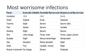
Most worrisome infections
• Clostridial Myonecrosis :
Radical débridement of involved tissue, high doses of intravenous
penicillin and hyperbaric oxygen , open amputation one joint above the
affected compartment.
• Streptococcal Myonecrosis:
Debridement , penicillin treatment , usually allow preservation of
the limb.
TUMOURS
• Would survival be affected by the treatment choice?
• Multimodal treatment - osteosarcoma patients has improved from
approximately 20% to approximately 70%.
• Osteosarcoma of the distal femur : recurrence is 5% to 10%
• Salvageable s better
How do short-term and long-term morbidity
compare?
• Amputation for malignancy may be technically demanding -
nonstandard flaps, bone graft, or prosthetic augmentation ,
periprosthetic fractures, prosthetic loosening
• Limb salvage involves a more extensive surgical procedure ,infection,
wound dehiscence, flap necrosis, blood loss, and deep venous
thrombosis , multiple subsequent operations , amputation
• Amputation s better
How would the function of a salvaged limb
compare with that of a prosthesis?
• The location most important factor
• Upper extremity lesion - limb salvage provides better function than
amputation
• Resection of a proximal femoral or pelvic lesion , better function than hip
disarticulation or hemipelvectomy
• Sarcomas around the ankle and foot frequently are treated with
amputation
Osteosarcoma around the knee
• Wide Resection with Prosthetic Knee Replacement
• Wide Resection with Allograft Arthrodesis
• Transfemoral Amputation
SURGICAL PRINCIPLES OF
AMPUTATION
DETERMINATION OF AMPUTATION LEVEL
• Level of amputation : increased function with a more distal level of
amputation and a decreased complication rate with a more proximal
level of amputation
• The energy required for walking is inversely proportionate to the
length of the remaining limb
• At self-selected walking velocities : tended to decrease their velocities
• Compensatory mechanism to conserve energy per unit time
• Anaerobic mechanisms to sustain muscle function: endurance is
greatly compromised
• Thus most distal level AMPUTATION : If ambulation is the chief
concern
• No ambulatory potential : wound healing chief concern
• Level for amputation : Best healing can be challenging
• Thermography or laser Doppler flowmetry , tissue clearance of
intradermally injected xenon-133 and Transcutaneous oxygen
TECHNICAL ASPECTS
Skin and Muscle Flaps
• Flaps should be kept thick :Unnecessary dissection avoided
• The scar should not be adherent to the underlying bone : prosthetic
fitting difficult and scar breaks down
• Redundant soft tissues or large “dog ears” : prosthetic fitting problem
‖
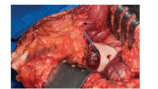
• Muscles divided at least 5 cm distal to bone resection
• Stabilized by Myodesis :
1. stronger insertion, help maximize strength, and minimize atrophy
2. counterbalance their antagonists, preventing contractures
Hemostasis
• Use of a tourniquet is desirable
• Larger vessels should be doubly ligated
• The tourniquet should be deflated before closure
• A drain should be used in most cases for 48 to 72 hours
Nerves
• Nerves isolated, pulled distally to the wound, and divided with a
sharp knife so that the cut end retracts well proximal to the level of
bone resection
• Strong tension on the nerve should be avoided
• Large nerves : contain relatively large arteries and should be ligated
• A neuroma always forms after a nerve has been divided
Bone
• Excessive periosteal stripping : Leads to ring sequestra or bony
overgrowth
• Bony prominences : well padded by soft tissue , remaining bone
rasped for smooth contour
OPEN AMPUTATION
• Skin is not closed at stump
• Indicated in infections and in severe traumatic wounds
• First operation is to remove devitalized structures
• vacuum-assisted closure : VAC is reapplied until the wound is ready
for closure
• Second sitting is to construct a satisfactory stump by secondary
closure, reamputation, revision, or plastic repair
POSTOPERATIVE CARE
• Multidisciplinary team approach
• Antibiotics , Analgesics ,DVT prophylaxis, pulmonary hygiene and
psychiatric help
• The stump is elevated by raising the foot of the bed : edema and
postoperative pain
• With transfemoral amputations : prevent flexion or abduction
contractures
• If non-weight bearing ambulation : rigid dressing ( POP ) applied.
• If weight bearing ambulation : true prosthetic cast with metal pylon
with a prosthetic foot
Advantages of rigid dressing
• Prevent edema at the surgical site
• Protect the wound from bed trauma
• Enhance wound healing
• Decrease postoperative pain
• Transtibial amputations : prevent knee flexion contractures
• Exercises, mobilized from bed to chair for the stump started on POD 1
• Regardless of when prosthetic ambulation is begun, the rigid dressing
should be removed and the wound inspected in 7 to 10 days
• Cast should be changed weekly
• rigid dressing is continued until the volume appears unchanged from
the previous week
• If the wound is progressing well, weight bearing can progress in 25-lb
increments each week
COMPLICATIONS
Hematoma
• Hemostasis before closure , use of a drain, rigid dressing has
minimized
• Delay wound healing and serve as a culture medium for bacterial
infection
• If it form : compressive dressing , evacuated if delaying wound healing
INFECTION
• More common in amputations for PVD, especially in diabetic patients.
• Deep wound infection : débridement and irrigation
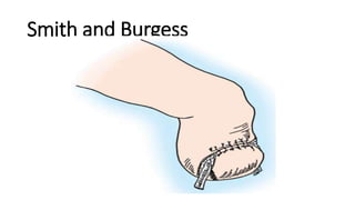
Smith and Burgess
WOUND NECROSIS
• Necrosis of the skin edges less than 1 cm can be treated
conservatively with open wound management
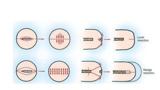
• Severe necrosis with poor coverage of the bone end, wedge resection
may be indicated.
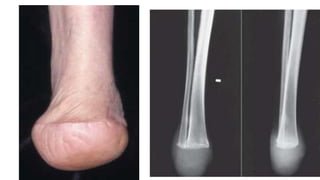
• Basic principle of wedge resection : reformation of the hemisphere,
while minimizing local pressures
CONTRACTURES
• Mild or moderate contractures of the joints of an amputation stump
treated by proper positioning of the stump, passive stretching, and
exercises
• Severe fixed contractures : wedging casts and surgical release
PAIN
• 1. Mechanical low back pain : more prevalent in amputees , proper
prosthetic ambulation to minimize abnormal stresses on the lumbar
spine.
• 2. Residual limb pain : a. Phantom Limb Pain
b. Poor fitting prosthesis
c. Painful neuroma
Phantom Limb Pain
• Silas Weir Mitchell (1829-1914) is credited with coining the term
phantom limb
• Caused by elimination or interruption of sensory nerve impulses
• PREVALENCE: 60% to 80%
• Not influenced by age in adults, gender, side, or level and cause of the
amputation
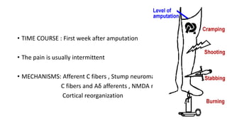
• TIME COURSE : First week after amputation
• The pain is usually intermittent
• MECHANISMS: Afferent C fibers , Stump neuromas
C fibers and Aδ afferents , NMDA receptor
Cortical reorganization
TREATMENT
• Amitriptyline: dose of 125 mg/day and tramadol
• Oral morphine: a significant reduction in phantom pain
• TNF – α antagonist : Etanercept
• Memantine at doses of 20 or 30 mg/day ????
• TENS , acupuncture , massage , Mirror therapy
Prevention
• Preoperative epidural blockade : lower incidence
• Catheter into the transected nerve sheath at the time of amputation
and infused bupivacaine for 72 hours
Poor fitting prosthesis
• Ill fitting with socket
• Stump should be evaluated for areas of abnormal pressure, especially
over bony prominences
• Distal stump edema, often called ―”choking” , may result if the end is
not completely seated in the prosthesis, and ulceration or gangrene
could result.
Painful neuroma
• Occurs when the nerve end is subjected to pressure or repeated
irritation
• A painful neuroma usually is easily palpable and often has a positive
Tinel sign
• Socket modification , neuroma excision or a more proximal
neurectomy
DERMATOLOGICAL
• Contact dermatitis : intense itching and burning
• Cause :
1.failure to rinse detergents from stump socks
2. nickel, chromates , antioxidants in rubber
Treatment : steroid cream , removal of the irritant
Bacterial folliculitis
• Areas of hairy, oily skin
• Exacerbate by poor hygiene.
• Improved hygiene and possibly socket modifications to relieve areas
of abnormal pressure
Verrucous hyperplasia
• Wartlike overgrowth of the skin at the end of the stump
• It is caused by proximal constriction that prevents the stump from
fully seating in the prosthesis
• This “choking”: causes distal stump edema followed by thickening of
the skin, fissuring, ulceration
• Salicylic acid to soften the keratin and Socket modification to prevent
recurrences
• Epidermoid cysts develop at the socket brim
• Treated with socket modification or Excision if required
AMPUTATION IN CHILDREN
• Divided into two general categories :
congenital (60%) and acquired (40%)
• Causes of congenital amputations:
Amniotic band syndrome
Exposure to teratogens ( thalidomide )
Polydactyly ,Syndactyly ,Macrodactyly
Congenital pseudoarthrosis of the tibia and fibula, radius and ulna
Congenital deficiencies of the long bones
Acquired amputations
• Secondary to trauma :
Power tool injuries are the most common
Motor vehicle accidents
• Neoplasm
• Infection
PRINCIPLES OF CHILDHOOD AMPUTATION
• Preserve length AND Preserve important growth plates
• Perform disarticulation rather than transosseous amputation
• Preserve the knee joint whenever possible
• Stabilize and normalize the proximal portion of the limb
Preserving length and growth plates
• 75% of the growth of the femur occurs at the distal growth plate :
transfemoral amputation performed in a young child would result in a
very short stump as an adult
• Conversely, even a very short transtibial stump in a young child may
result in a functional stump as an adult
Dysarticulation
• Provide well-balanced, sturdy stump capable of end weight bearing
• High mechanical demands that children place on their prostheses
• Secondary to preservation of the metaphyseal flares: Prosthetic
outcome is improved
TERMINAL BONE OVERGROWTH
• Appositional new bone formation and is unrelated to the growth of
the physis
• In a child amputee with a transosseous amputation
• More common in younger children : humerus and fibula
• Resulting bone is elongated and often pencil-shaped
• Swelling, edema, pain , may penetrate the skin
• It does not occur after disarticulation
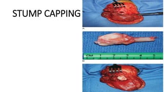
• Prevention of overgrowth : capping by epiphyseal graft OR capping
with tricortical iliac crest graft
• Treated effectively with surgical resection of the excess bone
STUMP CAPPING PROCEDURE
ADVANTAGES OF AMPUTATION IN
CHILDREN
• Children often can tolerate procedures on amputation stumps like
1. More forceful skin traction
2. Application of extensive skin grafts
3. Closure of skin flaps under moderate tension
Less severe Complications
• Painful phantom sensations do not develop
• Neuromas rare
• Extensive scars are tolerated well
• Psychological problems after amputation are rare in children
• Children use prostheses extremely well : High activity level
• Good growth potential: Prosthetic program should be designed that
parallels normal motor development
• Modifications may be made, such as the addition of a knee joint, a
mobile elbow joint
BELOW KNEE AMPUTATION
• Transtibial amputations are the most common amputations
performed for PVD
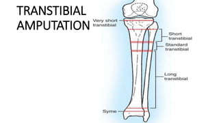
TRANSTIBIAL
AMPUTATION –
NONISCHEMIC LIMB AMPUTATION
• The optimal level of amputation determined by:
Level of trauma
clean end margins for tumor
Anatomical factors : stumps extending to the distal third of the leg
Less soft tissue for weight bearing
Distal third of the leg is relatively avascular and slower to heal
IDEAL
A stump length that allows a controlling lever arm for the prosthesis
i.e stump is to allow 2.5 cm of bone length for each 30 cm of body
height
Stumps lacking quadriceps function are not useful
Transecting the hamstring tendons
In a short stump whether fibula should be removed or preserved is
controversial as fitting of the prosthesis depends on it
Sufficient ―circulation‖ for healing and soft tissue‖ for protective end
weight bearing
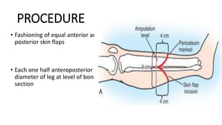
PROCEDURE
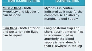
• Fashioning of equal anterior and
posterior skin flaps
• Each one half anteroposterior
diameter of leg at level of bone
section
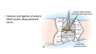
• Division and ligation of anterior
tibial vessels ,deep peroneal
nerve.
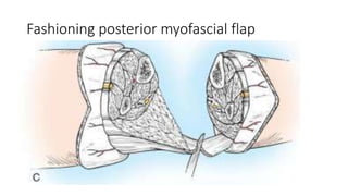
Fashioning posterior myofascial flap
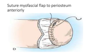
Suture myofascial flap to periosteum
anteriorly
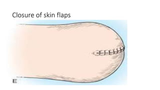
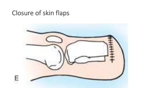
Closure of skin flaps
AFTERTREATMENT
• An immediate postoperative rigid dressing : control edema , limits
knee flexion contracture and trauma
• Mobilization with walker or crutches
• Cast can be changed every 5 to 7 days for skin care
• Within 3 to 4 weeks, prosthesis given
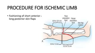
PROCEDURE FOR ISCHEMIC LIMB
• Fashioning of short anterior and
long posterior skin flaps
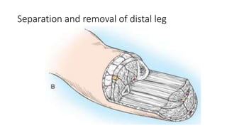
Separation and removal of distal leg
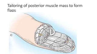
Tailoring of posterior muscle mass to form
flaps
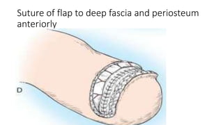
Suture of flap to deep fascia and periosteum
anteriorly
Closure of skin flaps
DISADVANTAGES OF LONG POSTERIOR FLAP
The stump, in the initial stages , unsuitable due to stump oedema :
delay in prosthetic fitting
Suture line passes over the distal end of the tibia: vulnerable to
trauma : due to the high pressure generated while using the
prosthesis
SKEW FLAP
The principle : thermographic mapping of the leg : higher
temperature on anteromedial AND posterolateral aspect
Indicates : Better blood flow
Anteromedial ( saphenous nerve artery)
Posterolateral (sural nerve artery)
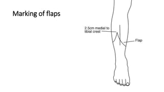
Marking of flaps
Advantages of skew flap
• Less of stump edema : NO Hazards of constricting bandages
• Early healing of the skin incision
• Early application and ambulation in a prosthetic cast
• Lessened risk of wound breakdown
ANKLE AMPUTATION
• Syme’s Amputation
• Modified Syme’s Amputation ( Sarmiento)
• Boyd Amputation
Syme’s Amputation
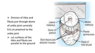
• Amputation at the distal tibia and fibula 0.6 cm proximal to the
periphery of the ankle joint and passing through the dome of the
ankle centrally
• The tough, durable skin of the heel flap provides normal weight
bearing skin
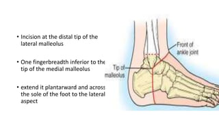
• Incision at the distal tip of the
lateral malleolus
• One fingerbreadth inferior to the
tip of the medial malleolus
• extend it plantarward and across
the sole of the foot to the lateral
aspect
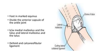
• Foot in marked equinus
• Divide the anterior capsule of
the ankle joint
• b/w medial malleolus and the
talus and lateral malleolus and
the talus
• Deltoid and calcaneofibular
ligament
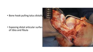
• Bone hook pulling talus distally,
• Exposing distal articular surface
of tibia and fibula
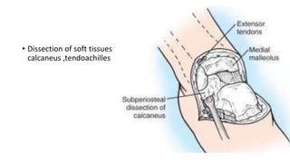
• Dissection of soft tissues from
calcaneus ,tendoachilles
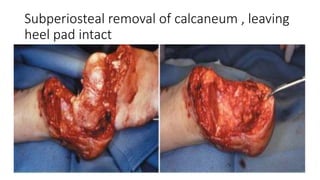
Subperiosteal removal of calcaneum , leaving
heel pad intact
 Division of tibia and
fibula just through dome
of ankle joint centrally
0.6 cm proximal to the
ankle joint
 cut surfaces of the
tibia and fibula are
parallel to the ground
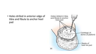
• Holes drilled in anterior edge of
tibia and fibula to anchor heel
pad
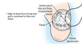
• Edge of deep fascia lining heel
pad is anchored to tibia and
fibula
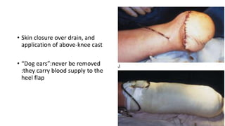
• Skin closure over drain, and
application of above-knee cast
• “Dog ears”:never be removed
:they carry blood supply to the
heel flap
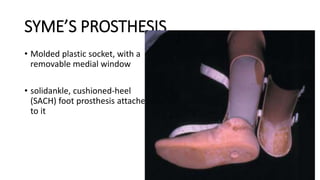
SYME’S PROSTHESIS
• Molded plastic socket, with a
removable medial window
• solidankle, cushioned-heel
(SACH) foot prosthesis attached
to it