CONGENITAL TALIPES EQUINO VARUS (CTEV)
•Download as PPTX, PDF•
2 likes•2,950 views

The document discusses congenital talipes equino varus (clubfoot). It is a birth deformity where the foot is twisted inward and downward. It involves muscle, tendon and bone abnormalities. Causes may be genetic or due to in-utero factors. Treatment involves manipulation, serial casting and sometimes surgery to correct the deformity. The goal is to fully correct the clubfoot early in life through non-surgical or surgical methods and maintain the correction through bracing and exercises.

Recommended
More Related Content
What's hot
What's hot (20)
Similar to CONGENITAL TALIPES EQUINO VARUS (CTEV)
Similar to CONGENITAL TALIPES EQUINO VARUS (CTEV) (20)
More from Ashish kumar Sharma
Recently uploaded
Recently uploaded (20)
CONGENITAL TALIPES EQUINO VARUS (CTEV)
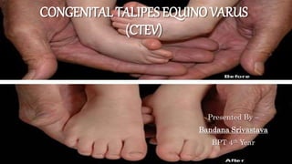
- 1. CONGENITAL TALIPES EQUINO VARUS (CTEV) Presented By – Bandana Srivastava BPT 4th Year
- 2. INTRODUCTION • Also known as clubfoot. • Congenital talipes equino varus describes a deformity noted at the birth and includes idiopathic as well as non-idiopathic talipes equino varus. • In non idiopathic group it is a manifestation of a systemic skeletal syndrome; the associated skeletal anomalies are due to the same etiological factor that caused failure of the normal development as in: i. From muscle imbalance e.g. neuromuscular disorders. ii. From fibrosis of soft tissue as in Arthrogryphosis iii. From bone and joint anomalies.
- 3. • Talipes Equinovarus comes from the following: 1. “Tali” means Ankle, 2. “Pes” means Foot 3. “Equinus” means foot pointing down (like a horse’s foot) 4. “Varus” means deviated towards midline
- 4. THEORIES Theories to explain 1. IDIOPATHIC CTEV - i. Mechanical pressure in utero e.g.: Oligohydraminos ii. Neuromuscular defect — Spina bifida, Weak peroneal muscles iii. Germ cell defect iv. Intrauterine arrest of the growth. v. Hereditary. vi. Multifactorial.
- 5. 2. NON-IDIOPATHIC CAUSES: i. Arthrogryphosis ii. Nail patella syndrome iii. Streeter syndrome iv. Muscular dystrophy v. Myelomeningocele, Spina bifida, Spinal cord defects
- 6. EPIDEMOLOGY • DEMOGRAPHICS - • Most common musculoskeletal birth defect • Overall incidence 1:1,000, though some populations 1:250 • Male: Female ratio approximately 2:1 • ANATOMIC LOCATION - • half of cases are bilateral • in 80%, clubfoot is an isolated deformity
- 7. ETIOLOGY • PATHOPHYSIOLOGY – 1. Muscle contractures contribute to the characteristic deformity that includes (CAVE): i. Cavus - (tight intrinsic, FHL, FDL) ii. Adductus of forefoot - (tight tibialis posterior) iii. Varus - (tight tendoachilles, tibialis posterior, tibialis anterior) iv. Equinus - (tight tendoachilles) 2. Bony deformity consists of medial spin of the midfoot and forefoot relative to the hindfoot: i. Talar neck is medially and plantarly deviated ii. Calcaneus is in varus and rotated medially around talus iii. Navicular and Cuboid are displaced medially
- 8. • GENETICS – 1. Genetic component is strongly suggested 2. Unaffected parents with affected child have 2.5% - 6.5% chance of having another child with a clubfoot 3. Familial occurrence in 25% 4. Recent link to PITX1, transcription factor critical for limb development
- 9. PATHOLOGICAL ANATOMY • The clubfoot deformity is due to the abnormal relationship of the tarsal bones: the navicular and calcaneus are displaced around the tarsus. • Correction of this abnormal tarsal relationship is resisted by pathological contracture of the associated softer parts. • The severity of the deformity depends on the degree of displacement, whereas the resistance to the treatment is determined by the rigidity of the soft tissue structures. • Two laws used for understanding are: 1. Wolf’s Law 2. Davis Law
- 10. 1. WOLF’S LAW: every change in the use of the static function of the bone causes a change in the internal form as well as the architecture and also the external form and function according to mathematical law. 2. DAVIS LAW: When ligaments and soft tissue in lax state they will shorten.
- 11. ANATOMICAL REGION WISE INVOLVEMENT 1. POSTERIOR CONTRACTURE: Tend Achilles, Tibiotalar capsule, talo calcaneal capsule, posterior talo fibular ligament, calcaneo fibulas ligament. These structures resist equinus correction. 2. MEDIAL: Most important and most resistant structures Tibialis posterior, deltoid, talonavicular capsule and spring ligament. 3. SUBTALAR: Talo calcaneal interosseousligament, bifurcated Y ligament. 4. PLANTAR CONTRACTURES: Abductor Hallucis, intrinsic flexors, quadratusplantae, plantar aponeurosis.
- 12. CLINICAL EXAMINATION • Smaller stubby feet with shortened first metatarsal ray. • Equinus deformity with inversion of the heel, adduction and varus of the fore foot. • Medial border of the foot is concave and elevated, its plantar surface face up ward. • Lateral border of the foot is convex and depressed down. • The posterior tuberosity of the heel is upwards, difficult to palpate and less visible. • Callosity on the dorsal aspect of the fifth metatarsal. • Boney prominence visible and palpable over the dorsolateral aspect of the foot represents the head and neck of the talus which are partially uncovered by the navicular
- 14. X-RAYS • A-P view 1. Tibio calcaneal angle normal range 20-40 degrees, abnormal if less than 20 degrees. • Lateral film in maximum dorsiflexion: 1. Talocalcaneal angle normal range 25-50 degrees, abnormal if less than 25 degrees. 2. Tibio calcaneal angle normal range 5-15 degrees, abnormal if less than 5 degrees or negative.
- 15. • KITES VIEW: AP view with foot flexed 30 degrees and tube angled 30 degree anteriorly in sagittal plane. Importance of x-ray on follow-up - Clinically the heel varus may appear to be corrected because manipulation may have displaced the heel pad laterally, but x-ray will demonstrate on abnormal tarsal relationship between talus and calcaneus confirming whether one is dealing with spurious correction.
- 16. MANAGEMENT OF CTEV • Aims – 1. To correct the deformity early 2. To correct the deformity fully 3. Hold the correction until growth stops
- 17. NON-OPERATIVE TREATMENT • Manipulation and serial casting • Stretching and adhesive strapping • Dennis - Browne splinting.
- 18. MANIPULATION AND SERIAL CASTING • Should begin in nursery ideally • Manipulation before the cast application is most important part of nonoperative treatment. The objective is to stretch the soft tissue contracture, the plaster of paris cast serves to maintain the correction obtained by manipulation. METHOD OF CASTING: • KITES - Each component of deformity corrected in the sequence. Kite believed that heel varus would correct simply by everting the heel. • PONSETTI - All component of the deformity must be corrected simultaneously, not in the sequence except for equinus, which should be corrected last. The cavus, which
- 19. arises from the pronation of the forefoot in relation to the hind foot is corrected by supinating the fore foot in proper alignment with the hind foot. With the arch well molded, the entire foot can be gently and gradually abducted under the talus, which is secured against rotation, in the ankle mortise by applying counter pressure with thumb against the lateral part of the talus. Heel varus will get corrected when the entire foot is entirely abducted. Finally equinus is corrected by dorsiflexing the foot, which can be facilitated by simple percutaneous tenotomy of the tend Achilles. Well molded plaster cast applied after manipulation is complete. FREQUENCY OF CAST CHANGE – • Ideally weekly but practically done fortnightly.
- 20. ON REASSESSMENT – • IF COMPLETELY CORRECTED: 1. Maintain in maximally corrected position for total of 6-8 months. 2. After 6- 8 months Dennis Browne bar with attached tarso pronator shoe for 24 hrs. 3. Checked at routine intervals for recurrence, mother also taught to look for heel cord shortening. 4. Once walking age attained only tarso pronator shoe with the Dennis Browne splint at night. 5. Night time splinting continued till 7 years of age. CTEV shoes used in day time. • PARTIALLY CORRECTED OR NO CORRECTION: 1. Observed for further 3 months with manipulation and casting. 2. If no correction, static deformity may require surgery at 10 months.
- 21. DENIS BROWNE SPLINT – • A dynamic splint in which the kicking movement of each leg exerts a corrective force on the counter part. RELAPSED FOOT – • The deformity recurred after Fair correction. RESISTANT FOOT – • Foot is considered resistant when the deformity shows no evidences of further improvement with manipulation the radiograph and the X rays confirming the persistence of equinovarus deformity.
- 23. OPERATIVE MANAGEMENT • INDICATIONS – 1. When a plateau has been reached in non operative treatment. 2. The child is old enough for the anatomy of foot to be recognized usually by ten months. • TREATMENT – 1. Soft tissue release 2. Osteotomy 3. Arthrodesis
- 24. • POTENTIAL COMPLICATIONS – 1. Infection and wound breakdown 2. AVN of talus 3. Overcorrection
- 25. RESIDUAL DEFORMITY • Must ensure that there is no neurologic cause. The residual deformity may be – 1. Dynamic - If unable to actively evert the foot. Consider SPLATT (Split ant. Tibialis transfer). 2. Fixed - Look for the uncorrected component and treat accordingly. • METATARSUS ADDUCTUS - after 5 year MT osteotomy • HIND FOOT VARUS – 1. < 2-3 year - complete subtalar release 2. 3-10 year - closed wedge or medial open edge osteotomy of calcaneum;
- 26. • EQUINUS - TA lengthening with posterior Capsulectomy of ankle, Subtalar joint. • All three deformities severe, resistant: TRIPLE ARTHRODESIS
- 27. PHYSIOTHERAPY MANAGEMENT • CORRECTION PHASE – 1. Daily corrective manipulations of the clubfoot are performed by an experienced physical therapist and the correction is held with elastic taping and splints until the next day's session. 2. Family participation is integral to the success of this treatment program as the family must be able to bring the infant to therapy during the week for 1-3 months. 3. Each session lasts approximately 30 mins per foot and manipulations are performed in a progressive gentle pattern.
- 28. 4. Begin with derotation of the calcaneopedal block and correction of forefoot adduction through massage of the Achilles tendon and gastrocnemius muscle. 5. Medial soft tissues are stretched to allow the navicular to move away from the medial malleolus and its medial position on the head of the talus. Distraction of the forefoot and midfoot helps to loosen the tightened structures, and derotation of the foot facilitates reduction of the talus 6. To maintain the gain achieved in passive range of motion, the toe extensors and peroneals are recruited by stimulating (tickling) the lateral border of the foot and leg and the tops of the toes. 7. Once the talonavicular joint has been reduced, attention is directed toward the correction of varus and equinus. With the valgus maneuver, the calcaneus gradually moves to a neutral and eventually valgus position. The ankle is externally rotated at the same time that the calcaneus is being mobilized into valgus. The knee should be kept at 90° during these maneuvers
- 29. 8. Equinus is corrected with gradual dorsiflexion of the foot. Correction of equinus can be augmented with a percutaneous heel cord tenotomy • MAINTENANCE PHASE – 1. Periodic follow-up is needed to monitor the range of motion of the foot and the development of the infant and to fabricate new splints. 2. Once the patient is walking, taping is discontinued and a resting ankle-foot orthosis is used during nighttime and naps until the age of two years. 3. Throughout this treatment program, the patient visits the physician every two to three months for evaluation of the foot.