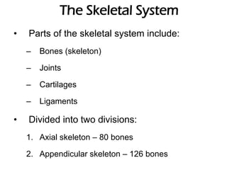
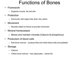
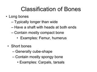
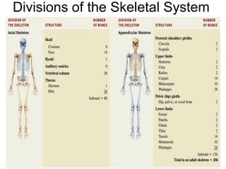
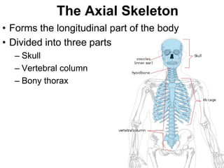
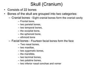
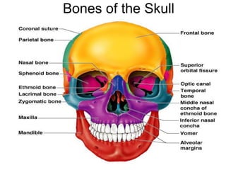
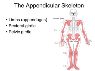
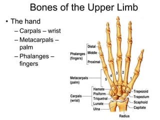
SKELETAL SYSTEM
Prepared By
Doppalapudi Sandeep
M. Pharmacy,
Assistant Professor
Department of Physiology & Pharmacology
Chebrolu Hanumaiah Institute of Pharmaceutical
Sciences, Chandramoulipuram, Chowdavaram,
Guntur, Andhra Pradesh, India – 522019
The Skeletal System
• Parts of the skeletal system include:
– Bones (skeleton)
– Joints
– Cartilages
– Ligaments
• Divided into two divisions:
1. Axial skeleton – 80 bones
2. Appendicular skeleton – 126 bones
Functions of Bones
• Framework
– Supports muscle, fat and skin
• Protection
– Surrounds vital organs like skull, ribs, pelvis
• Movement
– Muscles attach to bones to provide movement
• Mineral homeostasis
– Stores and maintain minerals (Calcium & phosphorus)
• Production of blood cells
– Red bone marrow – produes Red and white blood cells and platelets
• Storage
– Calcium
– Yellow bone marrow – has adipocytes – stores fat.
Classification of Bones
• Long bones
– Typically longer than wide
– Have a shaft with heads at both ends
– Contain mostly compact bone
• Examples: Femur, humerus
• Short bones
– Generally cube-shape
– Contain mostly spongy bone
• Examples: Carpals, tarsals
Classification of Bones
• Flat bones
– Thin and flattened, usually curved
– Thin layers of compact bone around a layer
of spongy bone
• Examples: Skull, ribs, sternum
• Irregular bones
– Irregular in shape
– Do not fit into other bone classification
categories
• Example: Vertebrae and hip
Gross Anatomy
of a Long Bone
• Diaphysis
– Shaft
– Composed of compact
bone
• Epiphysis
– Ends of the bone
– Composed mostly of
spongy bone
Divisions of the Skeletal System
The Axial Skeleton
• Forms the longitudinal part of the body
• Divided into three parts
– Skull
– Vertebral column
– Bony thorax
Skull (Cranium)
• Consists of 22 bones
• Bones of the skull are grouped into two categories:
– Cranial bones - Eight cranial bones form the cranial cavity
– Frontal bone,
– two parietal bones,
– two temporal bones,
– the occipital bone,
– the sphenoid bone,
– ethmoid bone
– Facial bones - Fourteen facial bones form the face
– Two nasal bones,
– two maxillae,
– two zygomatic bones,
– the mandible,
– two lacrimal bones,
– two palatine bones,
– two inferior nasal conchae and vomer
Skull
• The cranial and facial bones protect and support
special sense organs and the brain.
• Besides forming the large cranial cavity, the skull
also forms several smaller cavities
– Nasal cavity
– Orbits (eye sockets)
– Paranasal sinuses
– Small cavities which house organs involved in hearing
and equilibrium
Bones of the Skull
Skull
• Immovable joints called sutures fuse most of the skull
bones together.
• The skull provides large areas of attachment for
muscles that move various parts of the head.
• Skull and facial bones provide attachment for muscles
that produce facial expressions.
• The facial bones form the framework of the face and
provide support for the entrances to the digestive and
respiratory systems.
Skull (Cranial Bones)
• Frontal Bone
– Forms the forehead
• Parietal Bones
– Form the sides and roof of the cranial cavity
• Temporal Bones
– Form the lateral aspects and floor of the cranium
• Occipital Bone
– Forms the posterior part and most of the base of the cranium
• Sphenoid Bone
– Lies at the middle part of the base of the skull
• Ethmoid Bone
– Located on the midline in the anterior part of the cranial floor medial to
the orbits
– A major superior supporting structure of the nasal cavity
– Contain thin projections called conchae which are lined by mucous
membranes
– Increased surface area in the nasal cavity helps to humidify inhaled air
trapping inhaled particles
Posterior View
Skull (Facial Bones)
• Nasal Bones
– Form the bridge of the nose
• Maxillae
– Form the upper jawbone and most of the hard palate
– Separates the nasal cavity from the oral cavity
• Zygomatic Bones (Cheek bones)
– form the prominences of the cheeks
• Lacrimal Bones
– Form a part of the medial wall of each orbit
• Palatine Bones
– Form the posterior portion of the hard palate
• Inferior Nasal Conchae
– Form a part of the inferior lateral wall of the nasal cavity
Skull (Facial Bones)
• Vomer
– Forms the inferior portion of the nasal septum
• Mandible
– Lower jawbone and the only movable skull bone
– The largest, strongest facial bone
• Nasal Septum
– Divides the interior of the nasal cavity into right and left sides
• Orbits
– Eye socket
• Foramina
– Openings for blood vessels, nerves or ligaments of the skull
Sphenoid Bone
Ethmoid Bone & Vomer
Mandible
Paranasal Sinuses
• Hollow portions of bones surrounding the
nasal cavity
• Functions:
– Lighten the skull
– Give resonance and
amplification to voice
– Warm and moisten
air
The Hyoid Bone
• The only bone that
does not articulate
with another bone
• Serves as a
moveable base for
the tongue
• The hyoid bone also
helps to keep the
larynx (voice box)
open at all times
Vertebral Column
• Also called the spine, backbone, or spinal column
• Functions:
– Protect the spinal cord
– Support the head
– Serve as a point of attachment for the ribs, pelvic girdle, and
muscles
• The vertebral column is curved to varying degrees in
different locations
– Curves increase the column strength
– Help maintain balance in the upright position
– Absorb shocks during walking, and help protect the vertebrae from
fracture
Vertebral Column
• Composed of a series of bones called vertebrae
(Adult=26)
– 7 cervical are in the neck region
– 12 thoracic are posterior to the thoracic cavity
– 5 lumbar support the lower back
– 1 sacrum consists of five fused sacral vertebrae
– 1 coccyx consists of four fused coccygeal vertebrae
Cervical Vertebrae(7)
Thoracic Vertebrae (12)
Lumbar Vertebrae (5)
Sacrum
Coccyx
Vertebral Column
Intervertebral Discs
• Found between the bodies of adjacent vertebrae
• Functions to:
– Form strong joints
– Permit various movements of the vertebral column
– Absorb vertical shock
• Vertebrae typically consist of:
– A Body (weight bearing)
– A vertebral arch (surrounds the spinal cord)
– Several processes (points of attachment for muscles)
Vertebral Column (Regions)
• Cervical Region
– Cervical vertebrae (C1–C7)
– The atlas (C1) is the first cervical vertebra
– The axis (C2) is the second cervical vertebra
• Thoracic Region
– Thoracic vertebrae (T1–T12)
– Articulate with the ribs
• Lumbar Region
– Lumbar vertebrae (L1–L5)
– Provide for the attachment of the large back muscles
• Sacrum
– The sacrum is a triangular bone formed by the union of five
sacral vertebrae (S1–S5)
– Serves as a strong foundation for the pelvic girdle
• Coccyx
– The coccyx, like the sacrum, is triangular in shape
– It is formed by the fusion of usually four coccygeal vertebrae
Cervical
Vertebrae
Thoracic
Vertebrae
Comparison of Vertebrae
Sacrum & Coccyx
Thorax
• Thoracic cage is formed by the:
– Sternum
– Ribs
– Costal cartilages
– Thoracic vertebrae
• Functions to:
– Enclose and protect the organs in the thoracic and
abdominal cavities
– Provide support for the bones of the upper limbs
– Play a role in breathing
Thorax
• Sternum
– “Breastbone” located in the center of the thoracic wall
– Consists of the manubrium, body, xiphoid process
• Ribs
– Twelve pairs of ribs give structural support to the sides of the
thoracic cavity
– True ribs (7 pairs), False ribs (3 Pairs) and floating ribs(2 pairs).
• Costal cartilages
– Costal cartilages contribute to the elasticity of the thoracic cage
THORAX
Rib
Sternum
• Three parts
– Manubrium
– Body
– Xiphoid process
The Appendicular Skeleton
• Limbs (appendages)
• Pectoral girdle
• Pelvic girdle
Upper Limb
• The pectoral girdle consists of two bones,
the scapula and the clavicle
• The free part has 30 bones
• 1 humerus (arm)
• 1 ulna (forearm)
• 1 radius (forearm)
• 8 carpals (wrist)
• 19 metacarpal and phalanges (hand)
The Pectoral (Shoulder) Girdle
• Composed of two bones
– Clavicle – collarbone
– Scapula – shoulder blade
• These bones allow the upper limb to have
exceptionally free movement.
• The clavicle is convex in shape anteriorly
near the sternal junction
• The clavicle is concave anteriorly on its
lateral edge near the acromion
Bones of the Shoulder Girdle
Pectoral Girdle - Scapula
• Also called the shoulder blade
• Triangular in shape
• Most notable features include the spine,
acromion, coracoid process and the
glenoid cavity
Bones of the Shoulder Girdle
Features on the Scapula
• Spine - a large process on the posterior of
the scapula that ends laterally as the
acromion
• Acromion - the flattened lateral portion of
the spine of the scapula
• Coracoid process - a protruding projection
on the anterior surface just inferior to the
lateral aspect of the clavicle
• Glenoid cavity - shallow concavity that
articulates with the head of the humerus
Skeleton of the Arm - Humerus
• Longest and largest bone of the free part
of the upper limb
• The proximal ball-shaped end articulates
with the glenoid cavity of the scapula
• The distal end articulates at the elbow with
the radius and ulna
Bones of the Upper Limb
• The arm is formed by a single bone- Humerus
Skeleton of the Forearm - Ulna
• The longer of the two forearm bones
• Located medial to the radius
• Olecranon - the large, prominent proximal end, the “tip of
your elbow”
• Coronoid process - the anterior “lip” of the proximal ulna
• Trochlear notch - the deep fossa that receives the
trochlea of the humerus during elbow flexion
• Styloid process - the thin cylindrical projection on the
posterior side of the ulna’s head
Radius
• Lies lateral to the ulna (thumb side of the forearm)
• The head (disc-shaped) and neck are at the
proximal end
• The head articulates with the capitulum of the
humerus and the radial notch of the ulna
• Radial tuberosity - medial and inferior to neck,
attachment site for biceps brachii muscle
• Styloid process - large distal projection on lateral
side of radius
Radius & Ulna
Bones of the Upper Limb
• The hand
– Carpals – wrist
– Metacarpals –
palm
– Phalanges –
fingers
Skeleton of the Hand
• The carpus (wrist) consists of 8 small bones
(carpals)
• Two rows of carpal bones
• Proximal row - scaphoid, lunate, triquetrum, pisiform
• Distal row - trapezium, trapezoid, capitate, hamate
• Scaphoid - most commonly fractured
• Carpal tunnel - space between carpal bones and
flexor retinaculum
Metacarpals and Phalanges
• Five metacarpals - numbered I-V, lateral to
medial
• 14 phalanges - two in the thumb (pollex)
and three in each of the other fingers
• Each phalanx has a base, shaft, and head
• Joints - carpometacarpal,
metacarpophalangeal, interphalangeal
Bones of the Pelvic Girdle
• Hip bones
• Composed of three pair of fused bones
– Ilium
– Ischium
– Pubic bone
• The total weight of the upper body rests on the pelvis
• Protects several organs
– Reproductive organs
– Urinary bladder
– Part of the large intestine
The Pelvis
The Pelvis: Right Coxal Bone
The Ilium
• Largest of the three hip bones
• Ilium is the superior part of the hip bone
• Consists of a superior ala and inferior body which
forms the acetabulum (socket for head of the femur)
• Superior border - iliac crest
• Hip pointer - occurs at anterior superior iliac spine
• Greater sciatic notch - allows passage of sciatic nerve
Ischium and Pubis
• Ischium - inferior and posterior part of the hip
bone
• Most prominent feature is the ischial
tuberosity, it is the part that meets the chair
when you are sitting
• Pubis - inferior and anterior part of the hip
bone
• Superior and inferior rami and body
Comparing Male and Female Pelves
FEMUR
Skeleton of the Thigh - Femur and Patella
• Femur - longest, heaviest, and strongest bone in the
body
• Proximally, the head articulates with the acetabulum
of the hip bone forming the hip (coxal) joint
• Neck - distal to head, common site of fracture
• Distally, the medial and lateral condyles articulate
with the condyles of the tibia forming the knee joint
• Also articulates with patella
Patella
• Largest sesamoid bone in the body
• Forms the patellofemoral joint
• Superior surface is the base
• Inferior, narrower surface is the apex
• Thick articular cartilage lines the posterior surface
• Increases the leverage of the quadriceps femoris
muscle
TIBIA & FIBULA
Tibia (shin bone)
• The larger, medial weight-bearing bone of leg
• The lateral and medial condyles at the proximal
end articulate with the femur
• It articulates distally with the talus and fibula
• Tibial tuberosity - attachment site for the patellar
ligament
• Medial malleolus - medial surface of distal end
(medial surface of ankle joint)
Fibula
• The smaller, laterally placed bone of leg
• Non-weight bearing
• The head forms the proximal tibiofibular
joint
• Lateral malleolus - distal end, articulates
with the tibia and the talus at the ankle
Bones of Lower Limbs
• The foot
– Tarsus – ankle
– Metatarsals – sole
– Phalanges – toes
Skeleton of the Foot - Tarsals,
Metatarsals & Phalanges
• Seven tarsal bones - talus (articulates with
tibia and fibula), calcaneus (the heel bone,
the largest and strongest), navicular, cuboid
and three cuneiforms
• Five metatarsals - (I-V) base, shaft, head
• 14 phalanges (big toe is the hallux)
• Tarsus = ankle

Skeletal System presentation for college.pdf

  • 1.
    SKELETAL SYSTEM Prepared By DoppalapudiSandeep M. Pharmacy, Assistant Professor Department of Physiology & Pharmacology Chebrolu Hanumaiah Institute of Pharmaceutical Sciences, Chandramoulipuram, Chowdavaram, Guntur, Andhra Pradesh, India – 522019
  • 2.
    The Skeletal System •Parts of the skeletal system include: – Bones (skeleton) – Joints – Cartilages – Ligaments • Divided into two divisions: 1. Axial skeleton – 80 bones 2. Appendicular skeleton – 126 bones
  • 3.
    Functions of Bones •Framework – Supports muscle, fat and skin • Protection – Surrounds vital organs like skull, ribs, pelvis • Movement – Muscles attach to bones to provide movement • Mineral homeostasis – Stores and maintain minerals (Calcium & phosphorus) • Production of blood cells – Red bone marrow – produes Red and white blood cells and platelets • Storage – Calcium – Yellow bone marrow – has adipocytes – stores fat.
  • 4.
    Classification of Bones •Long bones – Typically longer than wide – Have a shaft with heads at both ends – Contain mostly compact bone • Examples: Femur, humerus • Short bones – Generally cube-shape – Contain mostly spongy bone • Examples: Carpals, tarsals
  • 5.
    Classification of Bones •Flat bones – Thin and flattened, usually curved – Thin layers of compact bone around a layer of spongy bone • Examples: Skull, ribs, sternum • Irregular bones – Irregular in shape – Do not fit into other bone classification categories • Example: Vertebrae and hip
  • 7.
    Gross Anatomy of aLong Bone • Diaphysis – Shaft – Composed of compact bone • Epiphysis – Ends of the bone – Composed mostly of spongy bone
  • 8.
    Divisions of theSkeletal System
  • 9.
    The Axial Skeleton •Forms the longitudinal part of the body • Divided into three parts – Skull – Vertebral column – Bony thorax
  • 10.
    Skull (Cranium) • Consistsof 22 bones • Bones of the skull are grouped into two categories: – Cranial bones - Eight cranial bones form the cranial cavity – Frontal bone, – two parietal bones, – two temporal bones, – the occipital bone, – the sphenoid bone, – ethmoid bone – Facial bones - Fourteen facial bones form the face – Two nasal bones, – two maxillae, – two zygomatic bones, – the mandible, – two lacrimal bones, – two palatine bones, – two inferior nasal conchae and vomer
  • 11.
    Skull • The cranialand facial bones protect and support special sense organs and the brain. • Besides forming the large cranial cavity, the skull also forms several smaller cavities – Nasal cavity – Orbits (eye sockets) – Paranasal sinuses – Small cavities which house organs involved in hearing and equilibrium
  • 12.
  • 13.
    Skull • Immovable jointscalled sutures fuse most of the skull bones together. • The skull provides large areas of attachment for muscles that move various parts of the head. • Skull and facial bones provide attachment for muscles that produce facial expressions. • The facial bones form the framework of the face and provide support for the entrances to the digestive and respiratory systems.
  • 14.
    Skull (Cranial Bones) •Frontal Bone – Forms the forehead • Parietal Bones – Form the sides and roof of the cranial cavity • Temporal Bones – Form the lateral aspects and floor of the cranium • Occipital Bone – Forms the posterior part and most of the base of the cranium • Sphenoid Bone – Lies at the middle part of the base of the skull • Ethmoid Bone – Located on the midline in the anterior part of the cranial floor medial to the orbits – A major superior supporting structure of the nasal cavity – Contain thin projections called conchae which are lined by mucous membranes – Increased surface area in the nasal cavity helps to humidify inhaled air trapping inhaled particles
  • 15.
  • 16.
    Skull (Facial Bones) •Nasal Bones – Form the bridge of the nose • Maxillae – Form the upper jawbone and most of the hard palate – Separates the nasal cavity from the oral cavity • Zygomatic Bones (Cheek bones) – form the prominences of the cheeks • Lacrimal Bones – Form a part of the medial wall of each orbit • Palatine Bones – Form the posterior portion of the hard palate • Inferior Nasal Conchae – Form a part of the inferior lateral wall of the nasal cavity
  • 17.
    Skull (Facial Bones) •Vomer – Forms the inferior portion of the nasal septum • Mandible – Lower jawbone and the only movable skull bone – The largest, strongest facial bone • Nasal Septum – Divides the interior of the nasal cavity into right and left sides • Orbits – Eye socket • Foramina – Openings for blood vessels, nerves or ligaments of the skull
  • 18.
  • 19.
  • 20.
  • 21.
    Paranasal Sinuses • Hollowportions of bones surrounding the nasal cavity • Functions: – Lighten the skull – Give resonance and amplification to voice – Warm and moisten air
  • 22.
    The Hyoid Bone •The only bone that does not articulate with another bone • Serves as a moveable base for the tongue • The hyoid bone also helps to keep the larynx (voice box) open at all times
  • 23.
    Vertebral Column • Alsocalled the spine, backbone, or spinal column • Functions: – Protect the spinal cord – Support the head – Serve as a point of attachment for the ribs, pelvic girdle, and muscles • The vertebral column is curved to varying degrees in different locations – Curves increase the column strength – Help maintain balance in the upright position – Absorb shocks during walking, and help protect the vertebrae from fracture
  • 24.
    Vertebral Column • Composedof a series of bones called vertebrae (Adult=26) – 7 cervical are in the neck region – 12 thoracic are posterior to the thoracic cavity – 5 lumbar support the lower back – 1 sacrum consists of five fused sacral vertebrae – 1 coccyx consists of four fused coccygeal vertebrae
  • 25.
    Cervical Vertebrae(7) Thoracic Vertebrae(12) Lumbar Vertebrae (5) Sacrum Coccyx Vertebral Column
  • 26.
    Intervertebral Discs • Foundbetween the bodies of adjacent vertebrae • Functions to: – Form strong joints – Permit various movements of the vertebral column – Absorb vertical shock • Vertebrae typically consist of: – A Body (weight bearing) – A vertebral arch (surrounds the spinal cord) – Several processes (points of attachment for muscles)
  • 27.
    Vertebral Column (Regions) •Cervical Region – Cervical vertebrae (C1–C7) – The atlas (C1) is the first cervical vertebra – The axis (C2) is the second cervical vertebra • Thoracic Region – Thoracic vertebrae (T1–T12) – Articulate with the ribs • Lumbar Region – Lumbar vertebrae (L1–L5) – Provide for the attachment of the large back muscles • Sacrum – The sacrum is a triangular bone formed by the union of five sacral vertebrae (S1–S5) – Serves as a strong foundation for the pelvic girdle • Coccyx – The coccyx, like the sacrum, is triangular in shape – It is formed by the fusion of usually four coccygeal vertebrae
  • 28.
  • 30.
  • 31.
  • 32.
    Thorax • Thoracic cageis formed by the: – Sternum – Ribs – Costal cartilages – Thoracic vertebrae • Functions to: – Enclose and protect the organs in the thoracic and abdominal cavities – Provide support for the bones of the upper limbs – Play a role in breathing
  • 33.
    Thorax • Sternum – “Breastbone”located in the center of the thoracic wall – Consists of the manubrium, body, xiphoid process • Ribs – Twelve pairs of ribs give structural support to the sides of the thoracic cavity – True ribs (7 pairs), False ribs (3 Pairs) and floating ribs(2 pairs). • Costal cartilages – Costal cartilages contribute to the elasticity of the thoracic cage
  • 34.
  • 35.
  • 36.
    Sternum • Three parts –Manubrium – Body – Xiphoid process
  • 37.
    The Appendicular Skeleton •Limbs (appendages) • Pectoral girdle • Pelvic girdle
  • 38.
    Upper Limb • Thepectoral girdle consists of two bones, the scapula and the clavicle • The free part has 30 bones • 1 humerus (arm) • 1 ulna (forearm) • 1 radius (forearm) • 8 carpals (wrist) • 19 metacarpal and phalanges (hand)
  • 39.
    The Pectoral (Shoulder)Girdle • Composed of two bones – Clavicle – collarbone – Scapula – shoulder blade • These bones allow the upper limb to have exceptionally free movement. • The clavicle is convex in shape anteriorly near the sternal junction • The clavicle is concave anteriorly on its lateral edge near the acromion
  • 40.
    Bones of theShoulder Girdle
  • 41.
    Pectoral Girdle -Scapula • Also called the shoulder blade • Triangular in shape • Most notable features include the spine, acromion, coracoid process and the glenoid cavity
  • 42.
    Bones of theShoulder Girdle
  • 43.
    Features on theScapula • Spine - a large process on the posterior of the scapula that ends laterally as the acromion • Acromion - the flattened lateral portion of the spine of the scapula • Coracoid process - a protruding projection on the anterior surface just inferior to the lateral aspect of the clavicle • Glenoid cavity - shallow concavity that articulates with the head of the humerus
  • 44.
    Skeleton of theArm - Humerus • Longest and largest bone of the free part of the upper limb • The proximal ball-shaped end articulates with the glenoid cavity of the scapula • The distal end articulates at the elbow with the radius and ulna
  • 45.
    Bones of theUpper Limb • The arm is formed by a single bone- Humerus
  • 46.
    Skeleton of theForearm - Ulna • The longer of the two forearm bones • Located medial to the radius • Olecranon - the large, prominent proximal end, the “tip of your elbow” • Coronoid process - the anterior “lip” of the proximal ulna • Trochlear notch - the deep fossa that receives the trochlea of the humerus during elbow flexion • Styloid process - the thin cylindrical projection on the posterior side of the ulna’s head
  • 47.
    Radius • Lies lateralto the ulna (thumb side of the forearm) • The head (disc-shaped) and neck are at the proximal end • The head articulates with the capitulum of the humerus and the radial notch of the ulna • Radial tuberosity - medial and inferior to neck, attachment site for biceps brachii muscle • Styloid process - large distal projection on lateral side of radius
  • 48.
  • 49.
    Bones of theUpper Limb • The hand – Carpals – wrist – Metacarpals – palm – Phalanges – fingers
  • 50.
    Skeleton of theHand • The carpus (wrist) consists of 8 small bones (carpals) • Two rows of carpal bones • Proximal row - scaphoid, lunate, triquetrum, pisiform • Distal row - trapezium, trapezoid, capitate, hamate • Scaphoid - most commonly fractured • Carpal tunnel - space between carpal bones and flexor retinaculum
  • 51.
    Metacarpals and Phalanges •Five metacarpals - numbered I-V, lateral to medial • 14 phalanges - two in the thumb (pollex) and three in each of the other fingers • Each phalanx has a base, shaft, and head • Joints - carpometacarpal, metacarpophalangeal, interphalangeal
  • 52.
    Bones of thePelvic Girdle • Hip bones • Composed of three pair of fused bones – Ilium – Ischium – Pubic bone • The total weight of the upper body rests on the pelvis • Protects several organs – Reproductive organs – Urinary bladder – Part of the large intestine
  • 53.
  • 54.
  • 55.
    The Ilium • Largestof the three hip bones • Ilium is the superior part of the hip bone • Consists of a superior ala and inferior body which forms the acetabulum (socket for head of the femur) • Superior border - iliac crest • Hip pointer - occurs at anterior superior iliac spine • Greater sciatic notch - allows passage of sciatic nerve
  • 56.
    Ischium and Pubis •Ischium - inferior and posterior part of the hip bone • Most prominent feature is the ischial tuberosity, it is the part that meets the chair when you are sitting • Pubis - inferior and anterior part of the hip bone • Superior and inferior rami and body
  • 57.
    Comparing Male andFemale Pelves
  • 58.
  • 59.
    Skeleton of theThigh - Femur and Patella • Femur - longest, heaviest, and strongest bone in the body • Proximally, the head articulates with the acetabulum of the hip bone forming the hip (coxal) joint • Neck - distal to head, common site of fracture • Distally, the medial and lateral condyles articulate with the condyles of the tibia forming the knee joint • Also articulates with patella
  • 60.
    Patella • Largest sesamoidbone in the body • Forms the patellofemoral joint • Superior surface is the base • Inferior, narrower surface is the apex • Thick articular cartilage lines the posterior surface • Increases the leverage of the quadriceps femoris muscle
  • 61.
  • 62.
    Tibia (shin bone) •The larger, medial weight-bearing bone of leg • The lateral and medial condyles at the proximal end articulate with the femur • It articulates distally with the talus and fibula • Tibial tuberosity - attachment site for the patellar ligament • Medial malleolus - medial surface of distal end (medial surface of ankle joint)
  • 63.
    Fibula • The smaller,laterally placed bone of leg • Non-weight bearing • The head forms the proximal tibiofibular joint • Lateral malleolus - distal end, articulates with the tibia and the talus at the ankle
  • 64.
    Bones of LowerLimbs • The foot – Tarsus – ankle – Metatarsals – sole – Phalanges – toes
  • 65.
    Skeleton of theFoot - Tarsals, Metatarsals & Phalanges • Seven tarsal bones - talus (articulates with tibia and fibula), calcaneus (the heel bone, the largest and strongest), navicular, cuboid and three cuneiforms • Five metatarsals - (I-V) base, shaft, head • 14 phalanges (big toe is the hallux) • Tarsus = ankle