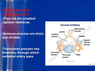
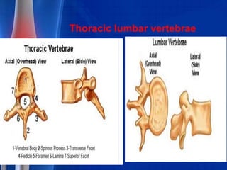
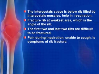
Downloaded 19 times


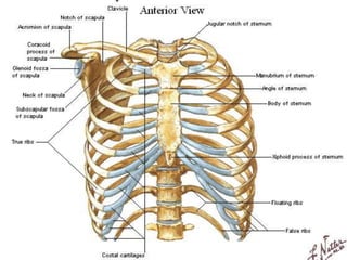
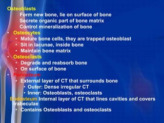
![Bony Thorax [thoracic cage]
Sternum, ribs and thoracic vertebrae form the
bony thorax.
Sternum
Formed by fusion of three
bones,(Manubrium,Body,and Xiphoid
process).
Attached to first seven pair of ribs.](https://image.slidesharecdn.com/t6skwtbarm2vehy77liu-signature-460517c25b85fc4e63c8080c3e27df73c8dfae9e0c6544cc7ea6d9e8b5e79cc7-poli-180213064029/85/Skeletal-system-pharma-36-320.jpg)


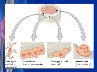


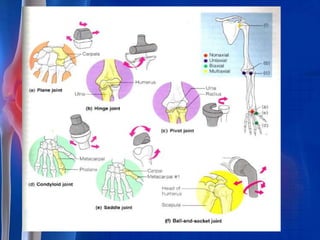

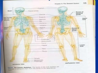
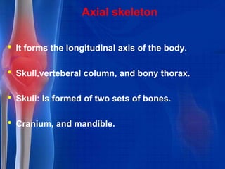
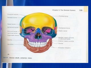
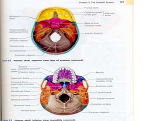
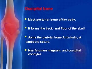
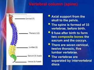
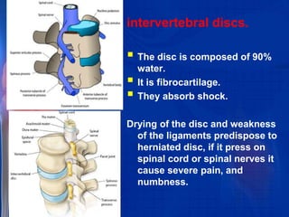
The skeletal system consists of bones, cartilages, and ligaments that provide structure, protection, movement, and blood cell formation. The axial skeleton forms the body's longitudinal axis and includes the skull, vertebral column, and thoracic cage. The appendicular skeleton includes the bones of the upper and lower limbs that attach to the axial skeleton. Long bones have a diaphysis shaft and epiphyses ends. The skull is formed from multiple flat bones that protect the brain and sense organs. The vertebral column consists of vertebrae separated by intervertebral discs that allow flexibility. The rib cage includes ribs and sternum and protects the heart and lungs.


