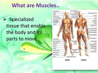
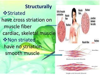
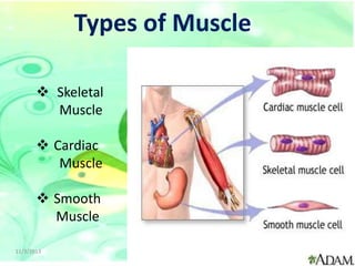
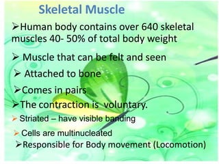
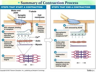
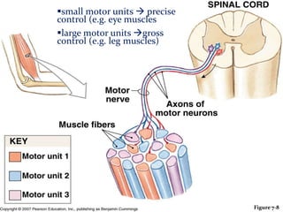
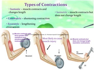
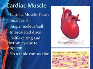
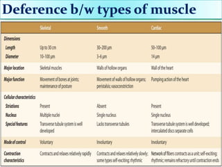
MUSCULAR SYSTEM
• Presented To :

SIR SHEERAZ
• Presented By:

ABBAS KHAN
What are Muscles..
 Specialized
tissue that enable
the body and its
parts to move.
Muscle
Derivation :

 Latin word mus
means mouse
 its due to many of
them resembles
mouse with tendon
representing tail

tendon

belly
Continue…
• A contractile
tissue which
bring about
movements
• Muscle can be
regarded as
motors of body
Properties of Muscle
• Excitability: capacity of muscle to respond
to a stimulus
• Contractility: ability of a muscle to shorten
and generate pulling force
• Extensibility: muscle can be stretched back
to its original length
• Elasticity: ability of muscle to recoil to
original resting length after stretched
Muscle Classification
Functionally
Voluntarily
can be moved at will
skeletal muscle
Involuntarily
can’t be moved at will
cardiac ,smooth muscle
Heart wall,Visceral organs
Structurally
Striated
have cross striation on
muscle fiber
cardiac, skeletal muscle
Non striated
have no striation
smooth muscle
Types of Muscle
 Skeletal
Muscle
 Cardiac
Muscle
 Smooth
Muscle
11/3/2013

9
Skeletal Muscle
Human body contains over 640 skeletal
muscles 40- 50% of total body weight

 Muscle that can be felt and seen
 Attached to bone
Comes in pairs.
The contraction is voluntary.
 Striated – have visible banding

 Cells are multinucleated

Responsible for Body movement (Locomotion)
Functions of muscle
 Body movement (Locomotion)
 Maintenance of posture
 Respiration
 Diaphragm and intercostals contractions

 Communication (Verbal and Facial)
 Constriction of organs and vessels
 Peristalsis of intestinal tract
 Vasoconstriction of b.v. and other structures

(pupils)

 Production of body heat (Thermo genesis)
11/3/2013

Jipmer Physiologist

12
11/3/2013

Jipmer Physiologist

13
Muscle ATTACHMENT
 Axial musculature
 Muscles with origins on the

axial skeleton that position
and move head, spine, rib
cage

 Appendicular
musculature
Muscles that stabilize or move
appendicular components

Copyright © 2007 Pearson Education, Inc., publishing as Benjamin Cummings
Muscle ATTACHMENT
Direct attachments

Fleshy part direct attached
e.g. intercostals muscle
 Indirect attachments

tendon or aponeurosis meet bones
Tubercles, trochanters, and crests
Movement of Muscles
 Origin: attachment
that remains stationary
or least mobile

origin
belly

 Insertion: attachment
of the muscle to the bone
that more mobile

 Belly: the fleshy part
of the muscle between
the tendons

insertion
Types of muscle fibers
SLOW FIBERS Type I
Half the diameter of
fast fibers
Take three times as
RESISTANT TO
FATIGUE
 Abundant

mitochondria
 Large capillary
supply
High concentrations
of myoglobin
11/3/2013

FAST FIBERS Type IIa
Large in diameter
Large glycogen
reserves
few mitochondria
Produce rapid,
powerful
contractions of short
duration

17
Intermediate type IIb
Similar to fast fibers
Greater resistance to fatigue

Activity of
type I
 e.g. neck,
trunk muscle
maintain
posture
11/3/2013

type II
leg
muscle
walking,running

Jipmer Physiologist

Type IIb
Shoulder
flexer,abbducter
wieght lifting
gymnastic Act
18
Fascicular architecture of muscle
 Parallel
 strap-like

Sartorius

 Fusiform
 spindle shaped

bicep brachii

 Convergent
 ex: pectoralis major

 Pennate
 "feather shaped”

 Unipennate
 ex: extensor digitorum

longus

 Bipennate
 ex: rectus femoris

 Circular
 sphincters
 ex: orbicularis oris

 Multipennate
 ex: deltoid
11/3/2013

Jipmer Physiologist

20
Naming Skeletal Muscles
 Location of the muscle
 Shape of the muscle

 Relative Size of the muscle
 Direction/Orientation of the muscle
fibers/cells
 Number of Origins
 Action of the muscle
Muscles Named by Location
 Epicranius

(around cranium)
 Tibialis anterior
(front of tibia)

tibialis
anterior
Name by shape
 Shape:
 deltoid (triangle)
 serratus (saw-toothed)

Deltoid

 rhomboideus

(rhomboid, 4 parallel
sides)
 orbicularis and

sphincters (circular)

Rhomboideus
major
Serratus anterior
Muscles Named by Size
 maximus (largest)

Psoas
 minimis (smallest) minor
 longus (longest)
 brevis (short)
Psoas
 major (large)
major
 minor (small)
Muscles Named by Direction of Fibers
 Rectus (straight)

–parallel to long
axis

Rectus
abdominis

 Transverse
 Oblique

External
oblique
Named for
Number of Origins
 Biceps (2)

 Triceps (3)
 Quadriceps

Biceps
brachii

(4)

Named for Action
•Abductor magnus
– abducts thigh

Adductor
magnus
Coordinated action of muscle
 Primary Action Categories

agonist

 Prime mover (agonist)


Main muscle in an action

 Synergist
 Helper muscle in an action

 Antagonist
 Opposed muscle to an action
 Fixater
muscle wich stablize proxmal joint
in movement of distal joint
Fixater
Copyright © 2007 Pearson Education, Inc., publishing as Benjamin Cummings

Antagonist
Connective Tissue of a Muscle
 Epimysium. Dense c.t. surrounding entire muscle

Separates muscle from surrounding tissue and
 Connected to the deep fascia
 Perimysium. Collagen and elastic fibers surrounding a
group of muscle fibers called a fascicle
 Contains b.v and nerves
 Endomysium. Loose connective tissue that surrounds
individual muscle fibers
 Also contains b.v., nerves, and satellite cells (embryonic
stem cells function in repair of muscle tissue
 Collagen fibers of all 3 layers come together at each end of
muscle to form a tendon or aponeurosis.

Anatomy of Skeletal Muscles
 The Organization of a Skeletal Muscle

Figure 7-1
Micro anatomy of Skeletal Muscles
 Microanatomy of a Muscle Fiber
 Sarcolemma


Muscle cell membrane

 Sarcoplasm
 Muscle cell cytoplasm
 Sarcoplasmic reticulum (SR)
 Like smooth ER
 Transverse tubules (T tubules)
 Myofibrils (contraction organelle)
 Sarcomeres

Copyright © 2007 Pearson Education, Inc., publishing as Benjamin Cummings
MicroAnatomy of Skeletal Muscles
 Sarcomere—Repeating structural unit of the

myofibril
 Components of a sarcomere




Myofilaments
 Thin filaments (mostly actin)
 Thick filaments (mostly myosin)
Z lines at each end
 Anchor for thin filaments

Copyright © 2007 Pearson Education, Inc., publishing as Benjamin Cummings
Anatomy of Skeletal Muscles
The Organization of a Single Muscle Fiber

Figure 7-2(a)
Anatomy of Skeletal Muscles
 The Organization of a
 Single Muscle Fiber

Figure 7-2(b)
Nerve and Blood Vessel Supply
Motor supply(60%)
 Alpha efferent fibers to extra fusel m.fibers
 gamma efferent supply intra fusal m.fiber

Sensory supply (40%)
 Mylinated fiber supply m.spindle for proprioception

and g.t.o

Capillary beds surround muscle fibers
 Muscles require large amounts of energy
 Extensive vascular network delivers necessary

oxygen and nutrients and
 carries away metabolic waste produced by muscle
fibers
Sensory supply
Muscle Spindle

+

+

+

Alpha Motor Neuron

Motor supply
Neuromuscular Junction
 Site where an axon
and muscle fiber meet

Copyright © The McGraw-Hill Companies, Inc. Permission required for reproduction or display.

 Parts to know:
• Motor neuron

Synaptic
vesicles
Mitochondria

• Motor end plate
• Synapse

Motor
neuron axon

Synaptic
cleft

Folded
sarcolemma

• Synaptic cleft
• Synaptic vesicles

Acetylcholine

Axon branches
Muscle fiber
nucleus

Motor
end plate
Myofibril of
muscle fiber

• Neurotransmitters
9
(a)

36
Control of Muscle Contraction
 Steps in Neuromuscular Transmission

 Motor neuron action potential
 Acetylcholine release and binding


Action potential in sarcolemma



T tubule action potential



Calcium release from SR



Ca2+ causes myosin binding sites changes on actin.



Myosin cross bridges can now attach and the cross
bridge cycle can take place.

Copyright © 2007 Pearson Education, Inc., publishing as Benjamin Cummings
 Summary of Contraction Process

Table 7-1
Cross bridging Process



Actin active sites and myosin cross-bridges
interact
Thin filaments slide past thick filaments


Cross-bridges undergo a cycle of movement
 Attach, pivot, detach, return

Copyright © 2007 Pearson Education, Inc., publishing as Benjamin Cummings
Calcium
ATP

Myosin head

Actin
11/3/2013

Jipmer Physiologist

40
Sarcomere

11/3/2013

Dark band

Jipmer Physiologist

41
Motor Units
A motor neuron and all the
muscle fiber it controls
 Small motor units = finer
control
 Motor units are intermixed in
the muscle to pull evenly on the
tendon


Copyright © 2007 Pearson Education, Inc., publishing as Benjamin Cummings
small motor units  precise
control (e.g. eye muscles
large motor units gross
 Motor Units
control (e.g. leg muscles)

Figure 7-8
Types of Contractions
• Isotonic – muscle contracts and
changes length

• Isometric – muscle contracts but
does not change length

• Concentric – shortening contraction

• Eccentric – lengthening
contraction
(a) Muscle

contracts with
Copyright © The McGraw-Hill Companies, Inc. Permission required for reproduction or display.
force greater than
resistance and
Most likely to cause
shortens (concentric
contraction)
muscle injury

(c) Muscle contracts but
does not change length
(isometric contraction)

No
movement
Movement

Movement

29
44
Smooth muscles

Muscle Mechanics
• Smooth muscles help
us• to move food
Muscle tone
• Tension digestive
through our in a “resting” muscle produced by a low
level of spontaneous
tract.(peristalsis)resting motor neuron activity.
• Distinct from
tension
•Nonstriated cells passive stretching
• produced by (no
sarcomeres)of muscle tone
• Function
 Stabilizes
Involuntarybones, joints
 Prevents atrophy are
• Smooth muscles(muscle wasting )
found in the digestive
system
Copyright © 2007 Pearson Education, Inc., publishing as Benjamin Cummings
Types of Smooth Muscle
Single unit Smooth Muscle
•Fibers held together by gap
junctions
•Independent of n.s
•Stimulated by stretch
• Exhibit rhythmicity
•Walls of most hollow organs
•stomach,intestine,uterus

11/3/2013

Jipmer Physiologist

Multi-unit Smooth
Muscle
• Less organized
• Function as separate
units
• dependent on n.s
• Iris of eye
• Walls of blood vessels

46
Cardiac Muscle
 Cardiac Muscle Tissue
 Small cells

 Single nucleus/cell
 Intercalated discs
 Self-exciting and

rhythmicy due to
c.system
 No tetanic contraction
Fibrillation
– ABNORMAL CONTRACTION OF CARDIADC
MUSCLE
– CARDIAC CHAMBER DON,T CONRACT
COMPLETELY
– RESULT IN DISRUPTION OF PUMPING
ACTION
– INAFFECTIVE PUMPING AND ABNORMAL
CONTRACTION OF ATRIA AND
VENTRICLEs

TWO TYPES
1.ATRIAL FIBRILLATION
2.VENTRICULER FIBRILLATION
VENTRICLE
FIVRILLATION
Muscle Strain






A muscle strain, or pulled muscle,
is when your muscle is
overstretched or torn.
can happen in any muscle, but
most common back, neck,
shoulder, and hamstring .
It due to high pressure on
muscles during normal daily
activities, with sudden heavy
lifting, during sports, or while
performing work tasks.
Muscle Hypertrophy
 Enlargement of a muscle
 More capillaries
 More mitochondria
 Caused by
 Strenuous exercise
 Steroid hormones
Muscle Atrophy
 Weakening and shrinking of a

muscle
 May be caused
 Immobilization
 Loss of neural stimulation
PARALYSIS
 LOSS OF MOTER POWER IS

CALLED PARALYSIS
 CAUSED BY DAMAGE TO
MOTER NEURAL
PATHWAYS
 OR it could be INHERIT
DESEASE OF MUSCLE
What is fatigue?
“loss in the capacity for

developing force and / or
velocity of a muscle, resulting
from muscular activity under
load and which is reversible by
rest”
Age-Related Reductions
 By age of 80 Muscle size,

half of muscle mass has
atrophied
 Muscle elasticity
 Muscle strength
 Exercise tolerance
 Injury recovery ability
Copyright © 2007 Pearson Education, Inc., publishing as Benjamin Cummings
56
For your knowledge
•

•

•

•

–
–

–
–

–
–

–
–

How many muscles are there in the human body?
Answer: 640 Muscles
The muscles make up about 40 % of the body mass.

What is the longest muscle in the body?
Answer: The Sartorius
The Sartorius runs from the outside of the hip, down and across to
the inside of the knee. It twists and pulls the thigh outwards.

What is the smallest muscle in the body?
Answer: The Stapedius
The Stapedius is located deep in the ear. It is only 5mm long and
thinner than cotton thread. It is involved in hearing.

What is the biggest muscle in the body?
Answer: The Gluteus Maximus
The Gluteus Maximus is located in the buttock. It pulls the leg
backwards powerfully for walking and running.
There are about 60 muscles in the face.

Smiling is easier than
frowning.

It takes 20 muscles to smile and over 40 to frown.

Smile and make someone happy.
11/3/2013

Jipmer Physiologist

59
Ppt mscler system
Ppt mscler system

Ppt mscler system

  • 2.
    MUSCULAR SYSTEM • PresentedTo : SIR SHEERAZ • Presented By: ABBAS KHAN
  • 3.
    What are Muscles.. Specialized tissue that enable the body and its parts to move.
  • 4.
    Muscle Derivation :  Latinword mus means mouse  its due to many of them resembles mouse with tendon representing tail tendon belly
  • 5.
    Continue… • A contractile tissuewhich bring about movements • Muscle can be regarded as motors of body
  • 6.
    Properties of Muscle •Excitability: capacity of muscle to respond to a stimulus • Contractility: ability of a muscle to shorten and generate pulling force • Extensibility: muscle can be stretched back to its original length • Elasticity: ability of muscle to recoil to original resting length after stretched
  • 7.
    Muscle Classification Functionally Voluntarily can bemoved at will skeletal muscle Involuntarily can’t be moved at will cardiac ,smooth muscle Heart wall,Visceral organs
  • 8.
    Structurally Striated have cross striationon muscle fiber cardiac, skeletal muscle Non striated have no striation smooth muscle
  • 9.
    Types of Muscle Skeletal Muscle  Cardiac Muscle  Smooth Muscle 11/3/2013 9
  • 10.
    Skeletal Muscle Human bodycontains over 640 skeletal muscles 40- 50% of total body weight  Muscle that can be felt and seen  Attached to bone Comes in pairs. The contraction is voluntary.  Striated – have visible banding  Cells are multinucleated Responsible for Body movement (Locomotion)
  • 12.
    Functions of muscle Body movement (Locomotion)  Maintenance of posture  Respiration  Diaphragm and intercostals contractions  Communication (Verbal and Facial)  Constriction of organs and vessels  Peristalsis of intestinal tract  Vasoconstriction of b.v. and other structures (pupils)  Production of body heat (Thermo genesis) 11/3/2013 Jipmer Physiologist 12
  • 13.
  • 14.
    Muscle ATTACHMENT  Axialmusculature  Muscles with origins on the axial skeleton that position and move head, spine, rib cage  Appendicular musculature Muscles that stabilize or move appendicular components Copyright © 2007 Pearson Education, Inc., publishing as Benjamin Cummings
  • 15.
    Muscle ATTACHMENT Direct attachments Fleshypart direct attached e.g. intercostals muscle  Indirect attachments tendon or aponeurosis meet bones Tubercles, trochanters, and crests
  • 16.
    Movement of Muscles Origin: attachment that remains stationary or least mobile origin belly  Insertion: attachment of the muscle to the bone that more mobile  Belly: the fleshy part of the muscle between the tendons insertion
  • 17.
    Types of musclefibers SLOW FIBERS Type I Half the diameter of fast fibers Take three times as RESISTANT TO FATIGUE  Abundant mitochondria  Large capillary supply High concentrations of myoglobin 11/3/2013 FAST FIBERS Type IIa Large in diameter Large glycogen reserves few mitochondria Produce rapid, powerful contractions of short duration 17
  • 18.
    Intermediate type IIb Similarto fast fibers Greater resistance to fatigue Activity of type I  e.g. neck, trunk muscle maintain posture 11/3/2013 type II leg muscle walking,running Jipmer Physiologist Type IIb Shoulder flexer,abbducter wieght lifting gymnastic Act 18
  • 19.
    Fascicular architecture ofmuscle  Parallel  strap-like Sartorius  Fusiform  spindle shaped bicep brachii  Convergent  ex: pectoralis major  Pennate  "feather shaped”  Unipennate  ex: extensor digitorum longus  Bipennate  ex: rectus femoris  Circular  sphincters  ex: orbicularis oris  Multipennate  ex: deltoid
  • 20.
  • 21.
    Naming Skeletal Muscles Location of the muscle  Shape of the muscle  Relative Size of the muscle  Direction/Orientation of the muscle fibers/cells  Number of Origins  Action of the muscle
  • 22.
    Muscles Named byLocation  Epicranius (around cranium)  Tibialis anterior (front of tibia) tibialis anterior
  • 23.
    Name by shape Shape:  deltoid (triangle)  serratus (saw-toothed) Deltoid  rhomboideus (rhomboid, 4 parallel sides)  orbicularis and sphincters (circular) Rhomboideus major Serratus anterior
  • 24.
    Muscles Named bySize  maximus (largest) Psoas  minimis (smallest) minor  longus (longest)  brevis (short) Psoas  major (large) major  minor (small)
  • 25.
    Muscles Named byDirection of Fibers  Rectus (straight) –parallel to long axis Rectus abdominis  Transverse  Oblique External oblique
  • 26.
    Named for Number ofOrigins  Biceps (2)  Triceps (3)  Quadriceps Biceps brachii (4) Named for Action •Abductor magnus – abducts thigh Adductor magnus
  • 27.
    Coordinated action ofmuscle  Primary Action Categories agonist  Prime mover (agonist)  Main muscle in an action  Synergist  Helper muscle in an action  Antagonist  Opposed muscle to an action  Fixater muscle wich stablize proxmal joint in movement of distal joint Fixater Copyright © 2007 Pearson Education, Inc., publishing as Benjamin Cummings Antagonist
  • 28.
    Connective Tissue ofa Muscle  Epimysium. Dense c.t. surrounding entire muscle Separates muscle from surrounding tissue and  Connected to the deep fascia  Perimysium. Collagen and elastic fibers surrounding a group of muscle fibers called a fascicle  Contains b.v and nerves  Endomysium. Loose connective tissue that surrounds individual muscle fibers  Also contains b.v., nerves, and satellite cells (embryonic stem cells function in repair of muscle tissue  Collagen fibers of all 3 layers come together at each end of muscle to form a tendon or aponeurosis. 
  • 29.
    Anatomy of SkeletalMuscles  The Organization of a Skeletal Muscle Figure 7-1
  • 30.
    Micro anatomy ofSkeletal Muscles  Microanatomy of a Muscle Fiber  Sarcolemma  Muscle cell membrane  Sarcoplasm  Muscle cell cytoplasm  Sarcoplasmic reticulum (SR)  Like smooth ER  Transverse tubules (T tubules)  Myofibrils (contraction organelle)  Sarcomeres Copyright © 2007 Pearson Education, Inc., publishing as Benjamin Cummings
  • 31.
    MicroAnatomy of SkeletalMuscles  Sarcomere—Repeating structural unit of the myofibril  Components of a sarcomere   Myofilaments  Thin filaments (mostly actin)  Thick filaments (mostly myosin) Z lines at each end  Anchor for thin filaments Copyright © 2007 Pearson Education, Inc., publishing as Benjamin Cummings
  • 32.
    Anatomy of SkeletalMuscles The Organization of a Single Muscle Fiber Figure 7-2(a)
  • 33.
    Anatomy of SkeletalMuscles  The Organization of a  Single Muscle Fiber Figure 7-2(b)
  • 34.
    Nerve and BloodVessel Supply Motor supply(60%)  Alpha efferent fibers to extra fusel m.fibers  gamma efferent supply intra fusal m.fiber Sensory supply (40%)  Mylinated fiber supply m.spindle for proprioception and g.t.o Capillary beds surround muscle fibers  Muscles require large amounts of energy  Extensive vascular network delivers necessary oxygen and nutrients and  carries away metabolic waste produced by muscle fibers
  • 35.
  • 36.
    Neuromuscular Junction  Sitewhere an axon and muscle fiber meet Copyright © The McGraw-Hill Companies, Inc. Permission required for reproduction or display.  Parts to know: • Motor neuron Synaptic vesicles Mitochondria • Motor end plate • Synapse Motor neuron axon Synaptic cleft Folded sarcolemma • Synaptic cleft • Synaptic vesicles Acetylcholine Axon branches Muscle fiber nucleus Motor end plate Myofibril of muscle fiber • Neurotransmitters 9 (a) 36
  • 37.
    Control of MuscleContraction  Steps in Neuromuscular Transmission  Motor neuron action potential  Acetylcholine release and binding  Action potential in sarcolemma  T tubule action potential  Calcium release from SR  Ca2+ causes myosin binding sites changes on actin.  Myosin cross bridges can now attach and the cross bridge cycle can take place. Copyright © 2007 Pearson Education, Inc., publishing as Benjamin Cummings
  • 38.
     Summary ofContraction Process Table 7-1
  • 39.
    Cross bridging Process   Actinactive sites and myosin cross-bridges interact Thin filaments slide past thick filaments  Cross-bridges undergo a cycle of movement  Attach, pivot, detach, return Copyright © 2007 Pearson Education, Inc., publishing as Benjamin Cummings
  • 40.
  • 41.
  • 42.
    Motor Units A motorneuron and all the muscle fiber it controls  Small motor units = finer control  Motor units are intermixed in the muscle to pull evenly on the tendon  Copyright © 2007 Pearson Education, Inc., publishing as Benjamin Cummings
  • 43.
    small motor units precise control (e.g. eye muscles large motor units gross  Motor Units control (e.g. leg muscles) Figure 7-8
  • 44.
    Types of Contractions •Isotonic – muscle contracts and changes length • Isometric – muscle contracts but does not change length • Concentric – shortening contraction • Eccentric – lengthening contraction (a) Muscle contracts with Copyright © The McGraw-Hill Companies, Inc. Permission required for reproduction or display. force greater than resistance and Most likely to cause shortens (concentric contraction) muscle injury (c) Muscle contracts but does not change length (isometric contraction) No movement Movement Movement 29 44
  • 45.
    Smooth muscles Muscle Mechanics •Smooth muscles help us• to move food Muscle tone • Tension digestive through our in a “resting” muscle produced by a low level of spontaneous tract.(peristalsis)resting motor neuron activity. • Distinct from tension •Nonstriated cells passive stretching • produced by (no sarcomeres)of muscle tone • Function  Stabilizes Involuntarybones, joints  Prevents atrophy are • Smooth muscles(muscle wasting ) found in the digestive system Copyright © 2007 Pearson Education, Inc., publishing as Benjamin Cummings
  • 46.
    Types of SmoothMuscle Single unit Smooth Muscle •Fibers held together by gap junctions •Independent of n.s •Stimulated by stretch • Exhibit rhythmicity •Walls of most hollow organs •stomach,intestine,uterus 11/3/2013 Jipmer Physiologist Multi-unit Smooth Muscle • Less organized • Function as separate units • dependent on n.s • Iris of eye • Walls of blood vessels 46
  • 47.
    Cardiac Muscle  CardiacMuscle Tissue  Small cells  Single nucleus/cell  Intercalated discs  Self-exciting and rhythmicy due to c.system  No tetanic contraction
  • 48.
    Fibrillation – ABNORMAL CONTRACTIONOF CARDIADC MUSCLE – CARDIAC CHAMBER DON,T CONRACT COMPLETELY – RESULT IN DISRUPTION OF PUMPING ACTION – INAFFECTIVE PUMPING AND ABNORMAL CONTRACTION OF ATRIA AND VENTRICLEs TWO TYPES 1.ATRIAL FIBRILLATION 2.VENTRICULER FIBRILLATION
  • 49.
  • 50.
    Muscle Strain    A musclestrain, or pulled muscle, is when your muscle is overstretched or torn. can happen in any muscle, but most common back, neck, shoulder, and hamstring . It due to high pressure on muscles during normal daily activities, with sudden heavy lifting, during sports, or while performing work tasks.
  • 51.
    Muscle Hypertrophy  Enlargementof a muscle  More capillaries  More mitochondria  Caused by  Strenuous exercise  Steroid hormones
  • 52.
    Muscle Atrophy  Weakeningand shrinking of a muscle  May be caused  Immobilization  Loss of neural stimulation
  • 53.
    PARALYSIS  LOSS OFMOTER POWER IS CALLED PARALYSIS  CAUSED BY DAMAGE TO MOTER NEURAL PATHWAYS  OR it could be INHERIT DESEASE OF MUSCLE
  • 54.
    What is fatigue? “lossin the capacity for developing force and / or velocity of a muscle, resulting from muscular activity under load and which is reversible by rest”
  • 55.
    Age-Related Reductions  Byage of 80 Muscle size, half of muscle mass has atrophied  Muscle elasticity  Muscle strength  Exercise tolerance  Injury recovery ability Copyright © 2007 Pearson Education, Inc., publishing as Benjamin Cummings
  • 56.
  • 57.
    For your knowledge • • • • – – – – – – – – Howmany muscles are there in the human body? Answer: 640 Muscles The muscles make up about 40 % of the body mass. What is the longest muscle in the body? Answer: The Sartorius The Sartorius runs from the outside of the hip, down and across to the inside of the knee. It twists and pulls the thigh outwards. What is the smallest muscle in the body? Answer: The Stapedius The Stapedius is located deep in the ear. It is only 5mm long and thinner than cotton thread. It is involved in hearing. What is the biggest muscle in the body? Answer: The Gluteus Maximus The Gluteus Maximus is located in the buttock. It pulls the leg backwards powerfully for walking and running.
  • 58.
    There are about60 muscles in the face. Smiling is easier than frowning. It takes 20 muscles to smile and over 40 to frown. Smile and make someone happy.
  • 59.