The Thoracic cage Anatomy.pptx
•Download as PPTX, PDF•
5 likes•702 views

To describe the structure of the thorax, cutaneous innervations of thorax (concept of the myotomes and dermatomes) and of bony framework that forms part of the thorax, and how it is adapted to their functions To define the thorax, rib cage and thoracic wall. To describe the structures that form the boundary of the rib cage i.e ribs, sternum, vertebrae. To outline the clinical importance of the structures that form the rib cage.

Recommended
More Related Content
What's hot
What's hot (20)
Similar to The Thoracic cage Anatomy.pptx
Similar to The Thoracic cage Anatomy.pptx (20)
More from Dr Ndayisaba Corneille
More from Dr Ndayisaba Corneille (20)
Recently uploaded
Recently uploaded (20)
The Thoracic cage Anatomy.pptx
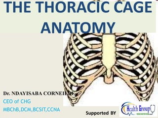
- 1. Dr. NDAYISABA CORNEILLE CEO of CHG MBChB,DCM,BCSIT,CCNA Supported BY THE THORACIC CAGE ANATOMY
- 2. objective • To describe the structure of the thorax, cutaneous innervations of thorax (concept of the myotomes and dermatomes) and of bony framework that forms part of the thorax, and how it is adapted to their functions • To define the thorax, rib cage and thoracic wall. • To describe the structures that form the boundary of the rib cage i.e ribs, sternum, vertebrae. • To outline the clinical importance of the structures that form the rib cage. Dr Ndayisaba Corneille
- 3. Introduction • The rib cage is a bony cartilaginous frame work that has protects the heart, lungs and other thoracic organs; provides attachment for muscles and aids in respiration. • It is flattened anteriorly and posteriorly but rounded on the sides • It is located in the thorax btn the neck and the abdomen Dr Ndayisaba Corneille
- 4. Boundaries • Anteriorly; the sternum and costal cartilages • Posteriorly: the thoracic vertebrae • Laterally: the ribs • Superiorly: the thoracic inlet • Inferiorly: the thoracic outlet Dr Ndayisaba Corneille
- 7. Sternum • A flat bone located in the anterior part of the rib cage. It is a midline bone • Has three parts: manubrium sternum, the body and the xiphoid process • The manubrium sternum lies opposite T3 and T4. It articulates with the first and second ribs(synovial joints). Meets the body at the sternal angle(angle of Luis), a fibrocartilaginous joint. Dr Ndayisaba Corneille
- 10. • Body of the sternum: forms middle part of the sternum • Articulates with 2nd to 7th coastal cartilages • Meets the manubrium at manubriosternal joint, and xiphoid process at xiphisternal joint.(opposite T9) • Xiphoid process: forms lower part. Doesn’t articulate with any ribs. Cartilaginous in young individuals but ossifies at proximal end in the elderly. CLINICAL USES OF THE STERNUM • Median thoracotomy • Bone marrow biopsies Dr Ndayisaba Corneille
- 11. Congenital anomalies: Pectus carinatum Anterior protrusion of the sternum – pigeon chest Dr Ndayisaba Corneille
- 12. A-Cup-shaped deformity B-Saucer-shaped C-Horns-of-steer deformity Dr Ndayisaba Corneille
- 14. Anatomical happenings at sternal angle 1) Lies btn manubrium sternum and body of the sternum 2) Lies btn T4 and T5 3) Its where the trachea ends 4) Its at the level of the second rib 5) At the level of ligamentum arteriosum 6) Its where the azygos vein drains into the SVC 7) Its where the ascending aorta ends and descending aorta starts 8) Where the left recurrent laryngeal nerve curves around the ligamentum arteriosum 9) separates the superior and inferior mediastinum Dr Ndayisaba Corneille
- 15. Ribs • Long horizontal bones located in the thoracic region • Have a number of functions • In man, main function is respiration, not so important in protection. Thoracic organs equally valunerable with or without ribs • In snakes, locomotion(inside feet) • In fish, protection against hydrostatic pressure • Muscle attachment Dr Ndayisaba Corneille
- 16. CATEGORIZATION ACCORDING TO FEATURES 1. Typical ribs: 3rd-9th. 2. Atypical ribs: 1st, 2nd, 10th, 11th, and 12th. The normal ribs have same general features, on the other hand the atypical ribs have special features and thus can be discerned from the rest of the ribs. ACCORDING TO RELATIONSHIP WITH THE STERNUM 1. True ribs: lst-7th (i.e., upper 7 ribs). 2. False ribs: 8th-12th (i.e., lower 5 ribs). True ribs articulate with the sternum anteriorly, on the other hand false ribs don’t articulate with the sternum anteriorly. ACCORDING TO ARTICULATION 1. Vertebrosternal ribs: lst-7th. The vertebrosternal ribs joint posteriorly with vertebrae and anteriorly with the sternum. 2. Vertebrochondral ribs: 8th-10th. The vertebrochondral ribs joint posteriorly with vertebrae and anteriorly their cartilages join the cartilage of the higher rib. 3. Vertebral (floating) ribs: 11th and 12th. The vertebral or floating ribs joint posteriorly with the vertebrae but their anterior ends are free.Dr Ndayisaba Corneille
- 17. ARRANGEMENT AND GENERAL OUTLINE The ribs are arranged one below the other and the gaps between the adjacent ribs are termed intercostals spaces. The length of ribs increases from 1st to 7th rib and after that slowly falls; therefore, seventh rib is the longest rib The transverse diameter of thorax increases progressively from 1st to 8th rib, thus 8th rib has the best lateral projection. The ribs are arranged obliquely, i.e., their anterior ends be located at lower level than their posterior ends. The obliquity of ribs rises progressively from 1st to 9th rib, for this reason 9th rib is most obliquely set. The width of ribs slowly reduced from above downward. Dr Ndayisaba Corneille
- 18. Classification of ribs 12 pairs of ribs, classified in 2 ways 1) true and false ribs 2) typical and atypical ribs True ribs: articulate with the sternum using their own coastal cartilages, 1 to 7 true False ribs: articulate with sternum using 7th coastal cartilage. E.g 8,9 and 10 Floating ribs: don’t articulate with the sternum. 11 and 12 Dr Ndayisaba Corneille
- 19. • Note: The 1st, 10th, 11th and 12th ribs have only one articular facet on their heads for articulation with the side of the body of the corresponding vertebra. • The tubercles of the 11th and 12th ribs make no synovial joints with the respective transverse processes, only being attached to it by ligaments Dr Ndayisaba Corneille
- 20. Typical vs atypical • Typical rib has the following features 1) A head with 2 articular facets, one for articulation with the side of the body of the corresponding vertebra and one for vertebra above 2) Neck, part below the head 3) Tubercle: an outer prominence for articulation with transverse process of corresponding vertebra 4) Angle: most curved part or rib 5) Shaft: smooth superiorly, sharp inferiorly with a costal groove for passage of intercostal vein, artery and nerve. Anterior end of shaft articulates with coastal cartilage Dr Ndayisaba Corneille
- 21. Typical ribs Dr Ndayisaba Corneille
- 22. • Atypical ribs: rib one atypical, shortest and most curved rib, flattened superiorly and inferiorly, has one facet on its head, has a scalene tubercle on medial surface for attachment of scalenous anterior muscle. Ant to the tubercle is a groove for subclavian vein. Post to the tubercle is a groove for the subclavian artery and the brachial plexus. Dr Ndayisaba Corneille
- 23. Atypical ribs Dr Ndayisaba Corneille
- 24. Other atypical ribs •2nd rib •Tuberosity for serratus anterior on external surface •10th One facet •11th and 12th One facet on head no neck or tubercle Dr Ndayisaba Corneille
- 25. Clinical notes on ribs 1) Flail chest: result from fractures of many ribs in more than one place. Part of chest is sucked in during inspiration and sucked out during expiration. 2) Rib grafts: can be used to replace mandible following mandibulectomy 3) Rib contusion: secondary to trauma. Small heamorrhage below peritoneum Dr Ndayisaba Corneille
- 26. Vertebrae • Irregular bones found in the back • Divided into different types: cervical 7, thoracic 12, lumbar 5, sacral 5, coccygeal 4 • Each vertebra has a an anterior arch, and a posterior body • Btn the arch and body are seven processes namely • One spine • 2 transverse processes • 2 superior articular facets • 2 inferior articular facets Dr Ndayisaba Corneille
- 27. Features of thoracic vertebrae • 12 in number • Body medium sized and heart shaped • Small circular vertebral foramen • Long spine that is inclined downwards • Have coastal facets on their transverse processes for articulation with tubercles of the ribs • Have articular facets on their bodies for articulation with heads of the ribs. Dr Ndayisaba Corneille
- 29. JOINTS OF STERNUM 1. MANUBRIOSTERNAL JOINT: . . cartilaginous joint, symphysis between Manubrium and body of Sternum 2. . . . XIPHISTERNAL JOINT cartilaginous joint between Xiphoid process and body of Sternum The Xiphoid process usually fuses with the body of the Sternum during middle age Dr Ndayisaba Corneille
- 30. JOINTS OF RIBS 1. COSTOVERTEBRAL JOINTS: 2 joints between heads of the Ribs and bodies of Vertebrae (corresponding and upper)- Synovial joints 1st, 10th, 11th 12th and rib has 1 synovial joint with the corresponding vertebra, the rest have 2 each; one for the corresponding vertebra and the other for the vertebra above it 1 joint between tubercle of Ribs and transverse process of Vertebra (corresponding) - Synovial joint (1st- 10th Rib) Intra articular ligament connects head of Rib to the intervertebral disc Dr Ndayisaba Corneille
- 31. .,., ,1.......-- body of ve T4 ib ..... 1---- interver head of rib ... sternum b facet for tuberde of rib ' rtebra · r . lubercle of tebral disc angle of rib • cross section of ri costal cartllago costal groove Dr Ndayisaba Corneille
- 33. 2. . COSTOCHONDRAL JOINTS: Joints of the Ribs with costal cartilages Cartilaginous joints 3. . . . STERNOCOSTAL JOINTS: Joints between Sternum and costal cartilages 1st : Cartilaginous joint 2nd 10th – : Synovial joints= 2nd-7th costal cartilages with Sternum 8th-10th costal cartilages with each other (11th and 12th costal cartilages are embedded in muscles) Dr Ndayisaba Corneille
- 34. ~ternal JOll'II l~l lnterchondral Joinfs -...!.--'---'--' Dr Ndayisaba Corneille
- 35. MOVEMENTS 1st . Cartilaginous joints are immobile (thus rib and all costochondral joints do not move during respiration) . Synovial joints are slightly mobile (due to movements in both the joints between head, tubercle and vertebrae, necks of Ribs rotate along their axis, helping in raising and lowering of ribs during respiration) Dr Ndayisaba Corneille
- 36. Intercoastal muscles Arranged in three groups: 1) External intercoastals 2) Internal intercoastals 3) Transversus thoracis group of muscles: Subcostales, Intercostales Intimi, Sternocostalis Dr Ndayisaba Corneille
- 38. External intercostal muscles • Fibers pass obliquely downwards and forwards from the lower border of the rib above to the upper border of the rib below. • The muscle extends from the superior costotransverse ligament at the back of the intercostal space as far as the costo chondral junction. Here it is replaced by the external intercostal membrane that extends as far as the sternum. Dr Ndayisaba Corneille
- 39. Internal intercoastal muscle • The fibers run downwards and backwards from the subcostal groove to the upper border of the rib below.it is replaced posteriorly by the internal intercoastal membrane which extends from the angle of the rib to the superior costal transverse ligament at the posterior limit of the space. Dr Ndayisaba Corneille
- 40. Transversus thoracis • Cross more than one intercostal space. • Poorly developed. • Action is depression of ribs Dr Ndayisaba Corneille
- 42. Anatomy of a typical intercostal space • Composed of an intercoastal nerve, intercostal arteries and interocstal veins. • Each space contains • 1 intercostal nerve • One posterior and 2 anterior intercostal veins • Corresponding intercostal arteries Dr Ndayisaba Corneille
- 43. Intercostal nerves • Mixed nerve, emerges from intervertebral foramen and enters intercostal space btn internal intercostal muscle and transversus thoracis gp • Gives off a collateral branch that supplies muscles in the particular space, the parietal pleura and periosteum of ribs • A lateral cutaneous branch that peirces muscles to supply overlying skin Dr Ndayisaba Corneille
- 44. • An anterior terminal branch that pieces muscles to reach the skin which it supplies • The lower five intercostals and the subcostal nerve slope downwards into the anterior abdominal wall which they supply. Dr Ndayisaba Corneille
- 45. Intercostal arteries Posterior intercostals: • Upper two spaces supplied by superior intercostal artery, branch of the costal cervical trunk from 2nd part of subclavian. • The remaining nine are branches of thoracic aorta. Anterior intercostals: upper 6 arise from internal thoracic, lower 6 from musculophrenc artery,branch of internal thoracic Dr Ndayisaba Corneille
- 47. Intercostal veins • Correspond to arteries • Each space has one posterior intercostal and 2 anterior intecostal veins • Anterior intercostal veins drain into internal thoracic and musculophrenic veins • 1st superior intercostal drains into vertebral or brachiocephalic, 2nd and 3rd to superior intercostal, rest to azygos superior and inferior hemiazygos veins Dr Ndayisaba Corneille
- 48. Thoracic inlet • An oblique space btn the neck and thorax • Allows entry of structures from the neck to the thorax Boundaries: • Anteriorly: sternal notch • Laterally: medial borders of first ribs • Posteriorly: upper border of T1 The inlet is covered by the suprapleural membrane. Dr Ndayisaba Corneille
- 49. Thoracic inlet syndrome • Presence of a cervical rib can compress on the subclavian artery, vein and brachial plexus • Patients present with ischeamic pain of the upper limb due to blockage of blood supply. Dr Ndayisaba Corneille
- 50. Suprapleural membrane • Dense fascial layer attached to the medial border of the first rib and costal cartilage. Not attached to neck of 1st rib. • Posterior attachment is to C7 • Medially it is thin and fades out into the mediastinal pleura. • It is flat and lies in the oblique plane of the thoracic inlet Dr Ndayisaba Corneille
- 51. • The cervical dome of pleura is attached to its undersurface • The subclavian vessels and related structures run on its outersurface. • Fxn: gives rigidity to the thoracic inlet preventing distortion during respiratory changes of intrathoracic pressure. Dr Ndayisaba Corneille
- 52. Thoracic outlet • Located on the inferior aspect of the rib cage • Lies btn the thorax and abdomen • Boundaries • Anteriorly: xiphoid process • Posteriorly: T12 • Laterally: subcostal margin • It is covered by the diaphragm that provides a passage for structures from the abdomen to the thorax and vice verser. Dr Ndayisaba Corneille
- 53. Diaphragm • A thin sheet of muscle found diatal to the lungs. • Found only in placentalia. • Composed of a peripheral muscle part and a central tendon. • Essential function is respiration. • Composed of a right and left domes, the former higher than the later due to larger size of right lobe of the liver. Dr Ndayisaba Corneille
- 56. Origin: • By means of right and left crura, and medial and lateral arcuate ligaments. • The right crus arises from L1, L2 and L3, plus intervening intervetebral discs. Left crus arises from L1 and L2 plus intervening intervertebral disc. • The medial arcuate ligament is a thickening of fascia over psoas muscle Dr Ndayisaba Corneille
- 57. • It extends from the body of L2 to the transverse process of L1, at the lateral margin of psoas. • The lateral arcuate ligament is a thickening of fascia over quadratus lumborum. • Insertion: in a central tendon. Shaped like a rounded leaf, nearer to the front than the back. Inseparable from fibrous pericardium. Btn right and left leaves is caval arpeture Dr Ndayisaba Corneille
- 59. Action • Respiration • Forcefull expiration: sneezing and coughing • Defeacation • Urination • Parturition • Weight lifting • Thoracoabdominal pump. • Nerve supply: phrenic nerve. Dr Ndayisaba Corneille
- 60. CLINICAL CORRELATES • ANOMALIES • TRAUMATIC TEAR • PARALYSIS Dr Ndayisaba Corneille
- 61. END Dr Ndayisaba Corneille THANKS FOR LISTENING By DR NDAYISABA CORNEILLE MBChB,DCM,BCSIT,CCNA Contact us: amentalhealths@gmail.com/ ndayicoll@gmail.com whatsaps :+256772497591 /+250788958241