Downloaded 412 times

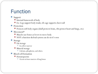
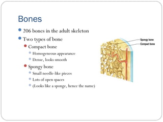
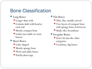
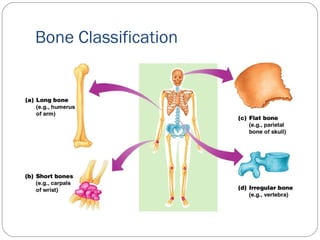
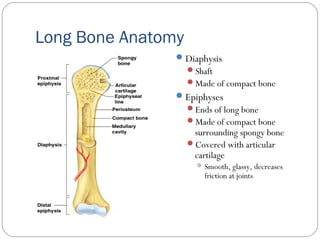
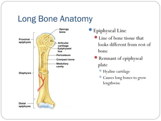
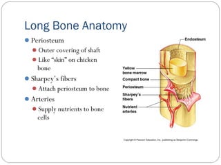
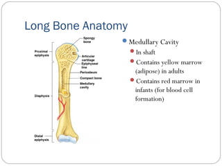
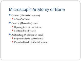
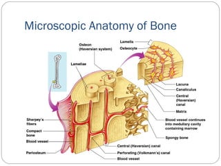
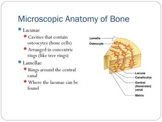
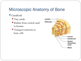
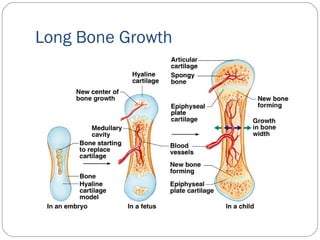
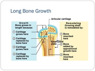
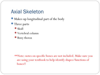
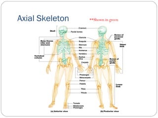
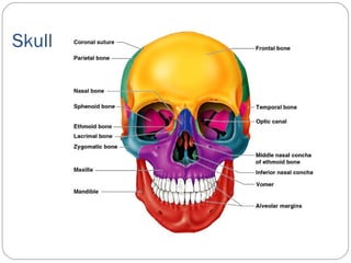
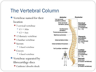
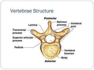
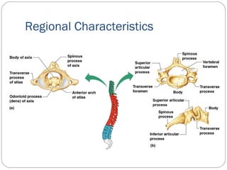
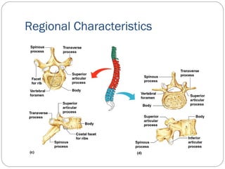
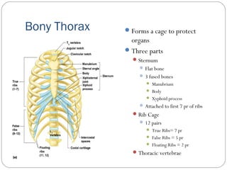
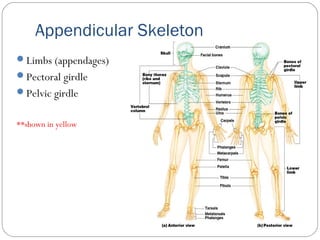
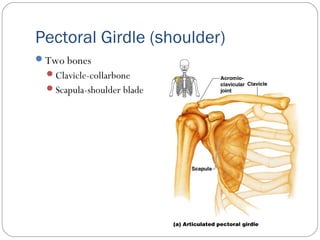
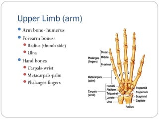
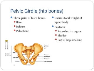
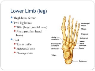
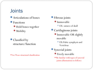
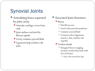
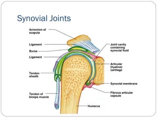
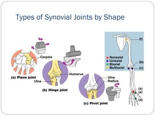
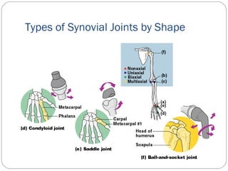

The skeletal system has several important functions including support, protection, movement, mineral storage, and blood cell formation. The skeletal system is made up of 206 bones that are classified as long, short, flat, or irregular. Long bones have a diaphysis, epiphyses, and contain red or yellow marrow. Bones are made up of compact and spongy bone and have microscopic structures including osteons, lacunae, and canaliculi. The skeletal system includes the axial skeleton which is made up of the skull, vertebral column, and rib cage, as well as the appendicular skeleton consisting of the pectoral girdle, upper limbs, pelvic girdle, and lower limbs. Joint


