
Recommended
More Related Content
What's hot
What's hot (20)
Similar to 2 anatomy of ls coccyx and sacrum
Similar to 2 anatomy of ls coccyx and sacrum (20)
More from DonBenny2
Recently uploaded
Recently uploaded (20)
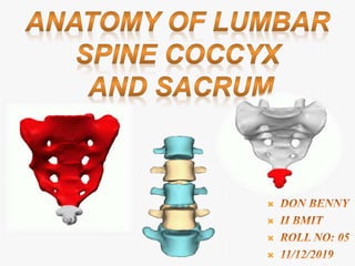
2 anatomy of ls coccyx and sacrum
- 1.
- 2. These are the five vertebrae between the rib cage and the pelvis. It starts about five or six inches below the shoulder blades, and connects with the Thoracic spine at the top and extends downward to the Sacral spine. Lumbar spine has 5 vertebrae, labeled L1-L5. T 12 S1 Scapula ( shoulder blades)
- 3. L1 to L4 are considered Typical Lumbar Vertebrae. The large bodies are oval and the vertebral foramen is triangular. The spinous process is stumpy and projects dorsally. L5 is considered Atypical Lumbar Vertebrae. L 5 Atypical Typical
- 4. Also known as Centrum (Corpus vertebrae) The bodies of the lumbar vertebrae are massive, sturdy, and designed to withstand vertical compression.
- 5. Also known as Spinal foramen. It is a large, triangular-shaped opening located posterior to the body. The spinal cord and several nerves pass through the foramen.
- 6. Also known as neural arch. It is formed by the two pedicles, two Laminae, and Spinous process. The arch encloses the posterior portion of the vertebral foramen. Vertebral Arch
- 7. Pedicles of the vertebral arch are posterior extensions from the lateral sides of the body. The two pedicles form the base of the vertebral arch.
- 8. Lamina of the vertebral arch are plates of bone that extend from the pedicles. The left and right lamina and the spinous process form the dorsal portion of the vertebral arch.
- 9. The Spinous process is a posterior projection from the junction of the two lamina. Its thick, broad, quadrilateral shape serves as an attachment point for ligaments and muscles that stabilize the back.
- 10. It is a long, thin, latero- posterior projection that originates near the junction of the pedicle, lamina, and superior articular process. It also serves as an attachment point for back muscles.
- 11. It is a superior projection near the junction of the pedicle and transverse process. The facet is concave, faces inward (medially), and articulates with the inferior articular facet on the vertebra above.
- 12. It is an inferior projection from the junction of the pedicle and lamina. The facet is convex, faces outward (laterally), and articulates with the superior - articular facet on the vertebra below.
- 13. It is a thick pad of connective tissue that helps hold adjacent vertebrae together and acts as a shock absorber. The lumbar vertebrae help support the weight of the body, and permit movement. Vertebrae articulate with one another via 2 ways ;
- 14. It is obliquely oriented, cylindrically-shaped articular surfaces. Inferior articular process of the vertebrae above always lies posterior to the superior articular process of the vertebrae below. Intervertebral joint
- 15. They can be divided into two groups 1) Present throughout Vertebral Column : Anterior Longitudinal Ligament. Posterior Longitudinal Ligament. Ligamentum Flavum. Interspinous Ligament. Supraspinous Ligament. Interansverse Ligament. A B C D E F
- 16. Iliolumbar ligaments: The lumbosacral joint (between L5 and S1 vertebrae) is strengthened by the Iliolumbar ligaments. These are fan-like ligaments radiating from the transverse processes of the L5 vertebra to the ilea of the pelvis 2) Unique to Lumbar Spine : Iliolumbar Ligament
- 17. Iliohypogastric Nerve. Ilioinguinal Nerve. Genitofemoral Nerve. Lateral Cutaneous Nerve of the Thigh. Obturator Nerve. Femoral Nerve. *Nerve plexus which provides motor and sensory nerves *Spinal cord ends at approximately L1-L2
- 19. SACRUM The sacrum is a large bone located at the terminal part of the vertebral canal, where it forms the posterior aspect of the pelvis. It is remarkably thick, which aids in supporting and transmitting the weight of the body. The sacrum is formed by the fusion of the five sacral vertebrae. It has an inverted triangular, concave shape. The bone consists of a base, apex and four surfaces:
- 20. Apex : narrow caudal (towards the tailbone) portion. Base : broad superior portion where there is an articulation with L5. sacral promontory : prominent bulge on anterior side of base. superior articular processes : articulates with inferior articular processes of L5. sacral canal : continuation of vertebral canal through fused arches of sacral vertebrae. median sacral crest : fused spinous processes of the first four sacral vertebrae.
- 21. sacral hiatus :opening that results from failure of laminae of S5 to form and fuse. lateral sacral crest : ridges on either side of median sacral crest. Sacral foraminae : Openings that correspond to intervertebral formina. There are anterior and posterior foramina. Ala : Wing-like extensions on either side of base. Auricular surface : Ear-shaped surface of the sacroiliac joint. Sacral tuberosity : Roughened area dorsal to auricular surface where ligaments that stabilize the sacroiliac joint attach.
- 23. 1 2 3 5 Base
- 24. ARTICULATIONS Lumbosacral Joint : The upper border articulates with the 5th Lumbar vertebra. Sacrococcygeal Joint : The inferior part articulates with the Coccyx Sacroiliac Joint : Lateral part articulates with the Hip bone(Ilium) SACROILIAC JOINT LUMBOSACRAL JOINT SACROCOCCYGEAL JO
- 25. It is the joint between the sacrum and the ilium bones of the pelvis, which are connected by strong ligaments. The joint is strong, supporting the entire weight of the upper body. It is a synovial plane joint with irregular elevations and depressions that produce interlocking of the two bones. Sacroiliac joints are paired C-shaped or L-shaped joints capable of a small amount of movement that are formed between the auricular surfaces of the sacrum and the ilium bones. Their main job is to carry the weight of your upper body when you stand or walk and shift that load to your legs.
- 26. LIGAMENTS Anterior longitudinal ligament Iliolumbar ligament. Lumbosacral ligament. Sacroiliac ligaments: Sacrospinous ligament Sacrotuberous ligament Sacrococcygeal ligaments:
- 28. SACRAL PLEXUS Superior & Inferior Gluteal Nerve. Nerve to the Quadratus Femoris. Nerve to the Obturator Internus . Post. Femoral Cutaneous Nerve. Perineal branches. Pudendal Nerve. Sciatic Nerve. Tibial Nerve.
- 29. The coccyx (also known as the tailbone) is the terminal part of the vertebral column. It is comprised of four vertebrae, which fuse to produce a triangular shape. The coccyx is formed from four rudimentary vertebrae and does not contain a spinal canal, pedicles, laminae or spinous processes it serves as the insertion site for the muscles of the pelvic floor and those that contribute to voluntary bowel control and supports the position of the anus.
- 30. The coccyx consists of an apex, base, anterior surface, posterior surface and two lateral surfaces. Base: It is located most superiorly, and contains a facet for articulation with the sacrum. Apex : It is situated inferiorly, at the terminus of the vertebral column. Lateral surfaces of the coccyx are marked by a small transverse process, which projects from Co1.
- 31. ARTICULATIONS Sacrococcygeal Joint: It is a fibrocartilaginous joint that connects the apex of the sacrum to the coccyx. Movement is passive minor flexion and extension and the joint typically fuses with age.
- 32. LIGAMENTS Anterior sacrococcygeal ligament : Deep posterior sacrococcygeal ligament. Superficial posterior sacrococcygeal ligament Lateral sacrococcygeal ligaments : Interarticular ligaments :
- 33. REFERENCE
- 34. Next : Jennifer D’S
- 35. The sacral plexus is a nerve plexus which provides motor and sensory nerves for the posterior thigh, most of the lower leg and foot, and part of the pelvis. It is part of the lumbosacral plexus and emerges from the lumbar vertebrae and sacral vertebrae (L4-S4)