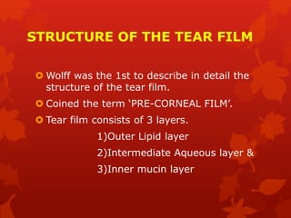
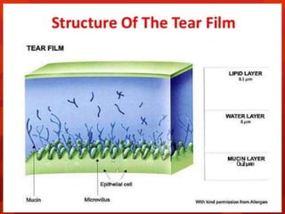
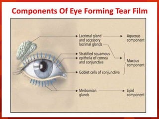
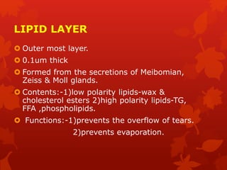
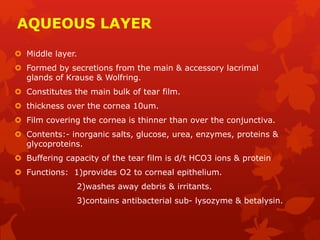
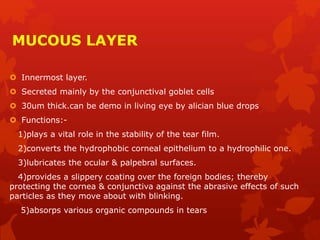
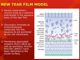
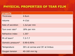
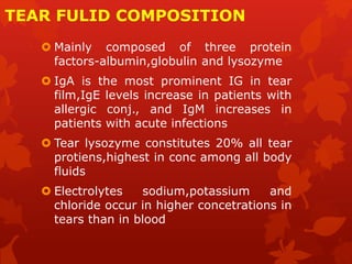
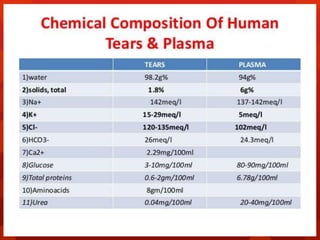
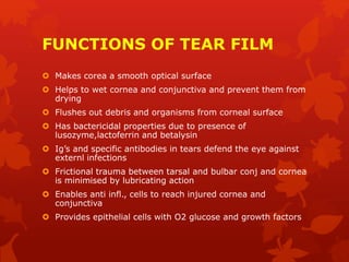
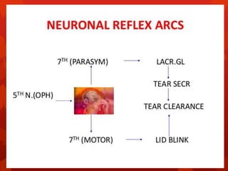
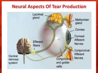
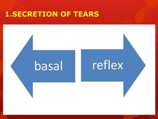
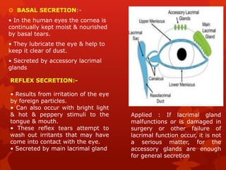
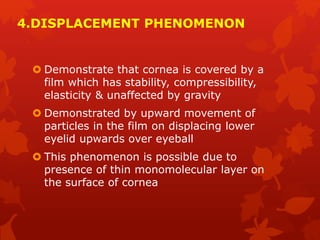
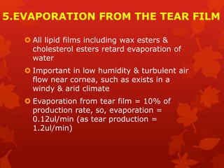
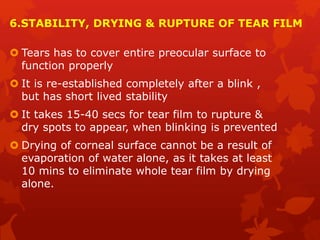
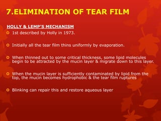
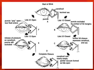
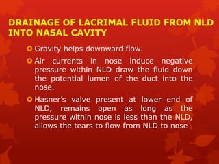
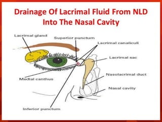
The document summarizes the structure and function of the tear film. It consists of three layers - an outer lipid layer, middle aqueous layer, and inner mucin layer. The lipid layer prevents evaporation and overflow of tears. The aqueous layer hydrates the cornea and contains nutrients. The mucin layer lubricates the eye surface. Tears are produced through basal and reflex secretion and drained through the lacrimal system into the nose. Blinking helps spread and replenish the tear film layers, which must be continuously renewed to maintain a smooth optical surface and protect the cornea.
The tear film is a complex mixture of substances secreted from multiple sources on the ocular surface, including the lacrimal gland, the accessory lacrimal glands, the meibomian glands, and the goblet cells.
LIMBUS… • The limbus forms the border between the transparent cornea and opaque sclera, contains the pathways of aqueous humour outflow, and is the site of surgical incisions for cataract and glaucoma
2. Anatomical Limbus: Circumcorneal transitional zone of the conjunctivocorneal & corneoscleral junction Conjunctivo-corneal junction: • Bulbar conjunctiva is firmly adherent to underlying structures • Substantia propria of the conjunctiva stops here but its epithelium continues with that of the cornea. Sclero-corneal junction: • Transparent corneal lamellae become continuous • With the oblique, circular and opaque fibres of sclera
3. CONTINUE…. • In the area near limbus, the conjunctiva, tenon’s capsule & the episcleral tissue are fused into a dense tissue which is strongly adherent to corneo scleral junction.It is preferred site for obtaining a firm hold of the eyeball during ocular surgery. • The limbus is a common site for the occurrence of corneal epithelial neoplasm. • The Limbus contains radially oriented fibrovascular ridge known as the palisades of Vogt that may harbour a stem cell population. The palisades of Vogt are more common in the superior and inferior quadrants around the eye
The tear film constitutes Three layers :- An outermost lipid (oily) layer An aqueous (watery) layer that makes up 90% of the tear film volume; and A mucin layer that coats the corneal surface.
3. To form smooth optical surface on cornea. To keep the surface of cornea & conjunctiva moist It serve as lubricant It transfer oxygen Provide antibacterial action Wash debris out It provides a pathway for WBC in case of injury
4. Functions of lipid layer Retards evaporation of tear film Prevents the overflow of tears
5. Function of Aqueous Layer Flushes, buffers and lubricates the corneal surface Delivers oxygen and other nutrients to the corneal surface Wash out debris Delivers antibacterial enzymes and antibodies such as lysozyme.
6. Functions of Mucin Layer Spreads tears over corneal surface. Protects the cornea against foreign substances . Makes corneal surface smooth by filling in surface irregularities
Cornea is the clear front surface of the eye. It lies directly in front of the iris and pupil, and it allows light to enter the eye.
Cornea forms the transparent and anterior 1/6th of the external fibrous coat of the globe of the eyeball.
The cornea is the eye's most powerful structure for focusing light that provides approximately 65 to 75 percent of the focusing power of the eye.
The cornea has unmyelinated nerve endings sensitive to touch, temperature and chemicals; a touch of the cornea causes an involuntary reflex to close the eyelid.
It explains the secretion of the tear film,its importance and the pathologies that can happen when its not being secreted well and as well as the pathophysiology of.It also addresses the different layers of the tear film and the various ways that it can lead to the different diseases of the eye
the paper addresses the different scretory pathways and it speaks about the regulation of the production of the tear film in that the various
The lacrimal apparatus (Tear System)
It's a group of glands, sacs and ducts that makes new tears and drains old ones away. consists of two divisions. The first is the secretory part, which is responsible for the production of tears, and the second is the drainage part. The secretory part contains the main lacrimal gland, which is lodged in a fossa in the lateral part of the roof of the orbit, and two accessory lacrimal glands of Krause and Wolfring. The latter are present near the fornix.
Dry eye disease is a common condition that occurs when your tears aren't able to provide adequate lubrication for your eyes. Tears can be inadequate and unstable for many reasons. For example, dry eyes may occur if you don't produce enough tears or if you produce poor-quality tears. This tear instability leads to inflammation and damage of the eye's surface.
Dry eyes feel uncomfortable. If you have dry eyes, your eyes may sting or burn. You may experience dry eyes in certain situations, such as on an airplane, in an air-conditioned room, while riding a bike or after looking at a computer screen for a few hours
Main physiologic function of cornea is to act as a major refracting medium, so that a clear retinal image is formed. • Normal corneal transparency is result of • 1.anatomical factor such as uniform and regular arrangement of corneal epithelium, peculiar arrangement of corneal lamella and corneal vascularity 2.Physiological factor [ie] relative state of corneal dehydration.
3. • Therefore, any process which upsets the anatomy or physiology of cornea will cause LOSS OF TRANSPARENCY to some degree.
4. FACTORS AFFECTING CORNEAL TRANSPARENCY • CORNEAL EPITHELIUM &TEAR FLIM • ARRANGEMENT OF STROMAL LAMELLA • CORNEAL VASCULARIZATION • CORNEAL HYDRATION • CELLULAR FACTORS AFFECTING TRANSPARENCY
Acute scrotum is a general term referring to an emergency condition affecting the contents or the wall of the scrotum.
There are a number of conditions that present acutely, predominantly with pain and/or swelling
A careful and detailed history and examination, and in some cases, investigations allow differentiation between these diagnoses. A prompt diagnosis is essential as the patient may require urgent surgical intervention
Testicular torsion refers to twisting of the spermatic cord, causing ischaemia of the testicle.
Testicular torsion results from inadequate fixation of the testis to the tunica vaginalis producing ischemia from reduced arterial inflow and venous outflow obstruction.
The prevalence of testicular torsion in adult patients hospitalized with acute scrotal pain is approximately 25 to 50 percent
ARTIFICIAL INTELLIGENCE IN HEALTHCARE.pdfAnujkumaranit
Artificial intelligence (AI) refers to the simulation of human intelligence processes by machines, especially computer systems. It encompasses tasks such as learning, reasoning, problem-solving, perception, and language understanding. AI technologies are revolutionizing various fields, from healthcare to finance, by enabling machines to perform tasks that typically require human intelligence.
Title: Sense of Taste
Presenter: Dr. Faiza, Assistant Professor of Physiology
Qualifications:
MBBS (Best Graduate, AIMC Lahore)
FCPS Physiology
ICMT, CHPE, DHPE (STMU)
MPH (GC University, Faisalabad)
MBA (Virtual University of Pakistan)
Learning Objectives:
Describe the structure and function of taste buds.
Describe the relationship between the taste threshold and taste index of common substances.
Explain the chemical basis and signal transduction of taste perception for each type of primary taste sensation.
Recognize different abnormalities of taste perception and their causes.
Key Topics:
Significance of Taste Sensation:
Differentiation between pleasant and harmful food
Influence on behavior
Selection of food based on metabolic needs
Receptors of Taste:
Taste buds on the tongue
Influence of sense of smell, texture of food, and pain stimulation (e.g., by pepper)
Primary and Secondary Taste Sensations:
Primary taste sensations: Sweet, Sour, Salty, Bitter, Umami
Chemical basis and signal transduction mechanisms for each taste
Taste Threshold and Index:
Taste threshold values for Sweet (sucrose), Salty (NaCl), Sour (HCl), and Bitter (Quinine)
Taste index relationship: Inversely proportional to taste threshold
Taste Blindness:
Inability to taste certain substances, particularly thiourea compounds
Example: Phenylthiocarbamide
Structure and Function of Taste Buds:
Composition: Epithelial cells, Sustentacular/Supporting cells, Taste cells, Basal cells
Features: Taste pores, Taste hairs/microvilli, and Taste nerve fibers
Location of Taste Buds:
Found in papillae of the tongue (Fungiform, Circumvallate, Foliate)
Also present on the palate, tonsillar pillars, epiglottis, and proximal esophagus
Mechanism of Taste Stimulation:
Interaction of taste substances with receptors on microvilli
Signal transduction pathways for Umami, Sweet, Bitter, Sour, and Salty tastes
Taste Sensitivity and Adaptation:
Decrease in sensitivity with age
Rapid adaptation of taste sensation
Role of Saliva in Taste:
Dissolution of tastants to reach receptors
Washing away the stimulus
Taste Preferences and Aversions:
Mechanisms behind taste preference and aversion
Influence of receptors and neural pathways
Impact of Sensory Nerve Damage:
Degeneration of taste buds if the sensory nerve fiber is cut
Abnormalities of Taste Detection:
Conditions: Ageusia, Hypogeusia, Dysgeusia (parageusia)
Causes: Nerve damage, neurological disorders, infections, poor oral hygiene, adverse drug effects, deficiencies, aging, tobacco use, altered neurotransmitter levels
Neurotransmitters and Taste Threshold:
Effects of serotonin (5-HT) and norepinephrine (NE) on taste sensitivity
Supertasters:
25% of the population with heightened sensitivity to taste, especially bitterness
Increased number of fungiform papillae
The prostate is an exocrine gland of the male mammalian reproductive system
It is a walnut-sized gland that forms part of the male reproductive system and is located in front of the rectum and just below the urinary bladder
Function is to store and secrete a clear, slightly alkaline fluid that constitutes 10-30% of the volume of the seminal fluid that along with the spermatozoa, constitutes semen
A healthy human prostate measures (4cm-vertical, by 3cm-horizontal, 2cm ant-post ).
It surrounds the urethra just below the urinary bladder. It has anterior, median, posterior and two lateral lobes
It’s work is regulated by androgens which are responsible for male sex characteristics
Generalised disease of the prostate due to hormonal derangement which leads to non malignant enlargement of the gland (increase in the number of epithelial cells and stromal tissue)to cause compression of the urethra leading to symptoms (LUTS
Title: Sense of Smell
Presenter: Dr. Faiza, Assistant Professor of Physiology
Qualifications:
MBBS (Best Graduate, AIMC Lahore)
FCPS Physiology
ICMT, CHPE, DHPE (STMU)
MPH (GC University, Faisalabad)
MBA (Virtual University of Pakistan)
Learning Objectives:
Describe the primary categories of smells and the concept of odor blindness.
Explain the structure and location of the olfactory membrane and mucosa, including the types and roles of cells involved in olfaction.
Describe the pathway and mechanisms of olfactory signal transmission from the olfactory receptors to the brain.
Illustrate the biochemical cascade triggered by odorant binding to olfactory receptors, including the role of G-proteins and second messengers in generating an action potential.
Identify different types of olfactory disorders such as anosmia, hyposmia, hyperosmia, and dysosmia, including their potential causes.
Key Topics:
Olfactory Genes:
3% of the human genome accounts for olfactory genes.
400 genes for odorant receptors.
Olfactory Membrane:
Located in the superior part of the nasal cavity.
Medially: Folds downward along the superior septum.
Laterally: Folds over the superior turbinate and upper surface of the middle turbinate.
Total surface area: 5-10 square centimeters.
Olfactory Mucosa:
Olfactory Cells: Bipolar nerve cells derived from the CNS (100 million), with 4-25 olfactory cilia per cell.
Sustentacular Cells: Produce mucus and maintain ionic and molecular environment.
Basal Cells: Replace worn-out olfactory cells with an average lifespan of 1-2 months.
Bowman’s Gland: Secretes mucus.
Stimulation of Olfactory Cells:
Odorant dissolves in mucus and attaches to receptors on olfactory cilia.
Involves a cascade effect through G-proteins and second messengers, leading to depolarization and action potential generation in the olfactory nerve.
Quality of a Good Odorant:
Small (3-20 Carbon atoms), volatile, water-soluble, and lipid-soluble.
Facilitated by odorant-binding proteins in mucus.
Membrane Potential and Action Potential:
Resting membrane potential: -55mV.
Action potential frequency in the olfactory nerve increases with odorant strength.
Adaptation Towards the Sense of Smell:
Rapid adaptation within the first second, with further slow adaptation.
Psychological adaptation greater than receptor adaptation, involving feedback inhibition from the central nervous system.
Primary Sensations of Smell:
Camphoraceous, Musky, Floral, Pepperminty, Ethereal, Pungent, Putrid.
Odor Detection Threshold:
Examples: Hydrogen sulfide (0.0005 ppm), Methyl-mercaptan (0.002 ppm).
Some toxic substances are odorless at lethal concentrations.
Characteristics of Smell:
Odor blindness for single substances due to lack of appropriate receptor protein.
Behavioral and emotional influences of smell.
Transmission of Olfactory Signals:
From olfactory cells to glomeruli in the olfactory bulb, involving lateral inhibition.
Primitive, less old, and new olfactory systems with different path
TEST BANK for Operations Management, 14th Edition by William J. Stevenson, Ve...kevinkariuki227
TEST BANK for Operations Management, 14th Edition by William J. Stevenson, Verified Chapters 1 - 19, Complete Newest Version.pdf
Pulmonary Thromboembolism - etilogy, types, medical- Surgical and nursing man...VarunMahajani
Disruption of blood supply to lung alveoli due to blockage of one or more pulmonary blood vessels is called as Pulmonary thromboembolism. In this presentation we will discuss its causes, types and its management in depth.
New Drug Discovery and Development .....NEHA GUPTA
The "New Drug Discovery and Development" process involves the identification, design, testing, and manufacturing of novel pharmaceutical compounds with the aim of introducing new and improved treatments for various medical conditions. This comprehensive endeavor encompasses various stages, including target identification, preclinical studies, clinical trials, regulatory approval, and post-market surveillance. It involves multidisciplinary collaboration among scientists, researchers, clinicians, regulatory experts, and pharmaceutical companies to bring innovative therapies to market and address unmet medical needs.
Lung Cancer: Artificial Intelligence, Synergetics, Complex System Analysis, S...Oleg Kshivets
RESULTS: Overall life span (LS) was 2252.1±1742.5 days and cumulative 5-year survival (5YS) reached 73.2%, 10 years – 64.8%, 20 years – 42.5%. 513 LCP lived more than 5 years (LS=3124.6±1525.6 days), 148 LCP – more than 10 years (LS=5054.4±1504.1 days).199 LCP died because of LC (LS=562.7±374.5 days). 5YS of LCP after bi/lobectomies was significantly superior in comparison with LCP after pneumonectomies (78.1% vs.63.7%, P=0.00001 by log-rank test). AT significantly improved 5YS (66.3% vs. 34.8%) (P=0.00000 by log-rank test) only for LCP with N1-2. Cox modeling displayed that 5YS of LCP significantly depended on: phase transition (PT) early-invasive LC in terms of synergetics, PT N0—N12, cell ratio factors (ratio between cancer cells- CC and blood cells subpopulations), G1-3, histology, glucose, AT, blood cell circuit, prothrombin index, heparin tolerance, recalcification time (P=0.000-0.038). Neural networks, genetic algorithm selection and bootstrap simulation revealed relationships between 5YS and PT early-invasive LC (rank=1), PT N0—N12 (rank=2), thrombocytes/CC (3), erythrocytes/CC (4), eosinophils/CC (5), healthy cells/CC (6), lymphocytes/CC (7), segmented neutrophils/CC (8), stick neutrophils/CC (9), monocytes/CC (10); leucocytes/CC (11). Correct prediction of 5YS was 100% by neural networks computing (area under ROC curve=1.0; error=0.0).
CONCLUSIONS: 5YS of LCP after radical procedures significantly depended on: 1) PT early-invasive cancer; 2) PT N0--N12; 3) cell ratio factors; 4) blood cell circuit; 5) biochemical factors; 6) hemostasis system; 7) AT; 8) LC characteristics; 9) LC cell dynamics; 10) surgery type: lobectomy/pneumonectomy; 11) anthropometric data. Optimal diagnosis and treatment strategies for LC are: 1) screening and early detection of LC; 2) availability of experienced thoracic surgeons because of complexity of radical procedures; 3) aggressive en block surgery and adequate lymph node dissection for completeness; 4) precise prediction; 5) adjuvant chemoimmunoradiotherapy for LCP with unfavorable prognosis.
Couples presenting to the infertility clinic- Do they really have infertility...Sujoy Dasgupta
Dr Sujoy Dasgupta presented the study on "Couples presenting to the infertility clinic- Do they really have infertility? – The unexplored stories of non-consummation" in the 13th Congress of the Asia Pacific Initiative on Reproduction (ASPIRE 2024) at Manila on 24 May, 2024.
micro teaching on communication m.sc nursing.pdfAnurag Sharma
Microteaching is a unique model of practice teaching. It is a viable instrument for the. desired change in the teaching behavior or the behavior potential which, in specified types of real. classroom situations, tends to facilitate the achievement of specified types of objectives.
NVBDCP.pptx Nation vector borne disease control programSapna Thakur
NVBDCP was launched in 2003-2004 . Vector-Borne Disease: Disease that results from an infection transmitted to humans and other animals by blood-feeding arthropods, such as mosquitoes, ticks, and fleas. Examples of vector-borne diseases include Dengue fever, West Nile Virus, Lyme disease, and malaria.
2. THE TEAR FILM
The main role of lacrimal system is to
establish & maintain a continuous tear
film over the ocular surface.
The presence of the pre-corneal tear film
was 1st demonstrated: by Fischer in 1928.
Rollet described it as the most superficial
6th layer of cornea.
3. STRUCTURE OF THE TEAR FILM
Wolff was the 1st to describe in detail the
structure of the tear film.
Coined the term ‘PRE-CORNEAL FILM’.
Tear film consists of 3 layers.
1)Outer Lipid layer
2)Intermediate Aqueous layer &
3)Inner mucin layer
4.
5.
6. LIPID LAYER
Outer most layer.
0.1um thick
Formed from the secretions of Meibomian,
Zeiss & Moll glands.
Contents:-1)low polarity lipids-wax &
cholesterol esters 2)high polarity lipids-TG,
FFA ,phospholipids.
Functions:-1)prevents the overflow of tears.
2)prevents evaporation.
7. AQUEOUS LAYER
Middle layer.
Formed by secretions from the main & accessory lacrimal
glands of Krause & Wolfring.
Constitutes the main bulk of tear film.
thickness over the cornea 10um.
Film covering the cornea is thinner than over the conjunctiva.
Contents:- inorganic salts, glucose, urea, enzymes, proteins &
glycoproteins.
Buffering capacity of the tear film is d/t HCO3 ions & protein
Functions: 1)provides O2 to corneal epithelium.
2)washes away debris & irritants.
3)contains antibacterial sub- lysozyme & betalysin.
8. MUCOUS LAYER
Innermost layer.
Secreted mainly by the conjunctival goblet cells
30um thick.can be demo in living eye by alician blue drops
Functions:-
1)plays a vital role in the stability of the tear film.
2)converts the hydrophobic corneal epithelium to a hydrophilic one.
3)lubricates the ocular & palpebral surfaces.
4)provides a slippery coating over the foreign bodies; thereby
protecting the cornea & conjunctiva against the abrasive effects of such
particles as they move about with blinking.
5)absorps various organic compounds in tears
9. NEW TEAR FILM MODEL
Recent observation--
mucins exist as a network
distributed in the aqueous
body of the tear film.
Glycocalyx emanate as
transmembrane
molecules into the
aqueous & are anchored
at the cell membrane.
Membrane associated
proteins-MUC1,4&16 as
well as secretory mucins-
MUC5AC &MUC7 have
been identified at the
ocular surface.
10. PHYSICAL PROPERTIES OF TEAR FILM
property
Thickness 4-8um
Volume 4-13ul
Rate of secretion 1.2ul per min
Turn over rate* 18% per min
Refractive index 1.357
Ph of tears* 7.3-7.7
Osmotic pressure* 0.90-0.95%
Temperature 30’c at cornea and 35’ at limbus
Oxygen tension 40-160 mm hg
11. TEAR FULID COMPOSITION
Mainly composed of three protein
factors-albumin,globulin and lysozyme
IgA is the most prominent IG in tear
film,IgE levels increase in patients with
allergic conj., and IgM increases in
patients with acute infections
Tear lysozyme constitutes 20% all tear
protiens,highest in conc among all body
fluids
Electrolytes sodium,potassium and
chloride occur in higher concetrations in
tears than in blood
12.
13. FUNCTIONS OF TEAR FILM
Makes corea a smooth optical surface
Helps to wet cornea and conjunctiva and prevent them from
drying
Flushes out debris and organisms from corneal surface
Has bactericidal properties due to presence of
lusozyme,lactoferrin and betalysin
Ig’s and specific antibodies in tears defend the eye against
externl infections
Frictional trauma between tarsal and bulbar conj and cornea
is minimised by lubricating action
Enables anti infl., cells to reach injured cornea and
conjunctiva
Provides epithelial cells with O2 glucose and growth factors
14. NEURAL ASPECTS OF TEAR PRODUCTION
The trigeminal v1 (fifth cranial) nerve bears
the sensory pathway(afferent) of the tear
reflexes.
The motor pathway is autonomic (involuntary), &,
in general, uses the pathway of the facial
(seventh) nerve in the parasympathetic division
via pterygopalatine palatine ganglion, as efferent
pathway.
Applied: A newborn infant has insufficient
development of nervous control, so she/he "cries
without weeping”.
15.
16.
17. APPLIED ASPECTS
Crocodile tears syndrome/ Bogorad's
syndrome" is an uncommon consequence of nerve
regeneration subsequent to Bell's palsy or other damage
to the facial nerve in which efferent fibers from the
superior salivary nucleus become improperly connected
to nerve axons projecting to the lacrimal glands (tear
ducts), causing one to shed tears (lacrimate) during
salivation while smelling foods or eating.
It is presumed that one would also salivate while crying
due to the inverse improper connection of the lacrimal
nucleus to the salivary glands, but this would be less
noticeable.
18. TEAR FILM DYNAMICS
Secretion of tears
Formation of tear film
Retention & redistribution of tear film
Displacement phenomenon
Evaporation from the tear film
Drying & break up of tear film
Dynamic events during blinking
Elimination of tears
20. BASAL SECRETION:-
• In the human eyes the cornea is
continually kept moist & nourished
by basal tears.
• They lubricate the eye & help to
keep it clear of dust.
• Secreted by accessory lacrimal
glands
REFLEX SECRETION:-
• Results from irritation of the eye
by foreign particles.
• Can also occur with bright light
& hot & peppery stimuli to the
tongue & mouth.
• These reflex tears attempt to
wash out irritants that may have
come into contact with the eye.
• Secreted by main lacrimal gland
Applied : If lacrimal gland
malfunctions or is damaged in
surgery or other failure of
lacrimal function occur, it is not
a serious matter, for the
accessory glands are enough
for general secretion
21. 2.FORMATION OF PRE OCULAR TEAR FILM
Conjunctival mucus spreads on to the cornea
by the action of the lids.
On this new surface- aqueous layer is spread
spontaneously.
Over this the superficial lipid layer spreads;
probably contributing to its stability &
retarding evaporation b/w blinks.
22. 3.RETENTION & REDISTRIBUTION
The outer most layer of corneal
epithelium+ mucopolysaccharides leads
to retention.
Precorneal film is stagnant.Redistribution
occurs in the form of bringing of new
tear fluid by way of marginal strip where
there is constant flow of tears
23. 4.DISPLACEMENT PHENOMENON
Demonstrate that cornea is covered by a
film which has stability, compressibility,
elasticity & unaffected by gravity
Demonstrated by upward movement of
particles in the film on displacing lower
eyelid upwards over eyeball
This phenomenon is possible due to
presence of thin monomolecular layer on
the surface of cornea
24. 5.EVAPORATION FROM THE TEAR FILM
All lipid films including wax esters &
cholesterol esters retard evaporation of
water
Important in low humidity & turbulent air
flow near cornea, such as exists in a
windy & arid climate
Evaporation from tear film = 10% of
production rate, so, evaporation =
0.12ul/min (as tear production =
1.2ul/min)
25. 6.STABILITY, DRYING & RUPTURE OF TEAR FILM
Tears has to cover entire preocular surface to
function properly
It is re-established completely after a blink ,
but has short lived stability
It takes 15-40 secs for tear film to rupture &
dry spots to appear, when blinking is prevented
Drying of corneal surface cannot be a result of
evaporation of water alone, as it takes at least
10 mins to eliminate whole tear film by drying
alone.
26. 7.ELIMINATION OF TEAR FILM
HOLLY & LEMP’S MECHANISM
1st described by Holly in 1973.
Initially all the tear film thins uniformly by evaporation.
When thinned out to some critical thickness, some lipid molecules
begin to be attracted by the mucin layer & migrate down to this layer.
When the mucin layer is sufficiently contaminated by lipid from the
top, the mucin becomes hydrophobic & the tear film ruptures
Blinking can repair this and restore aqueous layer
27.
28. 8.DYNAMIC EVENTS DURING BLINKING
Was 1st described by Holly in 1980.
As the upper lid moves downwards, the superficial lipid
layer is compressed b/w the lid edges
This will contaminate the mucus & this lipid contaminated
mucus is rolled up in a thread like shape & dragged into
lower fornix
When the eye opens, at 1st the lipid spreads in the form of
a monolayer against the upper eye lid
Then spreading of the excess lipid follows & in about 1 sec
multimolecular lipid layer is formed
The spreading lipid drags some aqueous tears with it
thereby thickening the tear film.
29.
30. 9.ELIMINATION OF TEARS
Lacrimal fluid over the preocular
surfacemarginal tear stripLacus
lacrimalisinner canthus lacrimal passages
nasal cavity
Lacrimal pump mechanism:- fibres of the
pretarsal & preseptal portion of the Orbicularis
which arise from the lacrimal fascia &
posterior lacrimal crest.
This LPM operates with the blinking
movements of the eyelids as follows:-
31.
32. DRAINAGE OF LACRIMAL FLUID FROM NLD
INTO NASAL CAVITY
Gravity helps downward flow.
Air currents in nose induce negative
pressure within NLD draw the fluid down
the potential lumen of the duct into the
nose.
Hasner’s valve present at lower end of
NLD, remains open as long as the
pressure within nose is less than the NLD,
allows the tears to flow from NLD to nose