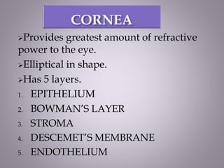
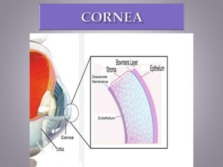
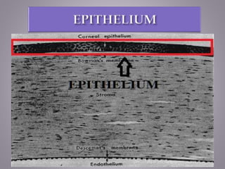
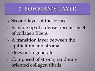
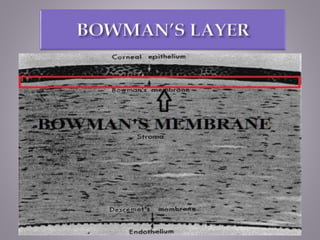
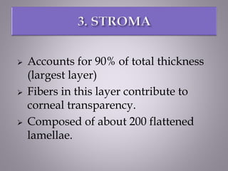
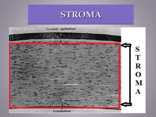
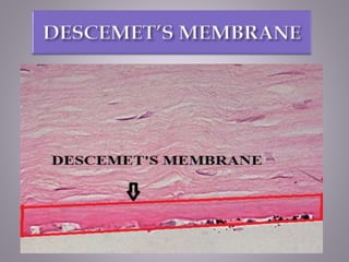
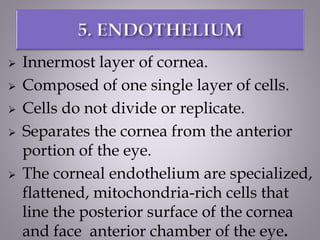
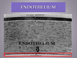
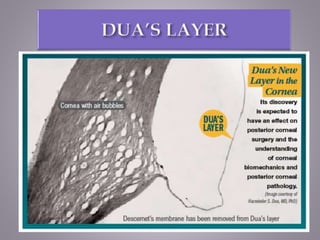
- Solanki Riddhi (B.Optom.)
Roll no : 05
Provides greatest amount of refractive
power to the eye.
Elliptical in shape.
Has 5 layers.
1. EPITHELIUM
2. BOWMAN’S LAYER
3. STROMA
4. DESCEMET’S MEMBRANE
5. ENDOTHELIUM
 Outermost layer of the cornea.
 Smooth corneal surface.
 Cells regenerate (only layer of the
cornea that does this)
 Made up of epithelial tissue.
 Cells of deepest layers are columnar,
known as basal cells.
 Middle layer cells are polyhedral
cells called wing cells.
 Lastly , there are three or four layers of
squamous cells, with flattened nuclei.
 Acts as a barrier to protect the cornea.
 Resisting the free flow of fluids from the tears.
 Prevents bacteria from entering the epithelium
& corneal stroma.
 Second layer of the cornea.
 Is made up of a dense fibrous sheet
of collagen fibers.
 A transition layer between the
epithelium and stroma.
 Does not regenerate.
 Composed of strong, randomly
oriented collagen fibrils .
 Act as a physical barrier to protect the
subepithelial nerve plexus.
 Serve as a barrier that prevents direct traumatic
contact with the corneal stroma & hence it is
highly involved in stromal healing.
 Associated with the corneal transparency at the
morphological level.
 Accounts for 90% of total thickness
(largest layer)
 Fibers in this layer contribute to
corneal transparency.
 Composed of about 200 flattened
lamellae.
 Secrete an extracellular matrix, which includes
collagen and proteoglycans.
 And also produce crystalline proteins to
maintain corneal transparency
 Considered basement membrane to the
Endothelium.
 Produces constantly, which means it
thickens over one’s lifetime (will
approximately triple in thickness over
the course of your life)
 It is composed of a different kind of
collagen (Type VIII) than the stroma.
 The endothelial layer is located at the
posterior of the cornea.
 This layer is important for the health of
endothelial cells.
 One of the leading needs for cornea transplant
is from a dystrophy of Descemet’s layer called
Fuch’s dystrophy.
 Innermost layer of cornea.
 Composed of one single layer of cells.
 Cells do not divide or replicate.
 Separates the cornea from the anterior
portion of the eye.
 The corneal endothelium are specialized,
flattened, mitochondria-rich cells that
line the posterior surface of the cornea
and face anterior chamber of the eye.
 Governs fluid and solute transport across the
posterior surface of the cornea.
 Actively maintains the cornea in the slightly
dehydrated state that is required for optical
transparency.
 Dua’s layer, according to a 2013 paper by Harminder
Singh Dua’s group at the University of Nottingham, is a
layer of the cornea that had not been detected previously.
 It is hypothetically 0.00059 inches thick, the fourth
caudal layer, and located between the corneal
stroma and Descemet’s membrane.
 Despite its thinness, the layer is very strong and
impervious to air. It is strong enough to withstand up to
2 bars (200 kPa) of pressure.
 While some scientists welcomed the announcement,
other scientists cautioned that time was needed for other
researchers to confirm the discovery and its
significance. Others have met the claim "with
incredulity". The choice of the name Dua’s layer has also
been criticized.
Layers of cornea
Layers of cornea

Layers of cornea

  • 1.
    - Solanki Riddhi(B.Optom.) Roll no : 05
  • 2.
    Provides greatest amountof refractive power to the eye. Elliptical in shape. Has 5 layers. 1. EPITHELIUM 2. BOWMAN’S LAYER 3. STROMA 4. DESCEMET’S MEMBRANE 5. ENDOTHELIUM
  • 4.
     Outermost layerof the cornea.  Smooth corneal surface.  Cells regenerate (only layer of the cornea that does this)  Made up of epithelial tissue.  Cells of deepest layers are columnar, known as basal cells.  Middle layer cells are polyhedral cells called wing cells.
  • 5.
     Lastly ,there are three or four layers of squamous cells, with flattened nuclei.
  • 6.
     Acts asa barrier to protect the cornea.  Resisting the free flow of fluids from the tears.  Prevents bacteria from entering the epithelium & corneal stroma.
  • 8.
     Second layerof the cornea.  Is made up of a dense fibrous sheet of collagen fibers.  A transition layer between the epithelium and stroma.  Does not regenerate.  Composed of strong, randomly oriented collagen fibrils .
  • 9.
     Act asa physical barrier to protect the subepithelial nerve plexus.  Serve as a barrier that prevents direct traumatic contact with the corneal stroma & hence it is highly involved in stromal healing.  Associated with the corneal transparency at the morphological level.
  • 11.
     Accounts for90% of total thickness (largest layer)  Fibers in this layer contribute to corneal transparency.  Composed of about 200 flattened lamellae.
  • 12.
     Secrete anextracellular matrix, which includes collagen and proteoglycans.  And also produce crystalline proteins to maintain corneal transparency
  • 14.
     Considered basementmembrane to the Endothelium.  Produces constantly, which means it thickens over one’s lifetime (will approximately triple in thickness over the course of your life)  It is composed of a different kind of collagen (Type VIII) than the stroma.  The endothelial layer is located at the posterior of the cornea.
  • 15.
     This layeris important for the health of endothelial cells.  One of the leading needs for cornea transplant is from a dystrophy of Descemet’s layer called Fuch’s dystrophy.
  • 17.
     Innermost layerof cornea.  Composed of one single layer of cells.  Cells do not divide or replicate.  Separates the cornea from the anterior portion of the eye.  The corneal endothelium are specialized, flattened, mitochondria-rich cells that line the posterior surface of the cornea and face anterior chamber of the eye.
  • 18.
     Governs fluidand solute transport across the posterior surface of the cornea.  Actively maintains the cornea in the slightly dehydrated state that is required for optical transparency.
  • 20.
     Dua’s layer,according to a 2013 paper by Harminder Singh Dua’s group at the University of Nottingham, is a layer of the cornea that had not been detected previously.  It is hypothetically 0.00059 inches thick, the fourth caudal layer, and located between the corneal stroma and Descemet’s membrane.  Despite its thinness, the layer is very strong and impervious to air. It is strong enough to withstand up to 2 bars (200 kPa) of pressure.  While some scientists welcomed the announcement, other scientists cautioned that time was needed for other researchers to confirm the discovery and its significance. Others have met the claim "with incredulity". The choice of the name Dua’s layer has also been criticized.