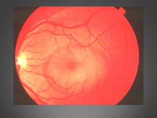
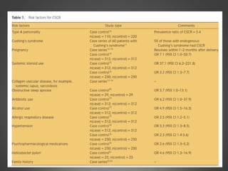
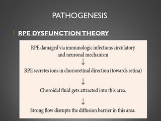
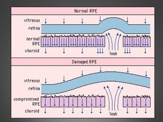
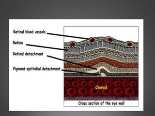
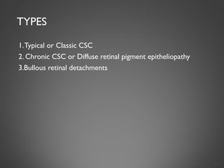
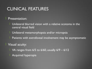
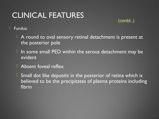
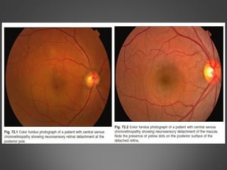
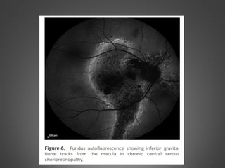
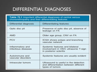
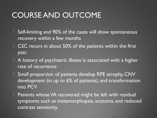
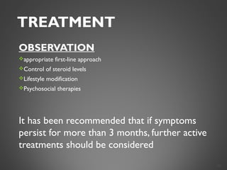
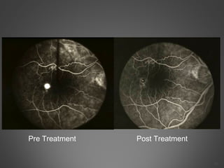
CENTRAL SEROUS CHORIORETINOPATHY
DR SIVATEJA CHALLA
1
INTRODUCTION
A Chorioretinal disorder characterized by an idiopathic localized serous
detachment of the neural retina in the macular region
 Usually unilateral
 May be associated with pigment epithelial detachment (PED)
 Relative preservation of visual function despite prolonged separation of neural retina from
the retinal pigment epithelium (RPE)
2
INTRODUCTION
 Initial description byVon Graefe in 1866 as relapsing central recurent
retinitis.
 1955 – Bennett – “central serous retinopathy”
 Synonyms:
 Central Serous Retinopathy
 Central Serous Pigment Epitheliopathy
 Central Serous Retinitis
3
(contd..)
4
EPIDEMIOLOGY
 Age: 30 – 50 years
 Gender: Male > female 6:1
 RACE: whites and asians>blacks
 Increased incidence in:
 Emotional stress
 Type A personality
 Physical Strains,pregnancy
 People engaged in visually demanding work
5
 Also found to be associated with vasoconstrictive agents,
endogenous hypercortisolism, Systemic corticosteroids, SLE,
Hypertension
 Helicobacter pylori infection has been reported to be associated
with CSC
 Smoking,
 Antibiotic use
 Antihistamine use
 Alcohol consumption and allergic respiratory diseases
 Obstructive sleep apnea
6
7
PATHOGENESIS
 RPE DYSFUNCTIONTHEORY
8
9
PATHOGENESIS
 CHOROID DYSFUNCTIONTHEORY
10
(contd..)
11
TYPES
1.Typical or Classic CSC
2. Chronic CSC or Diffuse retinal pigment epitheliopathy
3.Bullous retinal detachments
12
CLINICAL FEATURES
 Presentation:
 Unilateral blurred vision with a relative scotoma in the
central visual field
 Unilateral metamorphopsia and/or micropsia
 Patients with extrafoveal involvement may be asymptomatic
 Visual acuity:
 VA ranges from 6/5 to 6/60, usually 6/9 – 6/12
 Acquired hyperopia
13
CLINICAL FEATURES
 Fundus:
 A round to oval sensory retinal detachment is present at
the posterior pole
 In some small PED within the serous detachment may be
evident
 Absent foveal reflex
 Small dot like deposits in the posterior of retina which is
believed to be the precipitates of plasma proteins including
fibrin
14
(contd..)
15
CLINICAL FEATURES
 Other features:
 Impaired Dark adaptation
 Colour desaturation
 Patients may present as bullous inferior peripheral retinal
detachment (non-rhegmatogenous)
 In chronic form, diffuse retinal pigment epitheliopathy
progresses in conjunction with persistent or intermittent
SRF, and RDs tend to be shallower and more diffuse, and
visual prognosis is more guarded in such cases
 Chronic CSC may L/T CNV formation,CME,SR Lipid
deposition and chorio capillary atrophy
16
(contd..)
DIAGNOSIS
 Basically clinical, confirmed by FFA
 FFA critical to detect the extent of the retinal abnormalities
and to exclude the presence of other ocular pathology
17
DIAGNOSIS
 Fluoroscein Angiography: Characteristic features:
 Ink-blot appearance:
• Seen in 93% cases
• Leakage point/s with uniform dye filling is appreciated.
• Most common location – upper nasal quadrant.
• Least common location – lower temporal quadrant
18
(contd..)
19
DIAGNOSIS
 Fluoroscein Angiography: Characteristic features:
 Smoke-stack appearance:
 Small hyperfluorescent spot in the early phase due to
leakage of dye through the RPE
 Fluorescein passes into the subretinal space and ascends
vertically until the upper border, like a smoke-stack, in
the late venous phase
 The dye then spreads laterally, taking on a “mushroom”
or “umbrella” configuration until entire area of
detachment is filled
20
(contd..)
21
Optical CoherenceTomography(OCT):
IMPORTANCE
1.In diagonosis
2.Following the progress
3.Quantifying serous detachment
OCT reveals many aspects of pathophysiology of CSC,ranging from SRF,
PED,& retinal atrophy following chronic disease
OCT is especially helpful in identifying subtle, even subclinical,
neurosensory & macular detachments.
OCT is also helpful in identifying the dreaded complication of Choroidal
NeoVascular membrane
Increased choroidal thickening is a hallmark of CSC
22
 ACUTE CSR CHRONIC CSR
• NSD at macula 1.Foveal atrophy or thinning
• RPED 2.cystoid changes at fovea
• Combination of both
23
Indocyanin Green (ICG) Angiography:
ICGA is one of the most important investigations in CSC because
it demonstrates the choroidal vascular abnormalities and can act as a guide to
treatments such as photodynamic therapy.
Common findings in patients with CSC are multi focal areas of hyper fluorescence
in the early and midphases of the study,which then fade in the late phase of the
study
24
25
Fundus autofluorescence
 FAF typically shows hypofluorescence at the leakage point and
over the area of neurosensory detachment due to blockage by
subretinal fluid.
The subretinal yellow dots observed clinically might demonstrate
hyperfluorescence
In chronic-recurrent CSC,hyperfluorescence is common in areas
of residual neurosensory detachment.
Other tests
Multifocal ERG has been used to identify focal regions of
decreased retinal function, even in asymptomatic or clinically
inactive eyes.
Microperimetry has also shown that, despite clinical
resolution of CSR, there is lower retinal sensitivity in the macula
even once visual acuity returned to 20/20 26
27
DIFFERENTIAL DIAGNOSES
28
COURSE AND OUTCOME
 Self-limiting and 90% of the cases will show spontaneous
recovery within a few months
 CSC recurs in about 50% of the patients within the first
year.
 A history of psychiatric illness is associated with a higher
rate of recurrence
 Small proportion of patients develop RPE atrophy, CNV
development (in up to 6% of patients), and transformation
into PCV
 Patients whoseVA recovered might be left with residual
symptoms such as metamorphopsia, scotoma, and reduced
contrast sensitivity.
29
TREATMENT
OBSERVATION
appropriate first-line approach
Control of steroid levels
Lifestyle modification
Psychosocial therapies
It has been recommended that if symptoms
persist for more than 3 months, further active
treatments should be considered
30
LASER PHOTOCOAGULATION
Accelerates the resolution of the detachment
lowers the recurrence rate
MOA:
Beam destroys the cluster of diseased pigment epithelium cells,
thus stopping the secretion of fluid beneath the neurosensory
retina.
Resulting scar helps to transport fluid back into the
choriocapillaris.
31
Indications:
 Persistence of serous detachment beyond 3-4 months
 Recurrences in eyes with visual deficit from previous episodes
 Presence of permanent visual deficit from previous episodes in the
fellow eye
 Development of chronic signs such as cystic changes in the
neurosensory retina or widespread RPE abnormalities
 Occupational or other patient needs that require prompt
restoration of vision or stereopsis
32
 Technique:
 2 – 3 low to moderate intensity burns applied to the
leakage site (spot size of 200µm, for 0.1 seconds), to
produce mild graying of the RPE
 Decreases of duration of detachment.
33
Advantages
 Decreased duration
 Decreased recurrence
Complications
 Choroidal neovascularization
 Central scotoma
Should be avoided if the leak is within 500µm from the centre of the
foveal avascular zone
Careful follow up required as 2 – 5% of eyes treated with
photocoagulation develop CNV
34
35
Pre Treatment Post Treatment
PHOTODYNAMIC THERAPY
INDICATIONS
・ Juxtafoveal lesion.
・ Subfoveal lesion.
・ Lack of a clearly defined leakage hot spot.
. Concern about the potential induction of CNV.
MOA
choriocapillaris narrowing, choroidal hypoperfusion,
reduction of choroidal exudation and choroidal vascular
remodeling
36
 Safety-enhanced PDT was performed using half the normal dose of
verteporfin at 3 mg/m2.
 Infusion of verteporfin was performed over 8 minutes,
 delivery of laser at 692 nm 10 minutes afterwards
 CONCLUSIONThe modified safety enhanced PDT protocol with
half dose verteporfin appeared to be a beneficial treatment option for
patients with chronic CSC, especially in eyes without serous PED
37
OTHER RX MODALITIES
 ANTI-VEGF suggested that combined PDT and intravitreal anti-VEGF
has a beneficial role to play for CSC patients
  Small series exist to support a number of agents, including
mifepristone, ketoconazole, rifampin, finesteride, and eplerenone.
Patients with chronic severe CSC that is recalcitrant to local therapy
may consider these agents
38
THANKYOU
39

CENTRAL SEROUS CHORIO RETINOPATHY

  • 1.
  • 2.
    INTRODUCTION A Chorioretinal disordercharacterized by an idiopathic localized serous detachment of the neural retina in the macular region  Usually unilateral  May be associated with pigment epithelial detachment (PED)  Relative preservation of visual function despite prolonged separation of neural retina from the retinal pigment epithelium (RPE) 2
  • 3.
    INTRODUCTION  Initial descriptionbyVon Graefe in 1866 as relapsing central recurent retinitis.  1955 – Bennett – “central serous retinopathy”  Synonyms:  Central Serous Retinopathy  Central Serous Pigment Epitheliopathy  Central Serous Retinitis 3 (contd..)
  • 4.
  • 5.
    EPIDEMIOLOGY  Age: 30– 50 years  Gender: Male > female 6:1  RACE: whites and asians>blacks  Increased incidence in:  Emotional stress  Type A personality  Physical Strains,pregnancy  People engaged in visually demanding work 5
  • 6.
     Also foundto be associated with vasoconstrictive agents, endogenous hypercortisolism, Systemic corticosteroids, SLE, Hypertension  Helicobacter pylori infection has been reported to be associated with CSC  Smoking,  Antibiotic use  Antihistamine use  Alcohol consumption and allergic respiratory diseases  Obstructive sleep apnea 6
  • 7.
  • 8.
  • 9.
  • 10.
  • 11.
  • 12.
    TYPES 1.Typical or ClassicCSC 2. Chronic CSC or Diffuse retinal pigment epitheliopathy 3.Bullous retinal detachments 12
  • 13.
    CLINICAL FEATURES  Presentation: Unilateral blurred vision with a relative scotoma in the central visual field  Unilateral metamorphopsia and/or micropsia  Patients with extrafoveal involvement may be asymptomatic  Visual acuity:  VA ranges from 6/5 to 6/60, usually 6/9 – 6/12  Acquired hyperopia 13
  • 14.
    CLINICAL FEATURES  Fundus: A round to oval sensory retinal detachment is present at the posterior pole  In some small PED within the serous detachment may be evident  Absent foveal reflex  Small dot like deposits in the posterior of retina which is believed to be the precipitates of plasma proteins including fibrin 14 (contd..)
  • 15.
  • 16.
    CLINICAL FEATURES  Otherfeatures:  Impaired Dark adaptation  Colour desaturation  Patients may present as bullous inferior peripheral retinal detachment (non-rhegmatogenous)  In chronic form, diffuse retinal pigment epitheliopathy progresses in conjunction with persistent or intermittent SRF, and RDs tend to be shallower and more diffuse, and visual prognosis is more guarded in such cases  Chronic CSC may L/T CNV formation,CME,SR Lipid deposition and chorio capillary atrophy 16 (contd..)
  • 17.
    DIAGNOSIS  Basically clinical,confirmed by FFA  FFA critical to detect the extent of the retinal abnormalities and to exclude the presence of other ocular pathology 17
  • 18.
    DIAGNOSIS  Fluoroscein Angiography:Characteristic features:  Ink-blot appearance: • Seen in 93% cases • Leakage point/s with uniform dye filling is appreciated. • Most common location – upper nasal quadrant. • Least common location – lower temporal quadrant 18 (contd..)
  • 19.
  • 20.
    DIAGNOSIS  Fluoroscein Angiography:Characteristic features:  Smoke-stack appearance:  Small hyperfluorescent spot in the early phase due to leakage of dye through the RPE  Fluorescein passes into the subretinal space and ascends vertically until the upper border, like a smoke-stack, in the late venous phase  The dye then spreads laterally, taking on a “mushroom” or “umbrella” configuration until entire area of detachment is filled 20 (contd..)
  • 21.
  • 22.
    Optical CoherenceTomography(OCT): IMPORTANCE 1.In diagonosis 2.Followingthe progress 3.Quantifying serous detachment OCT reveals many aspects of pathophysiology of CSC,ranging from SRF, PED,& retinal atrophy following chronic disease OCT is especially helpful in identifying subtle, even subclinical, neurosensory & macular detachments. OCT is also helpful in identifying the dreaded complication of Choroidal NeoVascular membrane Increased choroidal thickening is a hallmark of CSC 22
  • 23.
     ACUTE CSRCHRONIC CSR • NSD at macula 1.Foveal atrophy or thinning • RPED 2.cystoid changes at fovea • Combination of both 23
  • 24.
    Indocyanin Green (ICG)Angiography: ICGA is one of the most important investigations in CSC because it demonstrates the choroidal vascular abnormalities and can act as a guide to treatments such as photodynamic therapy. Common findings in patients with CSC are multi focal areas of hyper fluorescence in the early and midphases of the study,which then fade in the late phase of the study 24
  • 25.
  • 26.
    Fundus autofluorescence  FAFtypically shows hypofluorescence at the leakage point and over the area of neurosensory detachment due to blockage by subretinal fluid. The subretinal yellow dots observed clinically might demonstrate hyperfluorescence In chronic-recurrent CSC,hyperfluorescence is common in areas of residual neurosensory detachment. Other tests Multifocal ERG has been used to identify focal regions of decreased retinal function, even in asymptomatic or clinically inactive eyes. Microperimetry has also shown that, despite clinical resolution of CSR, there is lower retinal sensitivity in the macula even once visual acuity returned to 20/20 26
  • 27.
  • 28.
  • 29.
    COURSE AND OUTCOME Self-limiting and 90% of the cases will show spontaneous recovery within a few months  CSC recurs in about 50% of the patients within the first year.  A history of psychiatric illness is associated with a higher rate of recurrence  Small proportion of patients develop RPE atrophy, CNV development (in up to 6% of patients), and transformation into PCV  Patients whoseVA recovered might be left with residual symptoms such as metamorphopsia, scotoma, and reduced contrast sensitivity. 29
  • 30.
    TREATMENT OBSERVATION appropriate first-line approach Controlof steroid levels Lifestyle modification Psychosocial therapies It has been recommended that if symptoms persist for more than 3 months, further active treatments should be considered 30
  • 31.
    LASER PHOTOCOAGULATION Accelerates theresolution of the detachment lowers the recurrence rate MOA: Beam destroys the cluster of diseased pigment epithelium cells, thus stopping the secretion of fluid beneath the neurosensory retina. Resulting scar helps to transport fluid back into the choriocapillaris. 31
  • 32.
    Indications:  Persistence ofserous detachment beyond 3-4 months  Recurrences in eyes with visual deficit from previous episodes  Presence of permanent visual deficit from previous episodes in the fellow eye  Development of chronic signs such as cystic changes in the neurosensory retina or widespread RPE abnormalities  Occupational or other patient needs that require prompt restoration of vision or stereopsis 32
  • 33.
     Technique:  2– 3 low to moderate intensity burns applied to the leakage site (spot size of 200µm, for 0.1 seconds), to produce mild graying of the RPE  Decreases of duration of detachment. 33
  • 34.
    Advantages  Decreased duration Decreased recurrence Complications  Choroidal neovascularization  Central scotoma Should be avoided if the leak is within 500µm from the centre of the foveal avascular zone Careful follow up required as 2 – 5% of eyes treated with photocoagulation develop CNV 34
  • 35.
  • 36.
    PHOTODYNAMIC THERAPY INDICATIONS ・ Juxtafoveallesion. ・ Subfoveal lesion. ・ Lack of a clearly defined leakage hot spot. . Concern about the potential induction of CNV. MOA choriocapillaris narrowing, choroidal hypoperfusion, reduction of choroidal exudation and choroidal vascular remodeling 36
  • 37.
     Safety-enhanced PDTwas performed using half the normal dose of verteporfin at 3 mg/m2.  Infusion of verteporfin was performed over 8 minutes,  delivery of laser at 692 nm 10 minutes afterwards  CONCLUSIONThe modified safety enhanced PDT protocol with half dose verteporfin appeared to be a beneficial treatment option for patients with chronic CSC, especially in eyes without serous PED 37
  • 38.
    OTHER RX MODALITIES ANTI-VEGF suggested that combined PDT and intravitreal anti-VEGF has a beneficial role to play for CSC patients   Small series exist to support a number of agents, including mifepristone, ketoconazole, rifampin, finesteride, and eplerenone. Patients with chronic severe CSC that is recalcitrant to local therapy may consider these agents 38
  • 39.