Downloaded 403 times


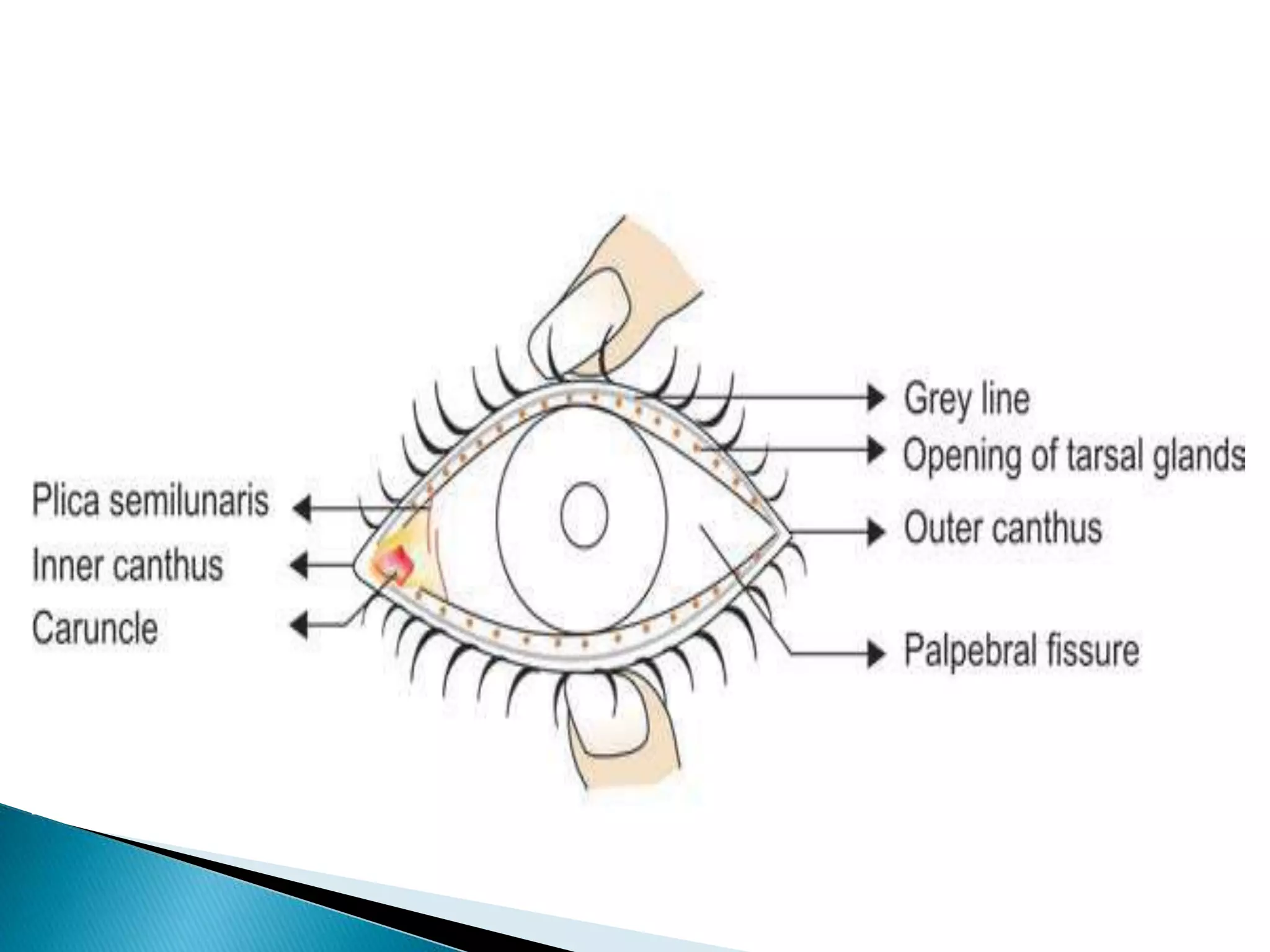

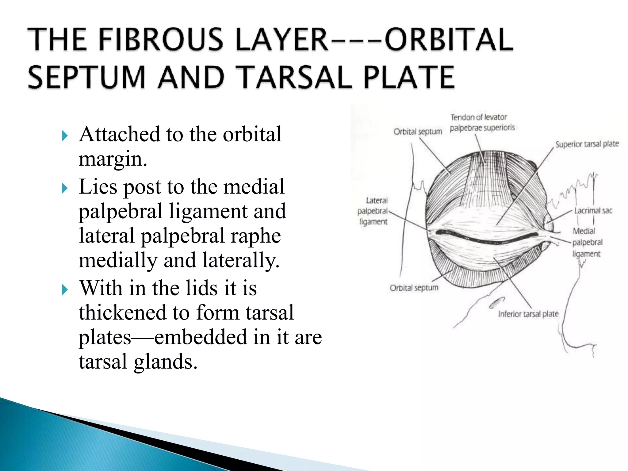


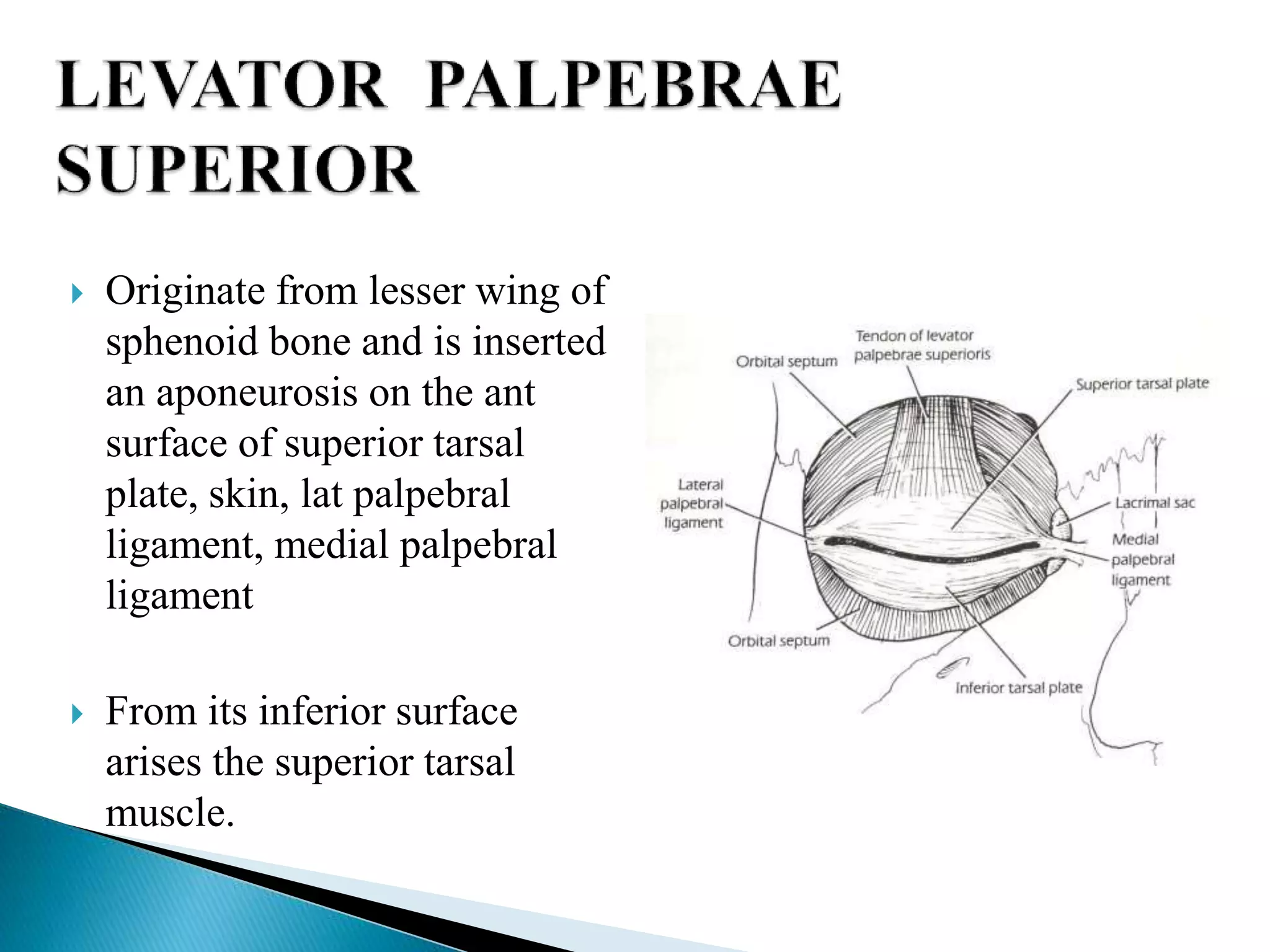
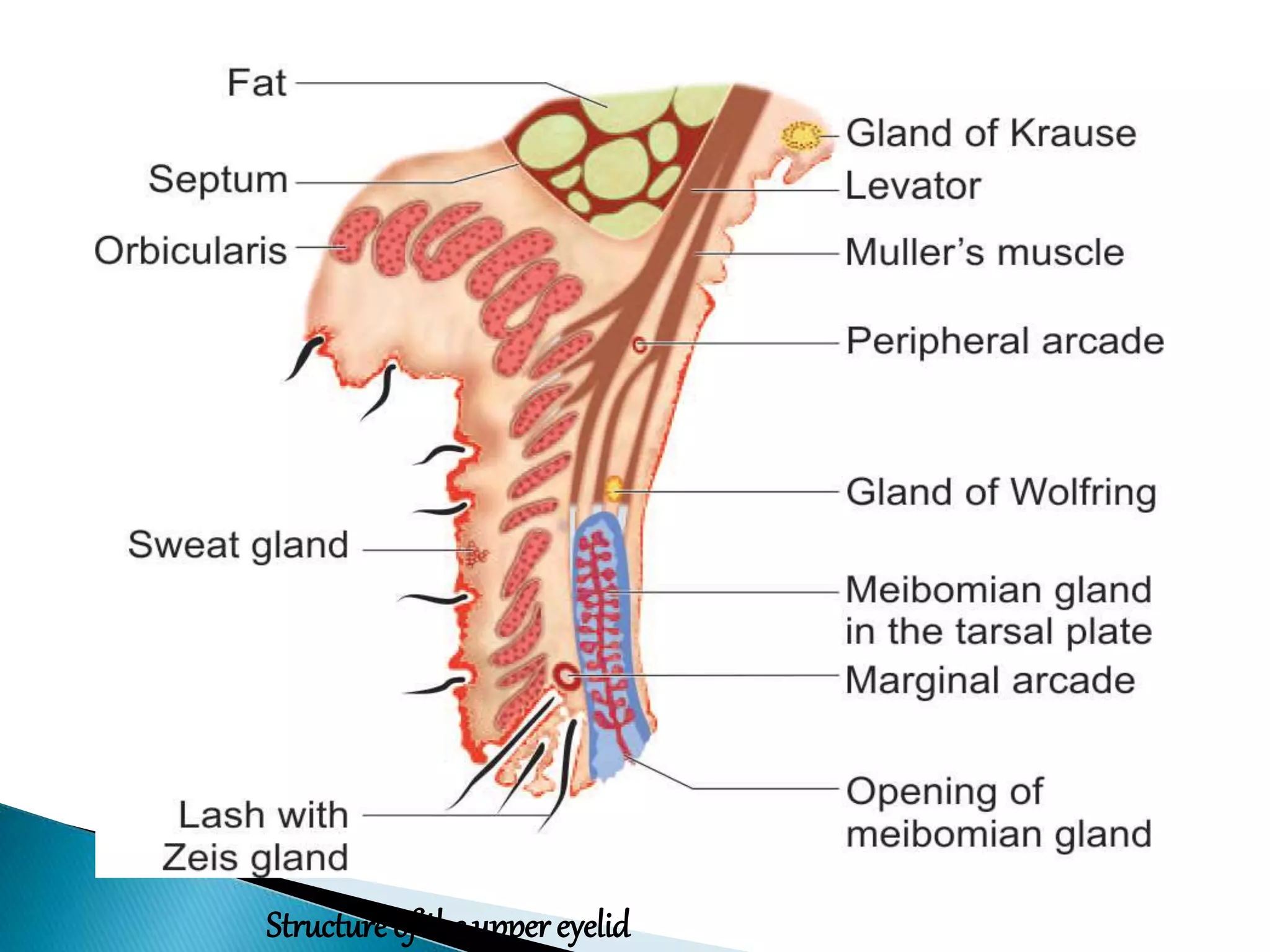


The eyelids are composed of several layers of tissue and perform important protective and lubricating functions for the eyes. They contain glands that secrete oils to form the outer layer of the tear film and help spread tears across the cornea. The eyelids are innervated by cranial nerves and contain muscles that open and close the palpebral fissure, protecting the eyes from damage and keeping them moist.