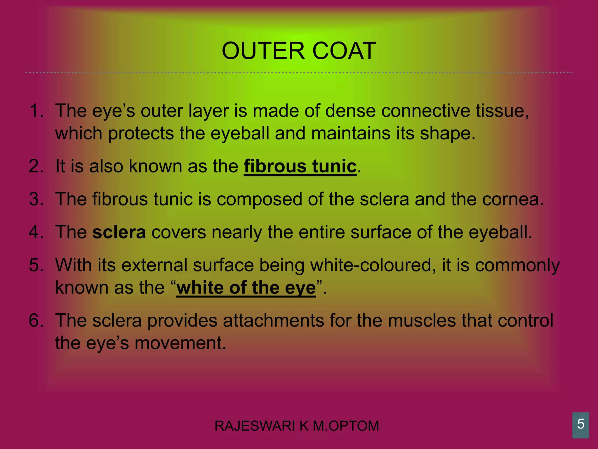
COATS OF
EYE
Unit I
https://www.kinderkrebsinfo.de/diseases/solid_tumours
pohretino_patinfo120120611/the_eye/index_eng.html
What do you mean
by a “COAT”
RAJESWARI K M.OPTOM
THREE COATS
1. Inner Coat - Nervous coat
2. Middle Coat - Vascular coat
3. Outer Coat - Fibrous coat
3
RAJESWARI K M.OPTOM 4
RAJESWARI K M.OPTOM
OUTER COAT
1. The eye’s outer layer is made of dense connective tissue,
which protects the eyeball and maintains its shape.
2. It is also known as the fibrous tunic.
3. The fibrous tunic is composed of the sclera and the cornea.
4. The sclera covers nearly the entire surface of the eyeball.
5. With its external surface being white-coloured, it is commonly
known as the “white of the eye”.
6. The sclera provides attachments for the muscles that control
the eye’s movement.
5
RAJESWARI K M.OPTOM
1. The transparent cornea occupies the front center part of the
external tunic.
2. It serves as the eye’s “window”, which lets the light in and
bends its rays, thereby providing most of the eye’s focusing
power.
3. The anterior, visible part of the sclera as well as the inner
surface of the eyelids are covered by the conjunctiva,
4. A mucous membrane that helps lubricating the eye together
with the tears
6
RAJESWARI K M.OPTOM
MIDDLE COAT
1. The middle layer of tissue surrounding the eye, also known as
the vascular tunic or “uvea“,
2. Formed by the choroid, the ciliary body, and the iris.
3. The choroid takes up the posterior five-sixths of the eye and
is mainly comprised of blood vessels.
4. Its major functions are oxygen supply and nutrition for the eye.
7
RAJESWARI K M.OPTOM
1. The anterior part of the choroid passes into the ciliary body,
one function of which is anchoring the lens in place.
2. The ciliary body contains a muscle (ciliary muscle), which can
change the shape of the lens for accommodation.
3. The iris, which is connected to the anterior part of the ciliary
body, covers the top of the lens.
4. The iris contains pigments, the amount of which determines a
person’s eye colour.
8
RAJESWARI K M.OPTOM
INNER COAT
1. The third and inner coat of the eye is the retina, which is
responsible for the perception of images – vision.
2. The retina is a light-sensitive layer of nervous tissue
composed of multiple nerve cells
3. For vision, there are two types of photoreceptor cells: rods
and cones. Rods provide the perception of vision, mostly in
dim light, whereas cones help to see colors in daylight.
9
RAJESWARI K M.OPTOM
THANK YOU…
10

Coats of eyeball

  • 1.
  • 2.
    What do youmean by a “COAT”
  • 3.
    RAJESWARI K M.OPTOM THREECOATS 1. Inner Coat - Nervous coat 2. Middle Coat - Vascular coat 3. Outer Coat - Fibrous coat 3
  • 4.
  • 5.
    RAJESWARI K M.OPTOM OUTERCOAT 1. The eye’s outer layer is made of dense connective tissue, which protects the eyeball and maintains its shape. 2. It is also known as the fibrous tunic. 3. The fibrous tunic is composed of the sclera and the cornea. 4. The sclera covers nearly the entire surface of the eyeball. 5. With its external surface being white-coloured, it is commonly known as the “white of the eye”. 6. The sclera provides attachments for the muscles that control the eye’s movement. 5
  • 6.
    RAJESWARI K M.OPTOM 1.The transparent cornea occupies the front center part of the external tunic. 2. It serves as the eye’s “window”, which lets the light in and bends its rays, thereby providing most of the eye’s focusing power. 3. The anterior, visible part of the sclera as well as the inner surface of the eyelids are covered by the conjunctiva, 4. A mucous membrane that helps lubricating the eye together with the tears 6
  • 7.
    RAJESWARI K M.OPTOM MIDDLECOAT 1. The middle layer of tissue surrounding the eye, also known as the vascular tunic or “uvea“, 2. Formed by the choroid, the ciliary body, and the iris. 3. The choroid takes up the posterior five-sixths of the eye and is mainly comprised of blood vessels. 4. Its major functions are oxygen supply and nutrition for the eye. 7
  • 8.
    RAJESWARI K M.OPTOM 1.The anterior part of the choroid passes into the ciliary body, one function of which is anchoring the lens in place. 2. The ciliary body contains a muscle (ciliary muscle), which can change the shape of the lens for accommodation. 3. The iris, which is connected to the anterior part of the ciliary body, covers the top of the lens. 4. The iris contains pigments, the amount of which determines a person’s eye colour. 8
  • 9.
    RAJESWARI K M.OPTOM INNERCOAT 1. The third and inner coat of the eye is the retina, which is responsible for the perception of images – vision. 2. The retina is a light-sensitive layer of nervous tissue composed of multiple nerve cells 3. For vision, there are two types of photoreceptor cells: rods and cones. Rods provide the perception of vision, mostly in dim light, whereas cones help to see colors in daylight. 9
  • 10.