
Download as PDF, PPTX


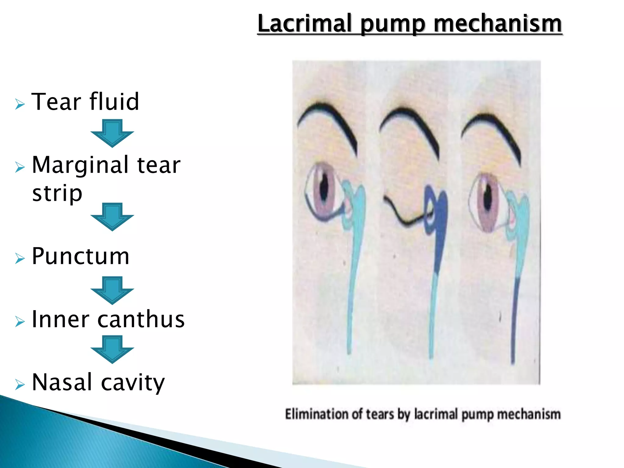
![Fluid flows over preocular surface & reaches
ciliary margin of each eyelid and collects in
the inner canthus.
Fluid is drained by lacrimal passage into nasal
cavity [“active lacrimal pump mechanism”]](https://image.slidesharecdn.com/physiologyoftearfilmitsdrainage-170122065759-190220165434/75/anatomy-And-Physiology-of-tear-film-19-2048.jpg)


The document details the structure and functions of the three layers of the tear film: the lipid layer, which prevents evaporation; the aqueous layer, which lubricates and nourishes the cornea; and the mucin layer, which spreads tears across the corneal surface. It explains the mechanisms of tear secretion—basal and reflex—and the dynamics involved in tear film stability, thinning, and redistribution during blinking. Additionally, it discusses factors affecting tear film integrity, such as environmental conditions and the role of the lacrimal pump mechanism in draining tears into the nasal cavity.


