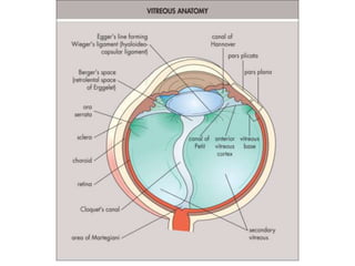
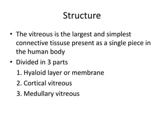
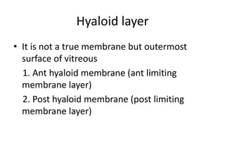
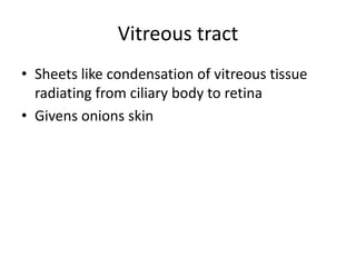
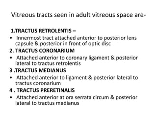
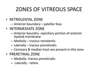
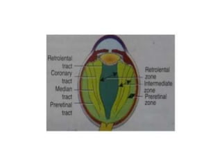
2. • Vitreous humour is an inert, transparent, jelly
–like, hydrophilic (dissolved in water) gel
structure
• Vitreous cpmposed of approximately 99%
water
• Vitreous cavity is bounded anteriorly by lens &
ciliary body & posteriorly by retina
3. • wt-4g
• Vol-4cc (2/3vol of entire globe)
• Approximately 99 % water , 1 % solid.
(0.9% salts ,0.08% protein, 0.02%
mucopolysaccharide)
5. Formation of Primary VITREOUS
• Primary vitreous appears at the time of
formation of the optic cup, is a fibrillated
secretion of the retinal cells, and fills in the
vitreous space with a feltwork of fine fibrils
• Primary cellular vitreous formation
• Vascularization of primary vitreous
• Its mixed ectodermal and mesenchymal origin
6. • Ingrowth of hyaloid artery through the embryonic
fissure. Ectoderm also contribute.
• Present in 1 st month of gestation.
• Regression of hyaloid artery by the end of 3 rd
month leads to retraction of primitive vitreous.
• Hyaloid artery is no longer patent and loses its
connection with disc- 7 th month.
• Remnants may remain as persistent pupillary
membrane.
7. FORMATION OF SECONDARY
VITREOUS:
• Sec avascular vitreous surrounds the primary
vitreous.
• Secreted by neuroectoderm of optic cup.
• Formation starts from 2 nd month and
completes by 5 th -6 th month.
• Marked by appearance of orderly array of fine
dense fibres.
8. FORMATION OF TERTIARY VITREOUS:
• Secreted by neuroectoderm in ciliary region.
• Secreted during 4 th month.
• Forms vitreous base & ciliary zonules.
9.
10.
11. Structure
• The vitreous is the largest and simplest
connective tissuse present as a single piece in
the human body
• Divided in 3 parts
1. Hyaloid layer or membrane
2. Cortical vitreous
3. Medullary vitreous
12. Hyaloid layer
• It is not a true membrane but outermost
surface of vitreous
1. Ant hyaloid membrane (ant limiting
membrane layer)
2. Post hyaloid membrane (post limiting
membrane layer)
13. ANTERIOR HYALOID MEMBRANE /
ANTERIOR LIMITING MEMBRANE
• Lies 1.5 mm anterior to oraserrata
• With other structure
• Hyalocilliary zonule- anterior hyloid membrane to
cilliary proccess
• Retrolental ligament – anterior hyaloid to lens
• Coronary ligament – anterior hyaloid to posterior
one-third of cilliary processes circum ferentially
• Median ligament – anterior hyaloid to pars plana
14. POSTERIOR HYALOID MEMBRANE /
POSTERIOR LIMITING MEMBRANE
• Extends back from vitreous based to optic disc
• Subhyaloid space: between internal limiting
membrane of retina and posterior hyaloid.
15.
16. Cortical vitreous
• It lie in peripheral zone appox 100 micron in width
• It is more condensed fibrillar vitreous
• It contain Type 2 collagen fibrils interspersed with the
sodium hyaluronate ,mucopolysaccharide molecules
which provide viscosity ,elasticity and tensile strenght
to it
• 2 percent of the total vitreous vol
• It is the metabolic centre of the vitreous body(vitreous
cells –hyalocytes)
• Vitreos cells synthesize the hyaluronic acid
17. Medullary vitreous
• Similar to cortical vitreous except it is less
fibrillar structure and cell free
• Forms major part of vitreous body.
• Homogenous but exhibit wavy lines.
• Watered silk appearance in slit lamp.
18. Vitreous tract
• Sheets like condensation of vitreous tissue
radiating from ciliary body to retina
• Givens onions skin
19. Vitreous tracts seen in adult vitreous space are-
1.TRACTUS RETROLENTIS –
• Innermost tract attached anterior to posterior lens
capsule & posterior in front of optic disc
2. TRACTUS CORONARIUM
• Attached anterior to coronary ligament & posterior
lateral to tractus retrolentis
3 .TRACTUS MEDIANUS
• Attached anterior to ligament & posterior lateral to
tractus coronarium
4 . TRACTUS PRERETINALIS
• Attached anterior at ora serrata circum & posterior
lateral to tractus medianus
20. ZONES OF VITREOUS SPACE
• RETROLENTAL ZONE
– Anterior boundary – patellar fosa
• INTERMEDIATE ZONE
– Anterior boundry –epiciliary portion of anterior
hyaloid membrane
– Medially – tractus retrolentis
– Laterally – tractus preretinalis
– Coronary & median tract are present in this zone
• PRERETINAL ZONE
– Medially- tractus preretinalis
– Laterally - retina
21.
22. BIOCHEMICAL COMPOSITION
• Composed of 3 major structural components
1. Water
2. Collagen-like fibres
3. Hyaluronic acid and glycosaminoglycans(GAGs)
• Macromolecules constituents are:
- Collagen: Type ii collagen :90%
Type ix collagen:10%
- Hyaluronic acid: N-acetyl glucosamine and glucoronic acid
- Soluble proteins: About 10% of serum conc. (low) And thus
does not scatter light
23. Vitreous expansion and contraction
• Vitreous has Na+ ions, NaCl and protein molecules
which neutralizes HA(hyaluronic acid)
→ expansion of vitreous
-vely charged HA molecules repel each other
Hyaluronic acid neutralization is disturbed
When Cationic molecules are removed(water)
Contraction of vitreous
If sodium ions /NaCl are replaced by
+ve protein molecules such as
protamine sulphate or cationic dyes
24. Physicochemical properties
Optical properties
• Transparency : Fine arrangement of vitreous
fibres and high water content
•Tyndall effect: Presence of low amount of
soluble proteins accounts for absence of light
scattering
• Light transmission: Transmits 90% of light
between 300-1400 nm
25. Plasticity: 3 D arrangement of collagen fibres
Viscoelasticity: Negatively charged hyaluronic
acid molecules precipitate extreme volume
changes with change in physicochemical
environment
Gel stability: collagen and hyaluronic acid interact
to stabilize the system termed as frictional
interaction
Molecular sieve effects: Selectively inhibit the
movement of water ,large molecules and cells