Siklus sel mitosis (Universitas Kuningan)
•Download as PPT, PDF•
14 likes•2,941 views

Berisi materi siklus sel mitosis yang berbentuk power point ppt

Recommended
More Related Content
What's hot
What's hot (20)
Viewers also liked
Similar to Siklus sel mitosis (Universitas Kuningan)
Similar to Siklus sel mitosis (Universitas Kuningan) (20)
More from Nursidiq 92
More from Nursidiq 92 (15)
Recently uploaded
Recently uploaded (20)
Siklus sel mitosis (Universitas Kuningan)
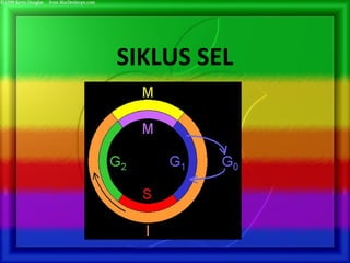
- 1. SIKLUS SEL
- 2. Figure 12.1 Kontinuitas kehidupan Didasarkan atas reproduksi sel /pembelahan sel
- 3. 100 µm (a) Reproduction. An amoeba, a single-celled eukaryote, is dividing into two cells. Each new cell will be an individual organism (LM).
- 4. • Organisme multiseluler tergantung pada pembelahan sel untuk: – Perkembangan (dari sel yang terfertilisasi)- reproduksi – Pertumbuhan – Repair 20 µm200 µm (b) Growth and development. This micrograph shows a sand dollar embryo shortly after the fertilized egg divided, forming two cells (LM). (c) Tissue renewal. These dividing bone marrow cells (arrow) will give rise to new blood cells (LM). Figure 12.2 B, C
- 5. •Semua organisme kompleks berasal dari a single fertilized egg. •Melalui pembelahan sel, jumlah sel meningkat •Sel kemudian terspesialisasi dan berubah menjadi fungsinya masing2
- 6. Tipe pembelahan sel • Mitosis: – Growth, development & repair – Asexual reproduction (yields identical cells) – Occurs in somatic (body) cells • Meiosis: – Sexual reproduction (yields different cells) – Occurs in specific reproductive cells
- 7. MITOSIS
- 10. The Cell Cycle 1. G1 Phase 1st growth phase 2. S Phase DNA duplicated 3. G2 Phase Final growth phase 4. Mitosis 5. Cytokinesis Purpose of the first three phases (Interphase) – to duplicate cell contents; 90% of the cell’s growth cycle (Sel tumbuh & menyalin kromosom dalam persiapan untuk pembelahan sel) Purpose of Mitosis – to divide the genetic material into exact two halves (pembelahan nukleus) Purpose of Cytokinesis – to divide all other contents (except nucleus) into two cells. (pembelahan sitoplasma) interphase
- 11. Interfase 1. G1 Phase 1st growth phase; sel tumbuh 2. S Phase DNA duplicated; kromosom diduplikasi 3. G2 Phase Final growth phase; tumbuh lagi sampai sel menyelesaikan persiapannya untuk pembelahan sel Selama 3 subfase ini, sel tumbuh dengan menghasilkan protein dan organel dalam sitoplasma
- 12. Phases of Mitosis (M) 1. Prophase 2. Prometaphase 3. Metaphase 4. Anaphase 5. Telophase • Pembelahan sel menghasilkan sel anak yang secara genetik identik/sama • Sel harus menduplikasikan material genetiknya – Before they divide, ensuring that each daughter cell receives an exact copy of the genetic material, DNA
- 13. Phases of Mitosis Hal-hal yang unik pada fase M 1.Kondensasi kromosom 2.Mitotic spindle mengatur kromosom yang telah direplikasi - di ekuator - ke kutub 3.Cincin kontraktil: filamen actin dan miosin tegak lurus spindle Menarik membran ke dalam
- 15. Distribution of Chromosomes During Cell Division • Pada persiapan cell division, DNA bereplikasi dan kromosom memadat • Tiap chromosome yang terduplikasi memiliki dua sister chromatids, yang berpisah selama cell division • Sentromer merupakan daerah ceking dari chromosome yang terduplikasi, dimana dua chromatids terikat dengan erat
- 16. Pembelahan sel mendistribusikan kumpulan sel yang identik ke sel anak • Sel menduplikasikan material genetik sebelum membelah, utk memastikan bahwa tiap sel anak menerima copy DNA dengan tepat • Karunia herediter keseluruhan, berisi DNA yang dimiliki oleh suatu sel disebut genome dari sel tersebut • Molekul DNA dalam sel ter-pack menjadi kromosom
- 17. • Setiap spesies eukariot memiliki sejumlah tertentu kromosom pada nukleus • Sel somatik (nonreproductive) memiliki 2 set kromosom • Gamet (reproductive cells: sperm and eggs) memiliki jumlah kromosom setengah jumlah kromosom sel somatik • Kromosom eukaryotik terdiri dari kromatin, sebuah komplex DNA dan protein yang memampat selama pembelahan sel
- 19. 0.5 µm Chromosome duplication (including DNA synthesis) Centromere Separation of sister chromatids Sister chromatids Centromeres Sister chromatids A eukaryotic cell has multiple chromosomes, one of which is represented here. Before duplication, each chromosome has a single DNA molecule. Once duplicated, a chromosome consists of two sister chromatids connected at the centromere. Each chromatid contains a copy of the DNA molecule. Mechanical processes separate the sister chromatids into two chromosomes and distribute them to two daughter cells. Figure 12.4
- 20. • Pembelahan sel pada eukariot terdiri dari: – Mitosis, the division of the nucleus – Cytokinesis, the division of the cytoplasm • Gamet diproduksi dalam pembelahan sel yang disebut meiosis • Meiosis menghasilkan sel anak yang tidak identik dengan induk yaitu hanya memiliki 1 set kromosom
- 22. Fase-fase dalam siklus sel • Siklus sel terdiri dari – Interphase – Fase mitosis INTERPHASE G1 S (DNA synthesis) G2 Cytokinesis M itosis MITOTIC(M) PHASE Figure 12.5 • Interphase – G1 phase – S phase – G2 phase • The mitotic phase – mitosis – cytokinesis
- 23. Phases of the Cell Cycle • Siklus sel terdiri dari – Interphase (cell growth and copying of chromosomes in preparation for cell division) – Mitotic (M) phase (mitosis and cytokinesis) • Interphase (terdiri dari sekitar 90% dari siklus sel) yang dapat dibagi dalam sub fase: – G1 phase (“first gap”) – S phase (“synthesis”) – G2 phase (“second gap”)
- 24. • Mitosis terdiri dari 5 phases – Prophase – Prometaphase G2 OF INTERPHASE PROPHASE PROMETAPHASE Centrosomes (with centriole pairs) ChromatinChromatin (duplicated)(duplicated) Early mitotic spindle Aster Centromere Fragments of nuclear envelope Kinetochore NucleolusNucleolus Nuclear envelope Plasma membrane Chromosome, consisting of two sister chromatids Kinetochore microtubule Nonkinetochore microtubules
- 25. – Metaphase – Anaphase – Telophase Centrosome at one spindle pole Daughter chromosomes METAPHASE ANAPHASE TELOPHASE AND CYTOKINESIS Spindle Metaphase plate Nucleolus forming Cleavage furrow Nuclear envelope forming
- 26. • The mitotic spindle – mikrotubul yang mengontrol pergerakan kromosom selama mitosis • Spindle muncul dari sentromer – spindle microtubules – asters
- 27. • Perakitan spindle microtubules dimulai dari sentrosom - microtubule organizing center • Sentrosom bereplikasi membentuk dua sentrosom yang bermigrasi ke kutub yang berlawanan, dan spindle microtubules tumbuh dari sentrosom • Aster (a radial array of short microtubules) muncul dari tiap sentrosom
- 28. The SpindleThe Spindle (benang gelendong)(benang gelendong) Spindle memiliki struktur seperti web terbuat dari microtubule . Sangat penting pada mitosis karena mengatur kromosom untuk berada pada posisi yang benar Mitotic center Microtubule A cell at metaphase a spindle
- 29. • Some spindle microtubules – Berikatan dengan kinetochores chromosomes CentrosomeAster Sister chromatids Metaphase Plate Kinetochores Overlapping nonkinetochore microtubules Kinetochores microtubules Centrosome ChromosomesMicrotubules 0.5 µm 1 µm Figure 12.7
- 30. Chromosomes attached to spindleChromosomes attached to spindle during nuclear divisionduring nuclear division
- 31. The kinds of microtubules • Kinetochore microtubules : berikatan dengan kinetochores chromosomes dan menggerakkan kromosom ke daerah metafase • Nonkinetochores: overlap satu sama lain tetapi tidak berikatan dengan chromosome 1 µm
- 32. Replikasi sentriol Pada G1 : 1 pasang sentriol terpisah S : sentriol anak tumbuh G2 : perpanjangan sentriol anak Awal fase M : kedua pasang sentriol masih dekat, kemudian terpisah
- 33. Siklus sentrosomSentrosom harus diduplikasi dan dipisahkan Fase S dan G2: 2 ps sentriol Profase awal: sentrosom membelah, tiap anak menjadi pusat mikrotubul astral Profase akhir: mikrotubul polar memanjang, 2 sentrosom terpisah Metafase: mikrotubul berinteraksi dg kromosom, tidak ada selubung nuklear
- 34. The Mitotic Spindle: How chromosomes moved?? The mitotic spindle (structure, shape name) – Is an apparatus of microtubules (composition name) that controls chromosome movement during mitosis – Usually called by Spindle mocrotuble
- 35. How kinetochore microtuble works • In anaphase, sister chromatids separate And move along the kinetochore microtubules toward opposite ends of the cell EXPERIMENT 1 The microtubules of a cell in early anaphase were labeled with a fluorescent dye that glows in the microscope (yellow). Spindle pole Kinetochore Figure 12.8
- 36. 2 A Laser was used to mark the kinetochore mircotubles by eliminating the fluorescnce in a region between one spindle pole and the chromosomes. As anaphase proceeded, researches monitored the changes in the lengths of the microtubles on either side of the mark. Mark RESULTS As the chromosomes moved toward the poles, the microtubule segments on the kinetochore side of the laser mark shortened, while those on the spindle pole side stayed the same length. Discuss about the results
- 37. CONCLUSION This experiment demonstrated that during anaphase, kinetochore microtubules shorten at their kinetochore ends, not at their spindle pole ends. This is just one of the experiments supporting the hypothesis that during anaphase, a chromosome tracks along a microtubule as the microtubule depolymerizes at its kinetochore end, releasing tubulin subunits Chromosome movement Microtubule Motor protein Chromosome Kinetochore Tubulin subunits
- 38. CONCLUSION dalam percobaan ini Selama anafase berlanjut dan kromosom berpindah ke arah kutub, segmen mikrotubul pada sisi kinetokor dari tanda laser memendek, sementara bagian sisi sentromer tetap. Percobaan ini mendukung hipotesis bahwa; kromosom menelusuri disepanjang mikrotubul pada saat mikrotubul berdepolarisasi pada ujung kinetokornya, sehingga terjadi pelepasan sub unit tubulin Chromosome movement Microtubule Motor protein Chromosome Kinetochore Tubulin subunits
- 39. Tiga kelas mikrotubul: 1.Mikrotubul polar : mendorong kutub spindle saling menjauh 2. Mikrotubul kinetokor: berikatan dengan kinetokor, mengatur kromosom 3. Mikrotubul astral: memancar ke seluruh arah dari sentrosom
- 40. Yang melekat pada sentrosom : ujung – ujung positif : dinamis, memanjang memendek overlap Protein yang berasosiasi dengan mikrotubul, mengikat mikrotubul
- 42. • Pada anafase, sister chromatid berpisah – Dan bergerak sepanjang kinetochore microtubules menuju arah berlawanan ujung sel Spindle pole Kinetochore Figure 12.8
- 45. Selubung nuklear: -fosforilasi lamina nuklear : penguraian dan pembangunan selubung nuklear -prometafase : meluruh -telofase : terbentuk kembali
- 46. CYTOKINESISCYTOKINESIS Division of the cytoplasm Mitosis is the splitting of the nucleus. Cytokinesis is the splitting of cytoplasm It usually begins during ANAPHASE
- 47. Sitokinesis • Saat anafase berlanjut ke telofase •Pada sel hewan : melekuknya selaput sel kontraksi cincin contractil • Pada sel tumbuhan : pembentukan dinding baru Sekat sel di antara 2 sel anak -sisa-sisa mikrotubul polar membentuk fragmoplast -vesikuli kecil dari kompleks golgi berisi prazat dinding sel • Tempat sekat : telah ditentukan oleh mikrotubul pada preprofase
- 48. • Pada sel hewan – Cytokinesis terjadi oleh proses yang disebut cleavage, membentuk sebuah a cleavage furrow Cleavage furrow Contractile ring of microfilaments Daughter cells 100 µm (a) Cleavage of an animal cell (SEM)
- 49. • Pada sel tumbuhan, selama cytokinesis – Terbentuk plat sel (cell plate) Daughter cells 1 µmVesicles forming cell plate Wall of patent cell Cell plate New cell wall (b) Cell plate formation in a plant cell (SEM)Figure 12.9 B
- 50. • Mitosis in a plant cell 1 Prophase. The chromatin is condensing. The nucleolus is beginning to disappear. Although not yet visible in the micrograph, the mitotic spindle is staring to from. Prometaphase. We now see discrete chromosomes; each consists of two identical sister chromatids. Later in prometaphase, the nuclear envelop will fragment. Metaphase. The spindle is complete, and the chromosomes, attached to microtubules at their kinetochores, are all at the metaphase plate. Anaphase. The chromatids of each chromosome have separated, and the daughter chromosomes are moving to the ends of cell as their kinetochore microtubles shorten. Telophase. Daughter nuclei are forming. Meanwhile, cytokinesis has started: The cell plate, which will divided the cytoplasm in two, is growing toward the perimeter of the parent cell. 2 3 4 5 Nucleus Nucleolus ChromosomeChromatine condensing Figure 12.10
- 51. • Mitosis in a plant cell a. Profase= kromatin memadat, nukleolus masih jelas terlihat tetapi akan segera menghilang, gelendong mitotik mulai terbentuk b. Prometafase= kromosom yang terpisah; masing-masing terdiri atas dua kromatid saudara yang identik yang salin melekat. Kemudiian seludang nukleus akan terfragmentasi dan mikrotubula gelendong akan melekat pada kinetokor kromosom c. Metafase= gelendong telah lengkap, dan kromosom yang ditarik sama kuat oleh mikrotubula kinetokor yang datang dari kutub sel yang berlawanan, berbaris pada pelat metafase d. Anafase= kromatid setiap kromosom telah terpisah dan kromosom anak berpindah ke kutub-kutub sel begitu mikrotubula kinetokornya memendek e. Telofase= nukleus anak terbentuk, sementara itu sitokinesis mulai terjadi; pelat sel , yang akan membagi sitoplasma menjadi 2, sedang tumbuh ke arah keliling sel induknya
- 52. In binary fission – The bacterial chromosome replicates – The two daughter chromosomes actively move apart Prokaryotes (bacteria and archaea) reproduce by a type of cell division called binary fission
- 53. LE 12-11_3 Origin of replication Cell wall Plasma membrane Bacterial chromosome E. coli cell Two copies of origin Chromosome replication begins. Soon thereafter, one copy of the origin moves rapidly toward the other end of the cell. Replication continues. One copy of the origin is now at each end of the cell. Origin Origin Replication finishes. The plasma membrane grows inward, and new cell wall is deposited. Two daughter cells result.
- 54. A comparison of mitosis and meiosis
- 55. A comparison of mitosis and meiosis: summary
- 56. Tugas Gambar Perbedaan fase mitosis dan miosis Setelah melihat FLASH mitosis dan Meiosis ini Contoh flash meiosis lainnya
Editor's Notes
- So far, I’ve been talking about mitosis only What? Somatic (body) cells Why? Growth & development, repair lost or injured cells Allows many organisms to reproduce asexually But there is a 2nd type of cell division - meiosis that occurs only in select cells within certain tissue at particular phases of an organism’s lifetime. meiosis is involved with organisms that undergo SEXUAL REPRODUCTION --means of reducing number of chromosomes in sperm or egg so when combined through fertilization, the original number is restored -- reduction of genetic state from diploidy to haploidy necessary --occurs in reproductive organs in humans -- ovary & testis -- produces haploid cells called gametes (sperm, egg) --completed with fertilization of male gamete & female gamete to produce diploid zygote
- Fig. 19.1, p. 345: cartoon (left), EM (right) Chromatin = helix wrapped around protein -- giving bead-like structure These proteins are called ‘histones’ Nucleosome = DNA + histone complex Chromatin = a string of these beads (thread-like) Chromosome = looped and compacted chromatin
- Click the return button to return to the prophase slide. Or the house button to return to the main menu. The purpose of the spindle is to organise the chromosomes during mitosis. It is a cradle of microtubule fibres which cause constriction around the centre of the cell, causing the cytoplasm to split.