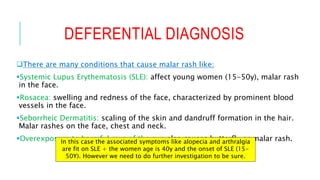
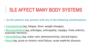
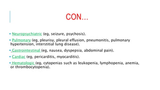
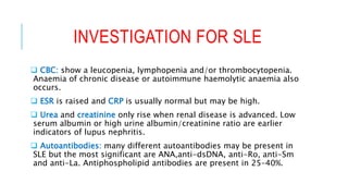
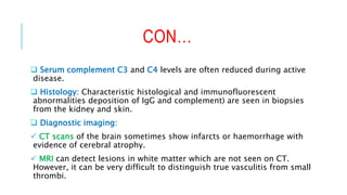
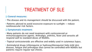
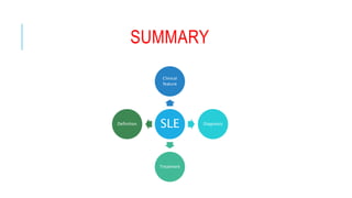
VIGNETTE B
Done by:
Arwa H. Al-
Onayzan.
ID: 215007943.
OUR CASE
 A 40 year-old women.
 Presented with a butterfly rash on her face.
 She does not use any medication.
 Other symptoms include arthralgia, alopecia and fatigue.
 However, there is no fever.
LEARNING OBJECTIVES
1. Which diagnosis is the most likely in view of the clinical
presentation?
2. Describe the extent to which other organs can be involved in this
disorder?
3. Which laboratory test would you order to establish the diagnosis in
this case, and to mentor the involvement of other organs than the
skin and joint?
4. What initial therapeutic measures do you advice?
5. Suppose there is no involvement of other organs initially. What
advice would you give the patient regarding check-up visits?
WHICH DIAGNOSIS IS THE MOST
LIKELY IN VIEW OF THE CLINICAL
PRESENTATION?
DEFERENTIAL DIAGNOSIS
There are many conditions that cause malar rash like:
Systemic Lupus Erythematosis (SLE): affect young women (15-50y), malar rash
in the face.
Rosacea: swelling and redness of the face, characterized by prominent blood
vessels in the face.
Seborrheic Dermatitis: scaling of the skin and dandruff formation in the hair.
Malar rashes on the face, chest and neck.
Overexposure to harmful rays of the sun also causes butterfly or malar rash.In this case the associated symptoms like alopecia and arthralgia
are fit on SLE + the women age is 40y and the onset of SLE (15-
50Y). However we need to do further investigation to be sure.
SYSTEMIC LUPUS ERYTHEMATOSIS
(SLE)
 Is Inflammatory autoimmune disorder that can affect multiple
systems like brain, heart, lungs, liver, kidneys, blood vessels, several
joints, the overall nervous system and the skin.
 Etiology:
 Unknown. However, some etiological factors may trigger
autoimmune response to a variety of tissue components. For
example:
 Sex hormone (mostly in female>>estrogen).
 UVR (in induce damage to DNA << enhance autoimmune).
 Drugs induce SLE (e.x hydrazine and procainamide).
DESCRIBE THE EXTENT TO WHICH
OTHER ORGANS CAN BE INVOLVED IN
THIS DISORDER?
SLE AFFECT MANY BODY SYSTEMS
 So the patients may present with any of the following manifestations :
 Constitutional (eg, fatigue, fever, weight changes).
 Musculoskeletal (eg, arthralgia, arthropathy, myalgia, frank arthritis,
avascular necrosis).
 Dermatologic (eg, malar rash, photosensitivity, discoid lupus).
 Renal (eg, acute or chronic renal failure, acute nephritic disease).
CON…
 Neuropsychiatric (eg, seizure, psychosis).
 Pulmonary (eg, pleurisy, pleural effusion, pneumonitis, pulmonary
hypertension, interstitial lung disease).
 Gastrointestinal (eg, nausea, dyspepsia, abdominal pain).
 Cardiac (eg, pericarditis, myocarditis).
 Hematologic (eg, cytopenias such as leukopenia, lymphopenia, anemia,
or thrombocytopenia).
WHICH LABORATORY TEST WOULD
YOU ORDER TO ESTABLISH THE
DIAGNOSIS IN THIS CASE, AND TO
MENTOR THE INVOLVEMENT OF OTHER
ORGANS THAN THE SKIN AND JOINT?
INVESTIGATION FOR SLE
 CBC: show a leucopenia, lymphopenia and/or thrombocytopenia.
Anaemia of chronic disease or autoimmune haemolytic anaemia also
occurs.
 ESR is raised and CRP is usually normal but may be high.
 Urea and creatinine only rise when renal disease is advanced. Low
serum albumin or high urine albumin/creatinine ratio are earlier
indicators of lupus nephritis.
 Autoantibodies: many different autoantibodies may be present in
SLE but the most significant are ANA,anti-dsDNA, anti-Ro, anti-Sm
and anti-La. Antiphospholipid antibodies are present in 25–40%.
CON…
 Serum complement C3 and C4 levels are often reduced during active
disease.
 Histology: Characteristic histological and immunofluorescent
abnormalities deposition of IgG and complement) are seen in biopsies
from the kidney and skin.
 Diagnostic imaging:
 CT scans of the brain sometimes show infarcts or haemorrhage with
evidence of cerebral atrophy.
 MRI can detect lesions in white matter which are not seen on CT.
However, it can be very difficult to distinguish true vasculitis from small
thrombi.
WHAT INITIAL THERAPEUTIC MEASURES
DO YOU ADVICE?
TREATMENT OF SLE
 General measures:
• The disease and its management should be discussed with the patient.
• Patients advised to avoid excessive exposure to sunlight + reduce
cardiovascular risk factors.
 Symptomatic treatment:
• Many patients do not need treatment with corticosteroid or
immunosuppressive agents. Arthralgia, arthritis, fever and serositis all
respond well to standard doses of NSAIDs.
• Topical corticosteroids are effective and widely used in cutaneous lupus.
• Antimalarial drugs (chloroquine or hydroxychloroquine) help mild skin
disease, fatigue and arthralgias that cannot be controlled with NSAIDs but
patients require regular eye checks.
CON…
 Corticosteroids and immunosuppressive drugs:
• Short courses of oral corticosteroids are useful in treating severe conditions.
• Renal or cerebral disease must be treated with high dose oral corticosteroids.
• Cyclophosphamide was most commonly used to achieve remission in severe
forms of lupus but is being replaced by mycophenolate mofetil, which has fewer
side-effects.
• Newer agents, which target cells or cytokines in the immune system, these
include:
• Rituximab(anti-CD20) and belimumab, both monoclonal antibodies acting
against B lymphocytes.
SUPPOSE THERE IS NO INVOLVEMENT OF
OTHER ORGANS INITIALLY. WHAT ADVICE
WOULD YOU GIVE THE PATIENT
REGARDING CHECK-UP VISITS?
CON…
 The disease and its management will be discussed with the patient.
 Particularly the effect upon the patient’s lifestyle e.g: debility due to
fatigue.
 Patients are advised to: Avoid excessive exposure to sunlight.
 Periodic follow up and blood tests are required for:
1. Detecting signs and symptoms of new organ-system involvement
in the patient.
2. Monitoring response and adverse reactions to therapies. (steroid
side effects).
Usual protocol is visits are arranged every 3
months (quarterly visits) at least 4 visits every
year.
SUMMARY
SLE
Clinical
feature
Diagnosis
Treatment
Definition
REFERENCE
 Kumar and Clark’s Clinical medicine, eight edition, page (536-
537).
 Shahedi, SLEWorkup: Laboratory Studies, Imaging Studies,
Procedures. [online] Emedicine.medscape.com.
https://www.hxbenefit.com/malar-rash-butterfly-rash-pictures-causes-
and-treatment.html

Systemic lupus erythematosus (SLE)

  • 1.
    VIGNETTE B Done by: ArwaH. Al- Onayzan. ID: 215007943.
  • 2.
    OUR CASE  A40 year-old women.  Presented with a butterfly rash on her face.  She does not use any medication.  Other symptoms include arthralgia, alopecia and fatigue.  However, there is no fever.
  • 3.
    LEARNING OBJECTIVES 1. Whichdiagnosis is the most likely in view of the clinical presentation? 2. Describe the extent to which other organs can be involved in this disorder? 3. Which laboratory test would you order to establish the diagnosis in this case, and to mentor the involvement of other organs than the skin and joint? 4. What initial therapeutic measures do you advice? 5. Suppose there is no involvement of other organs initially. What advice would you give the patient regarding check-up visits?
  • 4.
    WHICH DIAGNOSIS ISTHE MOST LIKELY IN VIEW OF THE CLINICAL PRESENTATION?
  • 5.
    DEFERENTIAL DIAGNOSIS There aremany conditions that cause malar rash like: Systemic Lupus Erythematosis (SLE): affect young women (15-50y), malar rash in the face. Rosacea: swelling and redness of the face, characterized by prominent blood vessels in the face. Seborrheic Dermatitis: scaling of the skin and dandruff formation in the hair. Malar rashes on the face, chest and neck. Overexposure to harmful rays of the sun also causes butterfly or malar rash.In this case the associated symptoms like alopecia and arthralgia are fit on SLE + the women age is 40y and the onset of SLE (15- 50Y). However we need to do further investigation to be sure.
  • 6.
    SYSTEMIC LUPUS ERYTHEMATOSIS (SLE) Is Inflammatory autoimmune disorder that can affect multiple systems like brain, heart, lungs, liver, kidneys, blood vessels, several joints, the overall nervous system and the skin.  Etiology:  Unknown. However, some etiological factors may trigger autoimmune response to a variety of tissue components. For example:  Sex hormone (mostly in female>>estrogen).  UVR (in induce damage to DNA << enhance autoimmune).  Drugs induce SLE (e.x hydrazine and procainamide).
  • 7.
    DESCRIBE THE EXTENTTO WHICH OTHER ORGANS CAN BE INVOLVED IN THIS DISORDER?
  • 8.
    SLE AFFECT MANYBODY SYSTEMS  So the patients may present with any of the following manifestations :  Constitutional (eg, fatigue, fever, weight changes).  Musculoskeletal (eg, arthralgia, arthropathy, myalgia, frank arthritis, avascular necrosis).  Dermatologic (eg, malar rash, photosensitivity, discoid lupus).  Renal (eg, acute or chronic renal failure, acute nephritic disease).
  • 9.
    CON…  Neuropsychiatric (eg,seizure, psychosis).  Pulmonary (eg, pleurisy, pleural effusion, pneumonitis, pulmonary hypertension, interstitial lung disease).  Gastrointestinal (eg, nausea, dyspepsia, abdominal pain).  Cardiac (eg, pericarditis, myocarditis).  Hematologic (eg, cytopenias such as leukopenia, lymphopenia, anemia, or thrombocytopenia).
  • 10.
    WHICH LABORATORY TESTWOULD YOU ORDER TO ESTABLISH THE DIAGNOSIS IN THIS CASE, AND TO MENTOR THE INVOLVEMENT OF OTHER ORGANS THAN THE SKIN AND JOINT?
  • 11.
    INVESTIGATION FOR SLE CBC: show a leucopenia, lymphopenia and/or thrombocytopenia. Anaemia of chronic disease or autoimmune haemolytic anaemia also occurs.  ESR is raised and CRP is usually normal but may be high.  Urea and creatinine only rise when renal disease is advanced. Low serum albumin or high urine albumin/creatinine ratio are earlier indicators of lupus nephritis.  Autoantibodies: many different autoantibodies may be present in SLE but the most significant are ANA,anti-dsDNA, anti-Ro, anti-Sm and anti-La. Antiphospholipid antibodies are present in 25–40%.
  • 12.
    CON…  Serum complementC3 and C4 levels are often reduced during active disease.  Histology: Characteristic histological and immunofluorescent abnormalities deposition of IgG and complement) are seen in biopsies from the kidney and skin.  Diagnostic imaging:  CT scans of the brain sometimes show infarcts or haemorrhage with evidence of cerebral atrophy.  MRI can detect lesions in white matter which are not seen on CT. However, it can be very difficult to distinguish true vasculitis from small thrombi.
  • 13.
    WHAT INITIAL THERAPEUTICMEASURES DO YOU ADVICE?
  • 14.
    TREATMENT OF SLE General measures: • The disease and its management should be discussed with the patient. • Patients advised to avoid excessive exposure to sunlight + reduce cardiovascular risk factors.  Symptomatic treatment: • Many patients do not need treatment with corticosteroid or immunosuppressive agents. Arthralgia, arthritis, fever and serositis all respond well to standard doses of NSAIDs. • Topical corticosteroids are effective and widely used in cutaneous lupus. • Antimalarial drugs (chloroquine or hydroxychloroquine) help mild skin disease, fatigue and arthralgias that cannot be controlled with NSAIDs but patients require regular eye checks.
  • 15.
    CON…  Corticosteroids andimmunosuppressive drugs: • Short courses of oral corticosteroids are useful in treating severe conditions. • Renal or cerebral disease must be treated with high dose oral corticosteroids. • Cyclophosphamide was most commonly used to achieve remission in severe forms of lupus but is being replaced by mycophenolate mofetil, which has fewer side-effects. • Newer agents, which target cells or cytokines in the immune system, these include: • Rituximab(anti-CD20) and belimumab, both monoclonal antibodies acting against B lymphocytes.
  • 16.
    SUPPOSE THERE ISNO INVOLVEMENT OF OTHER ORGANS INITIALLY. WHAT ADVICE WOULD YOU GIVE THE PATIENT REGARDING CHECK-UP VISITS?
  • 17.
    CON…  The diseaseand its management will be discussed with the patient.  Particularly the effect upon the patient’s lifestyle e.g: debility due to fatigue.  Patients are advised to: Avoid excessive exposure to sunlight.  Periodic follow up and blood tests are required for: 1. Detecting signs and symptoms of new organ-system involvement in the patient. 2. Monitoring response and adverse reactions to therapies. (steroid side effects). Usual protocol is visits are arranged every 3 months (quarterly visits) at least 4 visits every year.
  • 18.
  • 20.
    REFERENCE  Kumar andClark’s Clinical medicine, eight edition, page (536- 537).  Shahedi, SLEWorkup: Laboratory Studies, Imaging Studies, Procedures. [online] Emedicine.medscape.com. https://www.hxbenefit.com/malar-rash-butterfly-rash-pictures-causes- and-treatment.html