The document provides information on aortic valve disease including anatomy, etiology, and pathophysiology. It describes the key components of the aortic root including the aortic annulus, cusps, sinuses, and sinotubular junction. The three main causes of aortic stenosis are discussed as congenital bicuspid valve with calcification, calcification of a normal trileaflet valve, and rheumatic disease. The pathophysiology of aortic stenosis involves left ventricular pressure overload leading to hypertrophy and eventually decreased ejection fraction if severe stenosis is not corrected.
Kindly leave your comment if you found this helpful ;)
Some of the slides, i hide it from my real presentations for my own reference. Download to see all of them.
Kindly leave your comment if you found this helpful ;)
Some of the slides, i hide it from my real presentations for my own reference. Download to see all of them.
Ozempic: Preoperative Management of Patients on GLP-1 Receptor Agonists Saeid Safari
Preoperative Management of Patients on GLP-1 Receptor Agonists like Ozempic and Semiglutide
ASA GUIDELINE
NYSORA Guideline
2 Case Reports of Gastric Ultrasound
Factory Supply Best Quality Pmk Oil CAS 28578–16–7 PMK Powder in Stockrebeccabio
Factory Supply Best Quality Pmk Oil CAS 28578–16–7 PMK Powder in Stock
Telegram: bmksupplier
signal: +85264872720
threema: TUD4A6YC
You can contact me on Telegram or Threema
Communicate promptly and reply
Free of customs clearance, Double Clearance 100% pass delivery to USA, Canada, Spain, Germany, Netherland, Poland, Italy, Sweden, UK, Czech Republic, Australia, Mexico, Russia, Ukraine, Kazakhstan.Door to door service
Hot Selling Organic intermediates
Flu Vaccine Alert in Bangalore Karnatakaaddon Scans
As flu season approaches, health officials in Bangalore, Karnataka, are urging residents to get their flu vaccinations. The seasonal flu, while common, can lead to severe health complications, particularly for vulnerable populations such as young children, the elderly, and those with underlying health conditions.
Dr. Vidisha Kumari, a leading epidemiologist in Bangalore, emphasizes the importance of getting vaccinated. "The flu vaccine is our best defense against the influenza virus. It not only protects individuals but also helps prevent the spread of the virus in our communities," he says.
This year, the flu season is expected to coincide with a potential increase in other respiratory illnesses. The Karnataka Health Department has launched an awareness campaign highlighting the significance of flu vaccinations. They have set up multiple vaccination centers across Bangalore, making it convenient for residents to receive their shots.
To encourage widespread vaccination, the government is also collaborating with local schools, workplaces, and community centers to facilitate vaccination drives. Special attention is being given to ensuring that the vaccine is accessible to all, including marginalized communities who may have limited access to healthcare.
Residents are reminded that the flu vaccine is safe and effective. Common side effects are mild and may include soreness at the injection site, mild fever, or muscle aches. These side effects are generally short-lived and far less severe than the flu itself.
Healthcare providers are also stressing the importance of continuing COVID-19 precautions. Wearing masks, practicing good hand hygiene, and maintaining social distancing are still crucial, especially in crowded places.
Protect yourself and your loved ones by getting vaccinated. Together, we can help keep Bangalore healthy and safe this flu season. For more information on vaccination centers and schedules, residents can visit the Karnataka Health Department’s official website or follow their social media pages.
Stay informed, stay safe, and get your flu shot today!
Prix Galien International 2024 Forum ProgramLevi Shapiro
June 20, 2024, Prix Galien International and Jerusalem Ethics Forum in ROME. Detailed agenda including panels:
- ADVANCES IN CARDIOLOGY: A NEW PARADIGM IS COMING
- WOMEN’S HEALTH: FERTILITY PRESERVATION
- WHAT’S NEW IN THE TREATMENT OF INFECTIOUS,
ONCOLOGICAL AND INFLAMMATORY SKIN DISEASES?
- ARTIFICIAL INTELLIGENCE AND ETHICS
- GENE THERAPY
- BEYOND BORDERS: GLOBAL INITIATIVES FOR DEMOCRATIZING LIFE SCIENCE TECHNOLOGIES AND PROMOTING ACCESS TO HEALTHCARE
- ETHICAL CHALLENGES IN LIFE SCIENCES
- Prix Galien International Awards Ceremony
Tom Selleck Health: A Comprehensive Look at the Iconic Actor’s Wellness Journeygreendigital
Tom Selleck, an enduring figure in Hollywood. has captivated audiences for decades with his rugged charm, iconic moustache. and memorable roles in television and film. From his breakout role as Thomas Magnum in Magnum P.I. to his current portrayal of Frank Reagan in Blue Bloods. Selleck's career has spanned over 50 years. But beyond his professional achievements. fans have often been curious about Tom Selleck Health. especially as he has aged in the public eye.
Follow us on: Pinterest
Introduction
Many have been interested in Tom Selleck health. not only because of his enduring presence on screen but also because of the challenges. and lifestyle choices he has faced and made over the years. This article delves into the various aspects of Tom Selleck health. exploring his fitness regimen, diet, mental health. and the challenges he has encountered as he ages. We'll look at how he maintains his well-being. the health issues he has faced, and his approach to ageing .
Early Life and Career
Childhood and Athletic Beginnings
Tom Selleck was born on January 29, 1945, in Detroit, Michigan, and grew up in Sherman Oaks, California. From an early age, he was involved in sports, particularly basketball. which played a significant role in his physical development. His athletic pursuits continued into college. where he attended the University of Southern California (USC) on a basketball scholarship. This early involvement in sports laid a strong foundation for his physical health and disciplined lifestyle.
Transition to Acting
Selleck's transition from an athlete to an actor came with its physical demands. His first significant role in "Magnum P.I." required him to perform various stunts and maintain a fit appearance. This role, which he played from 1980 to 1988. necessitated a rigorous fitness routine to meet the show's demands. setting the stage for his long-term commitment to health and wellness.
Fitness Regimen
Workout Routine
Tom Selleck health and fitness regimen has evolved. adapting to his changing roles and age. During his "Magnum, P.I." days. Selleck's workouts were intense and focused on building and maintaining muscle mass. His routine included weightlifting, cardiovascular exercises. and specific training for the stunts he performed on the show.
Selleck adjusted his fitness routine as he aged to suit his body's needs. Today, his workouts focus on maintaining flexibility, strength, and cardiovascular health. He incorporates low-impact exercises such as swimming, walking, and light weightlifting. This balanced approach helps him stay fit without putting undue strain on his joints and muscles.
Importance of Flexibility and Mobility
In recent years, Selleck has emphasized the importance of flexibility and mobility in his fitness regimen. Understanding the natural decline in muscle mass and joint flexibility with age. he includes stretching and yoga in his routine. These practices help prevent injuries, improve posture, and maintain mobilit
Explore natural remedies for syphilis treatment in Singapore. Discover alternative therapies, herbal remedies, and lifestyle changes that may complement conventional treatments. Learn about holistic approaches to managing syphilis symptoms and supporting overall health.
These lecture slides, by Dr Sidra Arshad, offer a quick overview of physiological basis of a normal electrocardiogram.
Learning objectives:
1. Define an electrocardiogram (ECG) and electrocardiography
2. Describe how dipoles generated by the heart produce the waveforms of the ECG
3. Describe the components of a normal electrocardiogram of a typical bipolar leads (limb II)
4. Differentiate between intervals and segments
5. Enlist some common indications for obtaining an ECG
Study Resources:
1. Chapter 11, Guyton and Hall Textbook of Medical Physiology, 14th edition
2. Chapter 9, Human Physiology - From Cells to Systems, Lauralee Sherwood, 9th edition
3. Chapter 29, Ganong’s Review of Medical Physiology, 26th edition
4. Electrocardiogram, StatPearls - https://www.ncbi.nlm.nih.gov/books/NBK549803/
5. ECG in Medical Practice by ABM Abdullah, 4th edition
6. ECG Basics, http://www.nataliescasebook.com/tag/e-c-g-basics
TEST BANK for Operations Management, 14th Edition by William J. Stevenson, Ve...kevinkariuki227
TEST BANK for Operations Management, 14th Edition by William J. Stevenson, Verified Chapters 1 - 19, Complete Newest Version.pdf
TEST BANK for Operations Management, 14th Edition by William J. Stevenson, Verified Chapters 1 - 19, Complete Newest Version.pdf
The prostate is an exocrine gland of the male mammalian reproductive system
It is a walnut-sized gland that forms part of the male reproductive system and is located in front of the rectum and just below the urinary bladder
Function is to store and secrete a clear, slightly alkaline fluid that constitutes 10-30% of the volume of the seminal fluid that along with the spermatozoa, constitutes semen
A healthy human prostate measures (4cm-vertical, by 3cm-horizontal, 2cm ant-post ).
It surrounds the urethra just below the urinary bladder. It has anterior, median, posterior and two lateral lobes
It’s work is regulated by androgens which are responsible for male sex characteristics
Generalised disease of the prostate due to hormonal derangement which leads to non malignant enlargement of the gland (increase in the number of epithelial cells and stromal tissue)to cause compression of the urethra leading to symptoms (LUTS
MANAGEMENT OF ATRIOVENTRICULAR CONDUCTION BLOCK.pdfJim Jacob Roy
Cardiac conduction defects can occur due to various causes.
Atrioventricular conduction blocks ( AV blocks ) are classified into 3 types.
This document describes the acute management of AV block.
Ethanol (CH3CH2OH), or beverage alcohol, is a two-carbon alcohol
that is rapidly distributed in the body and brain. Ethanol alters many
neurochemical systems and has rewarding and addictive properties. It
is the oldest recreational drug and likely contributes to more morbidity,
mortality, and public health costs than all illicit drugs combined. The
5th edition of the Diagnostic and Statistical Manual of Mental Disorders
(DSM-5) integrates alcohol abuse and alcohol dependence into a single
disorder called alcohol use disorder (AUD), with mild, moderate,
and severe subclassifications (American Psychiatric Association, 2013).
In the DSM-5, all types of substance abuse and dependence have been
combined into a single substance use disorder (SUD) on a continuum
from mild to severe. A diagnosis of AUD requires that at least two of
the 11 DSM-5 behaviors be present within a 12-month period (mild
AUD: 2–3 criteria; moderate AUD: 4–5 criteria; severe AUD: 6–11 criteria).
The four main behavioral effects of AUD are impaired control over
drinking, negative social consequences, risky use, and altered physiological
effects (tolerance, withdrawal). This chapter presents an overview
of the prevalence and harmful consequences of AUD in the U.S.,
the systemic nature of the disease, neurocircuitry and stages of AUD,
comorbidities, fetal alcohol spectrum disorders, genetic risk factors, and
pharmacotherapies for AUD.
Ocular injury ppt Upendra pal optometrist upums saifai etawah
Aortic valve disease
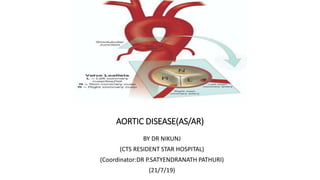
1. AORTIC DISEASE(AS/AR)
BY DR NIKUNJ
(CTS RESIDENT STAR HOSPITAL)
(Coordinator:DR P.SATYENDRANATH PATHURI)
(21/7/19)
2. ANATOMY
• The aortic valve is the last of four cardiac valves through which the blood is pumped before it goes to the
rest of the body.
• It separates the left ventricular outflow tract from the aorta.
• Its main function is to prevent backward blood flow from the aorta to the left ventricle, while allowing the
blood to flow forward during systole with minimal resistance.
3.
4. AORTIC ROOT
• The aortic root is the anatomic segment
between the left ventricle and the
ascending aorta. It contains the aortic valve
and other anatomic elements, which
function as a unit. The aortic root has
several anatomic components:
• subcommissural triangles,
• aortic annulus,
• aortic cusps,
• aortic sinuses or sinuses of Valsalva,
• sinotubular junction.
5.
6. THE SUBCOMMISSURAL TRIANGLES
• The subcommissural triangles are
part of the left ventricular out fow
tract,
• The subcommissural triangles of
the noncoronary aortic cusp are
fibrous extension of the
intervalvular fibrous body and
membranous septum, whereas the
subcommissural triangle beneath
the left and the right aortic cusps is
an extension of the muscular
interventricular septum.
7. THE AORTIC ANNULUS
• The aortic annulus, a fibrous structure with a scalloped shape, attaches the aortic
valve to the left ventricle.
• It is attached directly to the myocardium in approximately 45% of its circumference,
and to fibrous structures in the remaining 55%
• The diameter of the aortic annulus is 10% to 20% larger than the diameter of the
sinotubular junction of the aortic root in young patients . As the number of elastic
fibers in the arterial wall decreases with age, the sinotubular junction dilates, and
its diameter tends to become equal to that of the aortic annulus in older patients.
8. • With dilation of the aortic annulus, the subcommissural triangles of the noncoronary cusp tend to become
more obtuse as the crescent shape of the aortic annulus along its fibrous insertion flattens.
9.
10.
11. CUSPS
• The normal aortic valve has three cusps. Each cusp has a semilunar shape and has a
base and a free margin. The base is attached to the aortic annulus in a crescent
fashion. The point at which the free margin of a cusp joins its base is the
commissure, and
• the ridge in the aortic wall that lies immediately above the commissures is the
sinotubular junction.
• At the mid point of each free edge is fibrou nodulus arantii on either side of
nodulus is extremely thin.
• The free margin of an aortic cusp extends from one of its commissures to the other.
The length of the free margin of an aortic cusp is approximately 1.5 times the length
of its base.
12. CUSPS
• The three aortic cusps often have different sizes in
a person, and the right and noncoronary cusps are
usually larger than the left cusp.
• The same cusp may have different sizes in
individuals with the same body surface area
• During diastole, the free margins and part of the
body of the three cusps touch each other
approximately in the center of the aortic root to
seal the aortic orifice.
• Thus, the average length of the free margins of
three aortic cusps must exceed the diameter of
the sinotubular junction to allow the cusps to
coapt centrally and render the aortic valve
competent .
13. • The aortic leaflets display three layers of connective tissue: the ventriculosa on the ventricular side,
the fibrosa on the aortic side, and the spongiosa between them
• The fibrosa is mainly composed of collagen fibers while the ventriculosa mostly consists of elastic
fibers.
• Between these two layers, the spongiosa is composed of a mucopolysaccharide gel-like substance
that facilitates the motion of the ventriculosa and fibrosa.
14. • If a pathologic process causes shortening of the length of the free
margin of a cusp, or if the sinotubular junction dilates, the cusps cannot
coapt centrally, resulting in aortic insufciency .
• If the length of a free margin is elongated, the cusp prolapses, and
depending on the degree of prolapse, aortic insufficiency
15.
16. AORTIC SINUSES, OR SINUSES OF VALSALVA
• The spaces contained between the aortic annulus
and the sinotubular junction are the aortic
sinuses. There are three cusps and three sinuses:
• left cusp and sinus,
• right cusp and sinus,
• noncoronary cusp and sinus.
• The left main coronary artery arises from the left
aortic sinus, and the right coronary artery arises
from the right aortic sinus.
• There are three sinuses of the aortic valve, each
related to the valve’s corresponding cusps. Each
sinus is divided into three areas a central part and
two adjacent parts, which are named according
to the valve cusps they adjoin.
• The noncoronary sinus is also refferred to as the
posterior aortic sinus.
17. AORTIC SINUSES, OR SINUSES OF VALSALVA
• The aortic sinuses facilitate closure of the aortic valve by creating eddies and currents between the cusps
and arterial wall .
• They also prevent the cusps from occluding the coronary artery orifices during systole, thus guaranteeing
myocardial perfusion during the entire cardiac cycle.
18. RIGHT CORONARY SINUS
• entire right coronary sinus lies adjacent to
the RVOT.
• central part lies adjacent to the crista
supraventricularis,
• left part is adjacent to the area of the
RVOT in the angle between the crista
supraventricularis and the pulmonary
valve.
• posterior (noncoronary) part of the right
coronary sinus is related to the area of the
right ventricle posteroinferior to the crista
supraventricularis.
• Inferiorly, the entire right coronary sinus
is related to the interventricular septum;
the muscular septum lies under the
central and left parts, while either
membranous or muscular septum may lie
under the posterior part of the right
coronary sinus.
19. NONCORONARY SINUS
• The atrialchambers with the intervening
atrial septum lie adjacent to the
noncoronary sinus.
• right and central parts of the
noncoronary sinus are related to the
right atrium and the interatrial septum,
• left part is related to the left atrium.
• Inferiorly, the right part, like the
posterior part of the right coronary
sinus, may be related either to the
membranous or the muscular septum
depending on the size of the
membranous septum. However,
beneath the central part of the
noncoronary sinus, the membranous
septum is a constant structure. The left
part of the noncoronary sinus inserts
into the anterior mitral leaflet
20. LEFT CORONARY SINUS
• posterior part of the left coronary
• it is related to the left atrium
posteriorly and to the anterior mitral
leaflet inferiorly.
• central part of the left aortic sinus is
the only part of the aortic root that is
not related to a cardiac chamber; it is
adjacent to the epicardium only.
• right part of the left coronary sinus lies
adjacent to the pulmonary trunk at the
level of the left pulmonary sinus.
inferior to it lies the muscular
interventricular septum.
21. • The aortic root of young individuals is elastic and very compliant. It expands and contracts during the
cardiac cycle.
• The normal aortic root has a fairly consistent shape, and the sizes of the cusps, the aortic annulus, the aortic
sinuses, and the sinotubular junction are somewhat interdependent.
• Thus, large cusps have a proportionally large annulus, sinus, and sinotubular junction.
22.
23. AORTIC STENOSIS Epidemiology and Etiology
• Isolated aortic stenosis (AS) is more common in men than in women and is found in 2% of people 65 years of
age and older.
• The most common causes of AS include agerelated calcific degeneration, bicuspid aortic valve, and
rheumatic aortic valve.
• The distribution of these causes varies across age groups and geographic regions.
• Age related degeneration is the most common cause of AS in patients older than 70 years.
• In contrast, bicuspid aortic valve calcification accounts for most surgical cases in patients younger than 70
years.
24. • Valvular AS has three principle causes
1. Congenital bicuspid valve with superimposed calcification
2. Calcification of normal trileaflet valve
3. Rhumatic disease.
25. • Fixed obstruction to LVOT may occur above the valve(supravalvular stenosis)
• Below the valve (subvalvular stenosis)
26. CONGENITAL BICUSPID VALVE WITH SUPERIMPOSED CALCIFICATION
• Congenital malformation can be
• Unicuspid: infancy
• Bicuspid :
• Ttricuspid :
• Domeshaped diaphragm:
27. CALCIFIC AORTIC VALVE DISEASE
• Calcific aortic valve disease affecting a congenital bicuspid or normal trileaflet valve is the most commone
cause of AS in adults
28.
29. RHEUMATIC AORTIC STENOSIS
• It Results from adhesions and fusion of the comissures and cusps and vasclarazation of the leaflets of the
valve ring
• Leading to retraction and stiffening of the free borders of the cusps
• Calcific nodules deveope on bothe surface and orifice is reduced to small round or triangular opening.
• It can be regurgitant as well as stenotic.
30.
31. PATHOPHYSIOLOGY
• No appreciable gradient exists across the normal aortic valve during systole.
• In AS, gradual obstruction of the left ventricular outflow leads to an increased left ventricular afterload
and left ventricular wall stress, elevated left ventricular systolic and diastolic pressures, decreased
aortic pressure, and prolonged left ventricular ejection time.
• Over time, this results in compensatory concentric left ventricular hypertrophy (LVH) to maintain
ejection fraction.
• In patients with chronic severe AS, this compensatory mechanism may become insufficient, leading to
gradual dilation and thinning of the left ventricle, and result in a decrease in ejection fraction and in
congestive heart failure.
32. PATHOPHYSIOLOGY
• Myocardial oxygen supply and demand is also altered in AS.
• LVH, increased systolic pressure, and prolonged ejection time result in increased myocardial oxygen
demand.
• Increased diastolic pressure and prolonged systolic ejection time result in decreased diastolic and myocardial
perfusion time and hence myocardial oxygen supply.
• The alteration in myocardial oxygen supply and demand is the underlying mechanism behind myocardial
ischemia in patients with AS, even in the absence of coronary artery disease.
33. SYMPTOMS
• The classic symptoms of AS are angina, exertional syncope, and symptoms of congestive heart failure such as
shortness of breath.
• Angina: frequent symptom of pt with svere AS
• Syncope: reduced cerebral perfusion.
• GI Bleeding
34. PHYSICAL EXAMINATION
• palpation of carotid upstroke(Slow rising late peaking,low amplitude)
• Evaluation of systolic murmur
• Assessment of splitting of second heart sound
• Signs of HF
• crescendo-decrescendo systolic ejection murmur heard best at the second right intercostal space, which may
variably radiate to the carotid arteries.
• A palpable thrill may be present in severe cases of AS.
• Palpation of the arterial pulse may reveal a weak and delayed pulse known as pulsus parvus et tardus.
35. DIAGNOSIS AND GRADING
• Two-dimensional transthoracic echocardiography is the most common modality for the diagnosis and
grading of AS
36. • X ray chest
• EKG
• CARDIAC CT
• CARDIAC CATHETERIZATION
• TMT
37. NATURAL HISTORY
• Without intervention,
• Multiple studies consistently reported survivals of 3 years for angina and syncope
• 1.5 to 2 years for dyspnea and heart failure.
• These findings have driven recommendations for early surgical intervention in patients with symptomatic AS.
• One third of asymptomatic patients with severe AS will become symptomatic in 2 years with an estimated
cardiac death of less than 1% each year to 5% each year.
• Patients with higher grades of AS severity seem to progress at a faster rate than lower grades of AS.
• After AS becomes moderate, aortic valve area decreases on average by 0.1 cm2 per year, the pressure
gradient across the valve rises on average by 7 mm Hg per year, and the jet velocity increases by 0.3 m/sec
per year.
38. AORTIC REGURGITATION
• disturbance in any of the components of the functional unit of the aortic valve (e.g., cusps, sinuses of
Valsalva, sinotubular junction, ventriculoaortic junction).
• In general, the causes can be divided into
• affect the valve cusps (e.g., calcific degeneration, congenitally bicuspid valve, infective endocarditis,
rheumatic disease, myxomatous degeneration)
• affect the aortic root (e.g., aortic dissection, aortitis of various etiologies such as syphilis, connective tissue
disorders such as Marfan syndrome)
Primary Valve disease Primary Root disease
Congenital (bicuspid aortic valve)
Rheumatic fever
Infective endocarditis ,Collagen
vascular diseases ,Degenerative aortic
valve disease
Myxomatous (prolapse) Traumatic
Longstanding, uncontrolled hypertension
Marfan syndrome ,Idiopathic aortic dilation
Cystic medial necrosis, Senile aortic ectasia
and dilation
Aortic dissection, Marfan's syndrome
Syphilitic aortitis Giant cell arteritis,
Takayasu arteritis, Ankylosing spondylitis,
Whipple disease
39.
40. THE PATHOPHYSIOLOGY OF AR
• dependent on the acuity of onset and duration of the disease process.
• ACUTE AR, typically caused by aortic dissection, infective endocarditis, trauma, or valve prosthesis failure,
there is a sudden increase in left ventricular end-diastolic volume because of the regurgitation.
• Since the left ventricle has limited compliance and does not have time to adapt, the left ventricular end-
diastolic pressure (LVEDP) rises rapidly
41. • CHRONIC AR, there is a slow and insidious progression of left ventricular (LV) dilation and eccentric
hypertrophy because of an increase in left ventricular end-diastolic volume, LVEDP, and wall stress.
• Dilation of the LV, while maintaining normal systolic function, increases total stroke volume and maintains
forward flow.
• This increase in stroke volume coupled with an increase in LVEDP is associated with the wide pulse pressure
typical of chronic AR.
• Eventually, the hypertrophic response is exhausted, and LVEF drops as afterload increases, leading to heart
failure and its associated clinical presentation.
42.
43.
44. Valve Dysfunctions
Valve Dysfunctions
corresponding Lesions
Lesions
Type I: Normal leaflet motion Annular dilatation
Leaflet perforation
Vegetation
Type II: Leaflet prolapse Leaflet rupture, distension
Commissure detachment
Type IlIa: Restricted leaflet closure and opening Leaflet thickening Commissure fusion Calcification
Type Illb: Restricted leaflet closure only Sino-tubular dilatation
45. CLINICAL FEATURES
• ACUTE AR :usually exhibit sudden or rapidly progressive cardiovascular collapse, which can be life
threatening if not addressed promptly.
• They often exhibit ischemic symptoms because of the decrease in coronary blood flow and increased
myocardial oxygen demand.
• CHRONIC AR : asymptomatic for a long period of time because of the compensatory LV changes.
• Once the compensatory response is exhausted, the patients start having heart failure symptoms
1. PALPITATION - early symptom
2. HEAD POUNDING - on exertion
3. EXERTIONAL DYSPNOEA
4. ORTHOPNOEA
5. PAROXYSMAL NOCTURNAL DYSPNOEA
6. EXCESSIVE DIAPHORESIS
7. ANGINA - on exertion/ at rest - nocturnal
8. CCF - late
46. PULSE IN AR
• Corrigan’s pulse or
• Water hammer pulse or
• Collapsing pulse
- Rapid rise and rapid fall
• Bisferiens’s pulse -two peaks in systole
• A pulse that is bounding and forceful, rapidly increasing and subsequently collapsing, it resembles the strike
of waterhammer
47. • S1- may be soft due to premature closure of the mitral valve.
• A2 –normal or accentuated when AR is due to aortic root disease.
• S2- absent or single or exhibit narrow or paradoxical splitting
• S3 GALLOP - due to increased LV end diastolic volume or impaired LV function
• Systolic ejection sound - related to abrupt distention of the aorta by the augmented stroke volume.
48. • CHRONIC AORTIC REGURGITATION
• Early diastolic murmur
• High pitched, blowing decrescendo
Best heard in the 3rd left intercostal space with the patient sitting up and leaning forward breath held in
forced expiration
• Aortic root disorders murmur is best heard along right sternal border musical murmur Longer the duration
of murmur severer the aortic regurgitation
• Becomes short - cardiac failure
• Austin flint murmur
• Soft, low pitched rumbling mid diastolic murmur.
• Diastolic displacement of the anterior leaflet of the mitral valve by the aortic regurgitation stream.
49. • Classic signs of widened pulse pressure may also be found, including
1. Corrigan or water-hammer pulse,
2. De Musset sign (bobbing of the head with heart beats),
3. Quincke pulse (pulsations of the lip and fingers),
4. Traube sign (pistol shot sounds over the femoral artery),
5. Müller sign (pulsations of the uvula).
6. Becker sign - Visible systolic pulsations of the retinal arterioles
7. Light-house sign – Alternate flushing & blanching of forehead
8. Landolfi’s sign - Change in pupil size with each systole
9. Gerhardt’s sign - Visible systolic pulsations of spleen
10. Rosenbach’s sign - Visible systolic pulsations of liver
11. Corrigan’s sign – Dancing carotid in neck
12. Hill sign: Popliteal cuff systolic pressure exceeding brachial cuff systolic pressure by more than 20 mmHg.
50. DIAGNOSTIC CRITERIA
• Transthoracic echocardiography with Doppler colorflow is the most useful tool for the diagnosis of AR.
• The jet width and vena contracta width on Doppler color-flow are used to qualitatively assess the severity of
AR, whereas the regurgitant volume, regurgitant fraction, and regurgitant orifice area are used for the
quantitative assessment.
51.
52.
53.
54. CHEST X RAY
• acute AR, there may be minimal cardiac enlargement, but marked enlargement is a common finding in chronic AR.
55. ECG
•LV hypertrophy
•Left axis deviation
•Left atrial enlargement
•LV volume overload pattern - Prominent Q waves in leads I, aVL, and V3 to V6 and relatively small R waves in
V1
56. • AORTIC ANGIOGRAPHY
1. Mild (1+) - A small amount of contrast enters the LV during diastole and clears with each systole
2. Moderate AR (2+) - Contrast enters the LV with each diastole, but the LV chamber is less dense than the
aorta
3. Moderately severe AR (3+) - The LV chamber is equal in density to the ascending aorta.
4. Severe AR (4+) - Complete, dense opacification of the LV chamber occurs on the first beat, and the LV is
more densely opacified than the ascending aorta
• CARDIAC MRI :CMR provides accurate measurements of regurgitant volumes and the regurgitant orifice in
AR. It is the most accurate noninvasive technique for assessing LV end systolic volume, diastolic volume, and
mass
57. CARDIAC CATHETERIZATION
• Class I indications for cardiac catheterization under current ACC/AHA guidelines:
• Assessment of coronary anatomy prior to aortic valve surgery in patients with risk factors for coronary artery
disease
• Assessment of severity of AR, LV function, or aortic root size when noninvasive tests are inconclusive or are
discordant with clinical findings.